. 2018 Jul 5;8(1):10178.
doi: 10.1038/s41598-018-28471-1.

The frontal skull Hounsfield unit value can predict ventricular enlargement in patients with subarachnoid haemorrhage

Affiliations

The frontal skull Hounsfield unit value can predict ventricular enlargement in patients with subarachnoid haemorrhage

Yu Deok Won et al. Sci Rep. .

Abstract

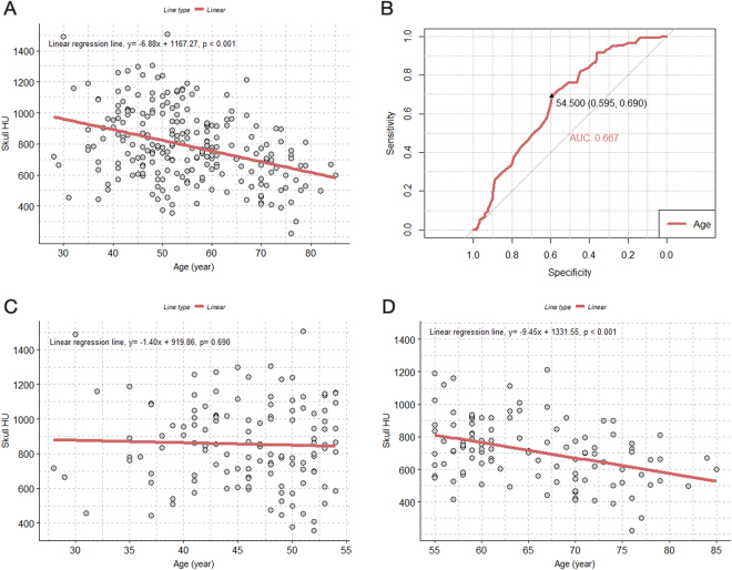
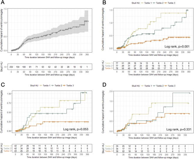
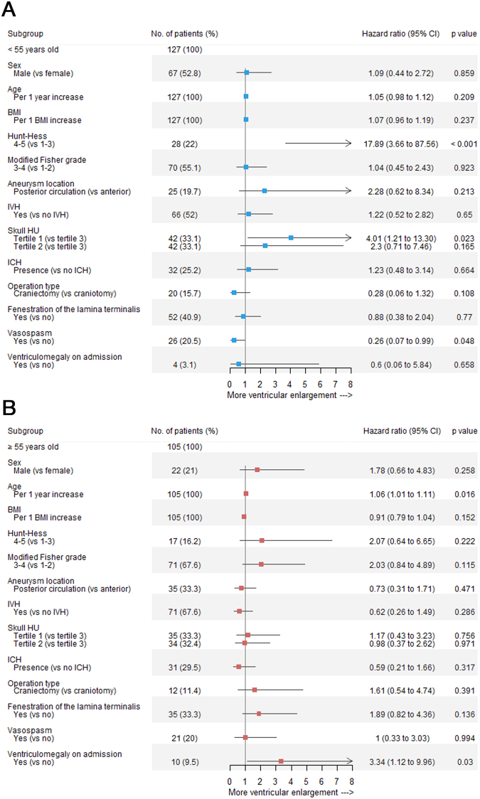
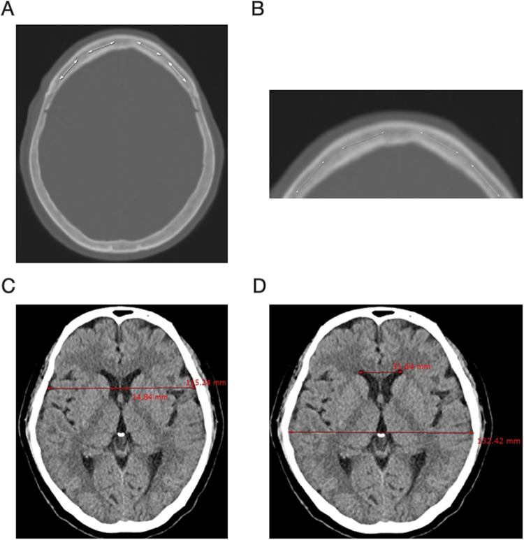
Hydrocephalus is a common complication following subarachnoid haemorrhage (SAH) arising from spontaneous aneurysm rupture. The Hounsfield unit (HU) value from computed tomography scans may reflect bone mineral density, which correlates with body mass index, which in turn is related to post-SAH ventricle size changes. We herein investigated potential associations between frontal skull HU values and ventricle size changes after SAH. HU values from four different areas in the frontal bone were averaged to minimize measurement errors. The bicaudate index and Evans ratio were measured using both baseline and follow-up CT images. CT images with bicaudate index >0.2 and Evans ratio >0.3 simultaneously were defined as indicating ventriculomegaly. We included 232 consecutive patients with SAH due to primary spontaneous aneurysm rupture, who underwent clipping over almost a 9-year period at a single institution. The first tertile of frontal skull HU values in older patients (≥55 years) was an independent predictor of ventriculomegaly after SAH, as compared to the third tertile in younger patients (hazard ratio, 4.01; 95% confidence interval 1.21-13.30; p = 0.023). The lower frontal skull HU value independently predicted ventricular enlargement post-SAH, due to the potential weak integrity of subarachnoid trabecular structures in younger patients.


Conflict of interest statement

The authors declare no competing interests.

Figures

Figure 1
Figure 1
Scatterplot with a linear regression line and receiver operating characteristic curve for evaluating the association between age and skull HU values. (A) Association between age and skull HU values in all patients; (B) receiver operating characteristic curve for the dependent variable of the skull HU value, divided by the median to determine the optimal cut-off age value; (C) association between age and skull HU value in the younger age group (under 55 years old); (D) association between age and skull HU values in patients who are 55-years-old or older. HU = Hounsfield unit.
Figure 2
Figure 2
Scatterplot with LOWESS curve showing the trend between time after SAH and the BCI and Evans ratio. (A) BCI and time after SAH in all patients. (B) BCI and time after SAH based on the age group (<55 years and ≥55 years). (C) Evans ratio and time after SAH in all patients. (D) Evans ratio and time after SAH based on the age group (<55 years and ≥55 years). BCI = bicaudate index; SAH = subarachnoid haemorrhage.
Figure 3
Figure 3
Kaplan–Meier curves showing the cumulative risk for ventriculomegaly. (A) Overall patients; (B) overall patients based on tertile groups of skull HU values; (C) younger age group based on tertile groups of skull HU values; (D) older age group based on tertile groups of skull HU values. HU = Hounsfield unit.
Figure 4
Figure 4
Forest plots of estimates from the multivariate Cox regression of ventriculomegaly according to the potential predictive factors (adjusted for sex, age [continuous variable], BMI [continuous variable], Hunt–Hess grade, modified Fisher grade, aneurysm location, IVH, skull HU values, ICH, operation type, fenestration of the lamina terminalis, vasospasm, and ventriculomegaly on admission). (A) Younger age group; (B) older age group. BMI = body mass index; IVH = intraventricular haemorrhage; HU = Hounsfield unit; ICH = intracerebral haemorrhage.
Figure 5
Figure 5
Measurement of the skull HU values, BCI, and Evans ratio: (A) measurement of HU values in four areas between the left and right coronal sutures of the cancellous bone of the frontal skull; (B) magnification of the frontal skull to measure HU values in cancellous bone, avoiding cortical bone; (C) measurement of the BCI by dividing the width of the frontal horns, at the level of the caudate nuclei, by the corresponding diameter of the brain at the level of the foramen Monro; (D) measurement of the Evans ratio with a linear ratio between the maximal width of the frontal horns in relation to the inner diameter of the cranium. HU = Hounsfield unit; BCI = bicaudate index.

Similar articles

Cited by

References

    1. Chen S, Luo J, Reis C, Manaenko A, Zhang J. Hydrocephalus after Subarachnoid Hemorrhage: Pathophysiology, Diagnosis, and Treatment. BioMed Res. Int. 2017;2017:8584753. - PMC - PubMed
    1. Bang, J. H. et al. Factors predicting ventricle volume increase after aneurysmal clipping in patients with subarachnoid hemorrhage. World Neurosurg., 10.1016/j.wneu.2017.08.076 (2017). - PubMed
    1. Lee S, Chung CK, Oh SH, Park SB. Correlation between Bone Mineral Density Measured by Dual-Energy X-Ray Absorptiometry and Hounsfield Units Measured by Diagnostic CT in Lumbar Spine. J. Korean Neurosurg. Soc. 2013;54:384–389. doi: 10.3340/jkns.2013.54.5.384. - DOI - PMC - PubMed
    1. Schreiber JJ, Anderson PA, Rosas HG, Buchholz AL, Au AG. Hounsfield units for assessing bone mineral density and strength: a tool for osteoporosis management. J. Bone Joint Surg. Am. 2011;93:1057–1063. doi: 10.2106/JBJS.J.00160. - DOI - PubMed
    1. Choi MK, Kim SM, Lim JK. Diagnostic efficacy of Hounsfield units in spine CT for the assessment of real bone mineral density of degenerative spine: correlation study between T-scores determined by DEXA scan and Hounsfield units from CT. Acta Neurochir. (Wien) 2016;158:1421–1427. doi: 10.1007/s00701-016-2821-5. - DOI - PubMed

MeSH terms