. 2018 Jul 5;8(1):10195.
doi: 10.1038/s41598-018-28420-y.

Rbpj direct regulation of Atoh7 transcription in the embryonic mouse retina

Affiliations

Rbpj direct regulation of Atoh7 transcription in the embryonic mouse retina

Joel B Miesfeld et al. Sci Rep. .

Abstract

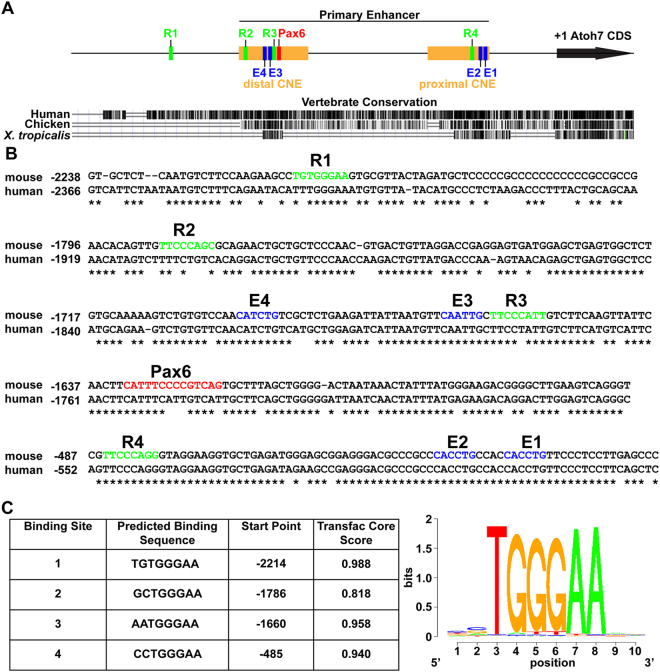
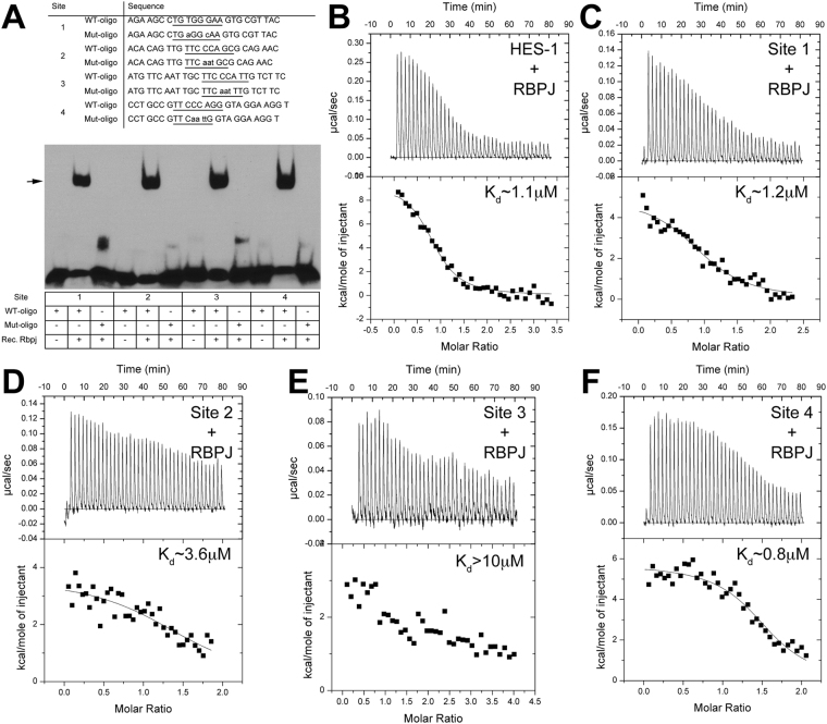
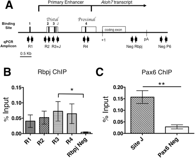
In vertebrate retinal progenitor cells, the proneural factor Atoh7 exhibits a dynamic tissue and cellular expression pattern. Although the resulting Atoh7 retinal lineage contains all seven major cell types, only retinal ganglion cells require Atoh7 for proper differentiation. Such specificity necessitates complex regulation of Atoh7 transcription during retina development. The Notch signaling pathway is an evolutionarily conserved suppressor of proneural bHLH factor expression. Previous in vivo mouse genetic studies established the cell autonomous suppression of Atoh7 transcription by Notch1, Rbpj and Hes1. Here we identify four CSL binding sites within the Atoh7 proximal regulatory region and demonstrate Rbpj protein interaction at these sequences by in vitro electromobility shift, calorimetry and luciferase assays and, in vivo via colocalization and chromatin immunoprecipitation. We found that Rbpj simultaneously represses Atoh7 transcription using both Notch-dependent and -independent pathways.


Conflict of interest statement

The authors declare no competing interests.

Figures

Figure 1
Figure 1
Predicted Rbpj CSL binding sites in mouse Atoh7 5′ regulatory DNA. (A) Diagram of the primary enhancer and the distal and proximal CNEs, indicating the position of four predicted Rbpj binding sites (R1–R4, green boxes), a previously characterized Pax6 binding site (red box) and four E-boxes binding sites (E1–E4, blue boxes). UCSC Genome browser view of a 3 kb 5′ noncoding region of mouse Atoh7 (+1 = A of ATG start codon) using mm10 assembly and vertebrate evolutionary conservation tracks, with mouse as the reference genome. (B) ClustalW alignments of mouse and human Atoh7 genomic sequence, with asterisks indicating nucleotide identity diagrammed in (A,C) The four predicted Rbpj binding site sequences, identified by the Transfac CSL consensus matrix.
Figure 2
Figure 2
Rbpj antibody specificity. (A,B) GFP-positive cells (green) indicate the E13.5 α-Cre retinal lineage. A-A”) Anti-Rbpj labeling of α-Cre;Z/EG;RbpjCKO/+ control cryosections shows ubiquitous expression of the nuclear Rbpj protein (red), including within GFP+ cells (arrow). (B-B”) Cell autonomous loss of Rbpj expression (red) in conditionally mutant α-Cre;Z/EG;RbpjCKO/CKO cells (arrow). n = 3 biologic replicates embryos/genotype. (C) Western blot of E14.5 lens protein extracts, collected from RbpjCKO/CKO, Le-Cre;RbpjCKO/+ and Le-Cre;RbpjCKO/CKO embryos, respectively. A single band of 56 kDa, the predicted size of Rbpj, is absent from mutant lenses, with β-actin loading control at bottom. The top panel is the uncropped blot ECL autorad exposure, which was stripped and reprobed with Actin and Jagged1 loading controls (see Supplemental Fig S3). (A-B”) apical is up. Bar in A for A,B = 20μm; in A’-for A’ to B” = 40 μm. White arrows point to GFP+ cells expressing Rbpj in controls (A-A”), but not in GFP+;Rbpj mutant cells (B-B”).
Figure 3
Figure 3
Colocalization of Rbpj and Atoh7 proteins during retinal neurogenesis. (AC) Double antibody labeling of retinal sections highlights essentially complete nuclear co-localization. The boxed area in each panel is shown at higher magnification to the right. (A-A’”) At E11.5 Atoh7 protein is restricted to a subset of central RPCs (A’). (B-B’”) By E13.5 the initial wave of neurogenesis has reached the periphery and Atoh7 is expressed more broadly throughout the apical neuroblast layer. (C-C’”) Consistent with mRNA expression studies, Atoh7+ cells are localized to the peripheral retina (C’). Panel C is a composite stitched together from 4 overlapping 10× image fields. Colocalization of Rbpj (green) and Atoh7 (purple) is shown as white. Rostral is up in all panels. L = lens; Bar in A,C,E = 100 μm; in B,D,F = 50 μm. In (A’-C”’) white arrows point to Atoh7+ cells that also contain high levels of Rbpj.
Figure 4
Figure 4
In vitro binding of Rbpj to CSL consensus sites in Atoh7 5′ DNA. (A) Oligonucleotides with CSL consensus sites (underlined) identified in 5′Atoh7 noncoding DNA, synthesized and used for testing Rbpj protein-DNA binding by EMSA (WT, wild type; Mut, mutated). Lower case indicates a mutated nucleotide. Recombinant Rbpj protein and biotin-labeled oligonucleotide complexes undergo specific mobility shifts (arrow). See uncropped autorad exposure in Supplemental Fig. 3. n = 4 independent assays. (BF) Representative thermograms (raw heat signal and nonlinear least squares fit to the integrated data) for Hes1 CSL consensus site (positive control in (B) and Atoh7 CSL sites R1–R4, all shown with their dissociation constants (Kd).
Figure 5
Figure 5
In vivo occupancy of Rbpj at Atoh7 locus in E14.5 retinal chromatin. (A) Diagram of mouse Atoh7 genomic locus indicating CSL binding sites and qPCR amplicons evaluated, with Pax6 site J site serving as positive control. (B,C). Real-time PCR analyses of DNA fragments amplified after Rbpj or Pax6 ChIP, displayed as the mean ± SEM of three biologic replicate assays, performed in PCR duplicate, with the preimmune IgG values subtracted. Only Rbpj site R3 is significantly enriched over negative control. *p ≤ 0.05; **p ≤ 0.01.
Figure 6
Figure 6
Differential Notch-mediated regulation of Atoh7 transcription. (A) Colocalization of transient mCherry transgenic expression, driven by a 2.4 kb mouse Atoh7 enhancer, to endogenous Atoh7 protein in the E13.5 retina (White arrows point to colabeled cells). Both cytoplasmic and nuclear Cherry expression are seen, presumably due to the inefficiency of a synthetic nuclear localization sequence and reporter antibody sensitivity, which detected both nascent Cherry protein in the cytoplasm and its accumulation in the nucleus. Scale bar = 100 μm (B) Comparison of mouse Atoh7 transcriptional activity in HEK293T cells following single versus quadruple CSL site mutation. Transcriptional activity of individual CSL site mutants, and quadruple mutant within Atoh7–2.6Kb luciferase/pGL2 construct. Only site R3 in the distal CNE is required to suppress Atoh7 transcription. However, loss of sites R1–4 caused significant downregulation of Atoh7 transcription. n = 9 biological replicates (each performed in technical triplicate). (C) Cotransfection of activated NICD1, NICD3, or both constructs with Atoh7 wild type or RΔ1–R4 mutant luciferase constructs. n ≥ 3 biological replicates (each in technical triplicate). All luciferase experiments were normalized to a co-transfected Renilla control. A two-tailed, unpaired t-test with equal standard deviation and Gaussian distribution was used to determine p-value. (D) RT-PCR analysis of E13.5 retinal cDNA showing expression of all four co-repressor gene mRNAs. Distinct PCR primers for the Fhl1 (KyoT) gene were used that amplified an exon common to all splice products (pan Fhl1), and the specific splice variants Fhl1b (KyoT3) and Fhl1c (KyoT2) that uniquely contain the Rbpj-interaction domain. All PCR reactions were run on a single gel, uncropped image provided in Supplemental Fig. S3. *p ≤ 0.05; **p ≤ 0.01; ***p ≤ 0.001; ****p < 0.0001 and Figure S2 ***p ≤ 0.001; ****p < 0.0001.

Similar articles

Cited by

References

    1. Ohsawa R, Kageyama R. Regulation of retinal cell fate specification by multiple transcription factors. Brain research. 2008;1192:90–98. doi: 10.1016/j.brainres.2007.04.014. - DOI - PubMed
    1. Brown NL, et al. Math5 encodes a murine basic helix-loop-helix transcription factor expressed during early stages of retinal neurogenesis. Development. 1998;125:4821–4833. - PubMed
    1. Brown NL, Patel S, Brzezinski J, Glaser T. Math5 is required for retinal ganglion cell and optic nerve formation. Development. 2001;128:2497–2508. - PMC - PubMed
    1. Kanekar S, et al. Xath5 participates in a network of bHLH genes in the developing Xenopus retina. Neuron. 1997;19:981–994. doi: 10.1016/S0896-6273(00)80391-8. - DOI - PubMed
    1. Kay JN, Finger-Baier KC, Roeser T, Staub W, Baier H. Retinal ganglion cell genesis requires lakritz, a Zebrafish atonal Homolog. Neuron. 2001;30:725–736. doi: 10.1016/S0896-6273(01)00312-9. - DOI - PubMed

Publication types

MeSH terms

Substances