. 2017 Nov;52(21):12574-12592.
doi: 10.1007/s10853-017-1229-9. Epub 2017 Jul 28.

A self-organising biomimetic collagen/nano-hydroxyapatite-glycosaminoglycan scaffold for spinal fusion

Affiliations

A self-organising biomimetic collagen/nano-hydroxyapatite-glycosaminoglycan scaffold for spinal fusion

Aman Sharma et al. J Mater Sci. 2017 Nov.

Abstract

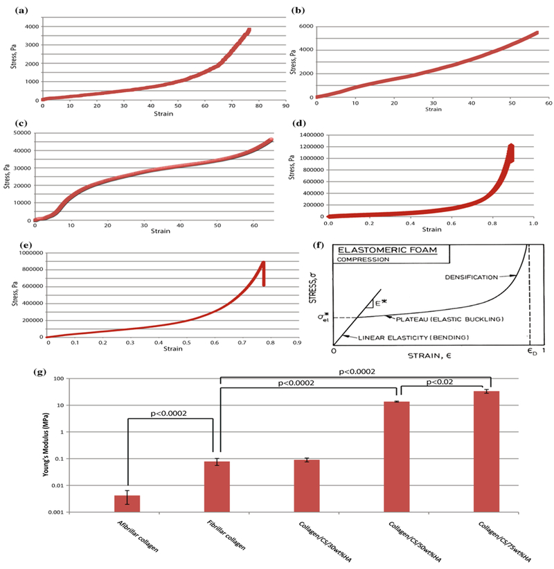
The use of spinal fusion surgery as a treatment for degenerative spinal conditions and chronic back pain is increasing. However, this technique requires use of a bone grafting material to fuse the vertebrae, traditionally autologous bone, which consists of an optimal combination of osteogenic cell precursors, extracellular matrix proteins and mineral components. To date, this remains the 'gold standard' material but its supply is limited and is associated with a number of clinical and ethical difficulties; consequently, various combinations of cells with biological scaffold materials have been tested but have failed to achieve fusion rates even comparable to autologous bone. We successfully fabricated a novel collagen-based scaffold using self-organising atelocollagen combined with nano-hydroxyapatite and chondroitin sulphate, cross-linked by microbial transglutaminase. The scaffold was characterised using a range of imaging, chemical composition and thermal analysis techniques. It was found to exhibit appropriate stiffness and suitable pore size for the adhesion, growth and differentiation of MSCs. The low toxicity makes it suitable for clinical application, and its slow degradation profile would enable the scaffold to promote bone growth over an extended period. This material therefore shows promise for clinical use in spinal fusion and other procedures requiring the use of bone grafts.


Conflict of interest statement

Compliance with ethical standards Conflicts of interest No conflict of interests to declare.

Figures

Figure 1
Figure 1
Scanning electron microscopy images show porous structure of cut surface of collagen and collagen/CS/HA scaffolds a Photograph of fibrillar collagen scaffold. SEM images of b afibrillar collagen, c fibrillar collagen, d collagen/CS 1wt%/HA 30wt%, e collagen/CS 1wt%/HA 50wt%, f collagen/CS 1wt%/HA 75wt%. All images were obtained using a JEOL scanning microscope 840F with a working distance of 10–30 mm, electron voltage of 5 kV and 50–10,000× magnification. CS chondroitin sulphate; HA hydroxyapatite.
Figure 2
Figure 2
SEM images show D-bandmg spacing of collagen, with agglomerates of nanoHA interwoven throughout the fibrils and calculated pore size a Fibrillar collagen, b collagen/CS 1wt%/HA 50wt%, c collagen/CS 1wt%/HA 75wt%. Graphs represent average pore diameter (d) and area (e) of afibrillar and HA/CS collagen scaffolds. Experiments performed in triplicate; error bars show standard deviation. All SEM images were obtained using a JEOL scanning microscope 840F, working distance of 10–30 mm, electron voltage of 3–5 kV and 25,000× magnification. ImageJ software was used to calculate pore size of collagen scaffolds from SEM images. CS chondroitin sulphate; HA hydroxyapatite. Arrowhead, nanoHA.
Figure 3
Figure 3
Transmission electron microscopy (TEM) and atomic force microscopy (AFM) images show D-banding spacing of collagen, with agglomerates of nanoHA interwoven throughout the fibrils. TEM images of a afibrillar collagen, b and c fibrillary collagen/CS 1wt%/HA 50wt%, scale as shown. d AFM image of fibrillar collagen scaffold. CS chondroitin sulphate; HA hydroxyapatite.
Figure 4
Figure 4
Thermogravimetric analysis (TGA) confirms composition of scaffolds. a Summary table of TGA-derived wt% values of HA in collagen/CS/HA scaffolds. Data are shown for each sample in triplicate with mean values ± SD. TGA data for scaffolds as prepared with 30–75 wt% HA and heated from 25 to 1000 0C at a rate of 100Cmin−1 in nitrogen. Graphs show representative data for each scaffold prepared with desired weight percentage of HA b 30 c 50 and d 75. CS chondroitin sulphate; HA hydroxyapatite.
Figure 5
Figure 5
Energy dispersive x-ray (EDX) analysis of collagen scaffold with increasing wt% of hydroxyapatite (HA). Collagen scaffold containing a 30 wt% b 50 wt% and c 75 wt% HA was analysed by EDX. For each sample, SEM images are shown for the cross-sectional cut surface with and without EDX overlay, demonstrating the homogeneity of HA throughout the scaffold. Red, phosphorous; Green, calcium; Blue, oxygen. For each sample, graphs of elemental mapping are shown on the right. HA hydroxyapatite; SEM scanning electron microscopy.
Figure 6
Figure 6
X-ray diffraction (XRD) analysis of collagen scaffolds XRD was performed over a 2_ range of 10–800 with a step size of 0.05, dwell time of 12 s at RT and divergent slit of 0.5. Bruker analysis software was used to analyse the data. a ICDD standard diffraction pattern for HA (#09–432). XRD plots for collagen/CS scaffolds containing b 30 wt%, c 50 wt% and d 75 wt% HA. RT room temperature; XRD X-ray diffraction; ICDD International Centre for Diffraction Data; CS, chondroitin sulphate; HA hydroxyapatite.
Figure 7
Figure 7
Dynamic scanning calorimetry (DSC) shows thermal stability of scaffolds at body temperature. Graph of mean denaturation temperatures for each scaffold type, calculated from DSC data of peak temperature. Error bars standard deviation; *P < 0.02. CS chondroitin sulphate; HA hydroxyapatite.
Figure 8
Figure 8
Mechanical testing of collagen scaffolds Compression testing was performed. Graphs showing stress/strain curves for a afibrillar collagen, b fibrillar collagen and scaffolds containing collagen/CS with c 30, d 50 and e 75 wt% HA. f Schematic representation of calculation of Young’ modulus from stress/strain curves, Gibson, 2000. g Graph of Young’s moduli calculated using LINEST function in Microsoft Excel; log scale, average of triplicate samples. CS chondroitin sulphate; HA hydroxyapatite.
Figure 9
Figure 9
Viable human mesenchymal stem cells grow throughout 3-dimensional collagen-based scaffolds. a 3-D reconstruction of hMSCs in collagen scaffold. Representative multiphoton microscopy images of live hMSCs in b collagen, c collagen/CS/ HA30wt%, d collagen/CS/HA50wt%, e collagen/CS/HA75wt% and f collagen/mTG scaffolds. 100,000 hMSCs seeded per scaffold, all images obtained after 3 days of culture using a BioRad Radiance 2100 MP Multiphoton Microscope. Blue collagen; Green live cells; Red dead cells.
Figure 10
Figure 10
Collagen-based scaffolds support the growth and differentiation of human mesenchymal stem cells. a Graph showing relative number of hMSCs grown on collagen, collagen/CS/ HA30 wt%, collagen/CS/ HA50wt%, collagen/CS/ HA75 wt%, collagen/CS/ MTG or 2-D culture, compared to seeding density, using Alamar Blue assay. Values at 24, 48 and 72 h. b Graph showing relative number (shown as log scale) of hMSCs grown on collagen, collagen/CS/HA30 wt%, collagen/CS/HA50 wt%, collagen/CS/HA75 wt%, collagen/CS/MTG or 2-D culture, compared to seeding density, using Alamar Blue assay. Values at 0 and 14 days. Mean values, error bars represent standard deviation. *P < 0.05, unpaired t test with Welch’s correction.
Figure 11
Figure 11
Osteogenic gene expression is enhanced by the presence of collagen-based scaffolds. Quantitative gene expression in hMSCs harvested from either 2-D culture on Matrigel-coated dishes or 3-D collagen-based scaffolds (collagen, collagen/CS/ 30 wt% HA, collagen/CS/50 wt% HA, collagen/CS/75 wt% HA, collagen/mTG) using RT-PCR. Values shown for fold change in expression level of mRNA normalised to a panel of housekeeping genes (TBP, β-ACTIN), error bars represent standard deviation. Genes tested were a osterix, b osteocalcin, c TGF-βΙ, d RUNX2, e alkaline phosphatase, f BMP-2. *P < 0.05, unpaired t test using Welch’s correction.
Figure 12
Figure 12
Bone mineral density of collagen scaffolds is increased with the presence of HA. a Graph showing mean BMD of collagen scaffolds with and without hMSCs, after 7 days of culture. Error bars standard deviation. *P < 0.02. Representative micro-CT images of b collagen and c collagen/75wt% HA scaffolds, obtained using Skyscan 1174 (Bruker, Belgium) compact micro-CT scanner, at 50 kV, 800 μA.

Similar articles

Cited by

References

    1. Rajaee SS et al. (2012) Spinal fusion in the United States: analysis of trends from 1998–2008. Spine 37(1):67–76 - PubMed
    1. Vater CA, Harris ED, Siegel RC (1979) Native cross-links in collagen fibrils induce resistant to human synovial collagenase. Biochem J 181(3):639–645 - PMC - PubMed
    1. Zhang P et al. (2008) Clinical study of lumbar fusion by hybrid construct of stem cells technique and biodegradable material. Zhonghua wai ke za zhi Chin J Surg 46(7):493–496 - PubMed
    1. Neen D et al. (2006) Healos and bone marrow aspirate used for lumbar spine fusion: a case controlled study comparing healos with autograft. Spine 31(18):E636–E640 - PubMed
    1. Kuwahara K et al. (2011) Enzymatic crosslinking and degradation of gelatin as a switch for bone morphogenetic protein-2 activity. Tissue Eng Part A 17(23–24):2955–2964 - PubMed

LinkOut - more resources