. 2018 Jun 21:12:173.
doi: 10.3389/fncel.2018.00173. eCollection 2018.

C57BL/6 and Swiss Webster Mice Display Differences in Mobility, Gliosis, Microcavity Formation and Lesion Volume After Severe Spinal Cord Injury

Affiliations

C57BL/6 and Swiss Webster Mice Display Differences in Mobility, Gliosis, Microcavity Formation and Lesion Volume After Severe Spinal Cord Injury

Harun Najib Noristani et al. Front Cell Neurosci. .

Abstract

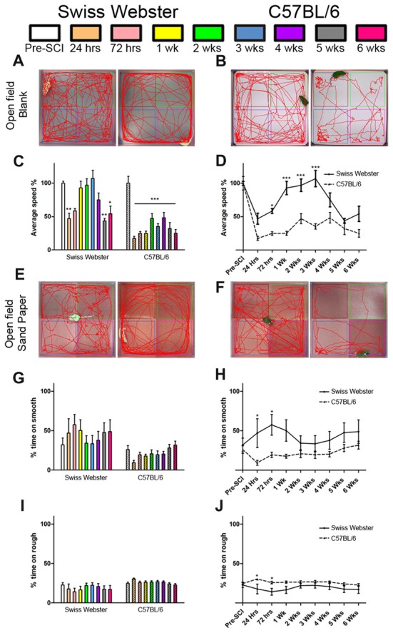
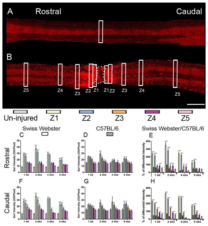
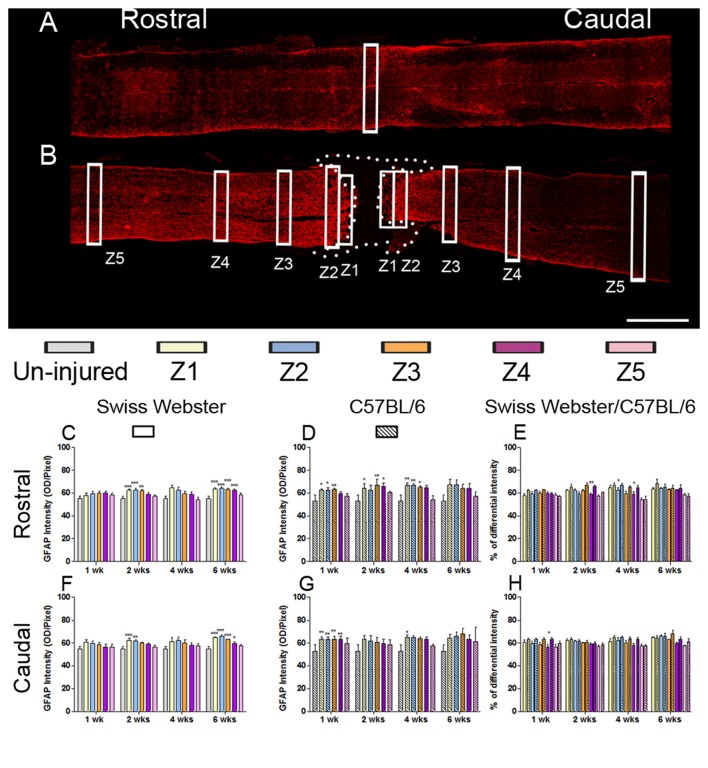
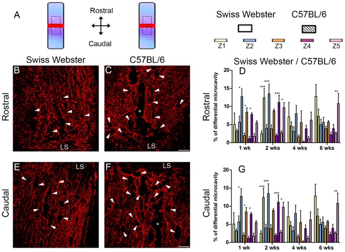
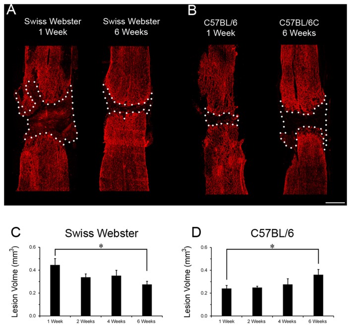
Spinal cord injuries (SCI) are neuropathologies causing enormous physical and emotional anguish as well as irreversibly disabilities with great socio/economic burdens to our society. The availability of multiple mouse strains is important for studying the underlying pathophysiological response after SCI. Although strain differences have been shown to directly affect spontaneous functional recovery following incomplete SCI, its influence after complete lesion of the spinal cord is unclear. To study the influence of mouse strain on recovery after severe SCI, we first carried out behavioral analyses up to 6 weeks following complete transection of the spinal cord in mice with two different genetic backgrounds namely, C57BL/6 and Swiss Webster. Using immunohistochemistry, we then analyzed glial cell reactivity not only at different time-points after injury but also at different distances from the lesion epicenter. Behavioral assessments using CatWalk™ and open field analyses revealed increased mobility (measured using average speed) and differential forelimb gross sensory response in Swiss Webster compared to C57BL/6 mice after complete transection of the spinal cord. Comprehensive histological assessment revealed elevated microglia/macrophage reactivity and a moderate increase in astrogliosis in Swiss Webster that was associated with reduced microcavity formation and reduced lesion volume after spinal cord transection compared to C57BL/6 mice. Our results thus suggest that increased mobility correlates with enhanced gliosis and better tissue protection after complete transection of the spinal cord.

Keywords: glial cells; microcavity; mobility; protection; spinal cord injury.


Figures

Figure 1
Figure 1
Swiss Webster display increased mobility after spinal cord transection as compared to C57BL/6 mice. Bar and line graphs displaying alterations in average speed using CatWalk™ analysis after spinal cord transection in Swiss Webster and C57BL/6 mice (A,B). Swiss Webster displayed increase average speed (A,B), no change in max contact (C,D) and augmented usage of their front paws (print area; E,F) compared to C57BL/6 mice. Conversely to Swiss Webster, C57BL/6 mice displayed increased max contact throughout the 6 weeks (C,D). One-way ANOVA with Tukey post hoc tests (A,C,E) and two-way ANOVA with Bonferroni post hoc (B,D,F). *P < 0.05, **P < 0.01, ***P < 0.001.
Figure 2
Figure 2
Swiss Webster display increased mobility and differential gross sensory response after spinal cord transection as compared to C57BL/6 mice. Photomicrographs indicating the experimental arrangement of the open field test used to measure average speed after spinal cord transection in Swiss Webster (A) and C57BL/6 mice (B). Bar and line graphs displaying alterations in average speed using open field analysis after spinal cord injuries (SCI) in Swiss Webster and C57BL/6 mice (C,D). Swiss Webster displayed increased average speed compared to C57BL/6 mice (D). Photomicrographs indicating the experimental arrangement of the sandpaper test used to measure sensory response after spinal cord transection (E,F). Bar and line graphs displaying alteration in sensory response after SCI in Swiss Webster and C57BL/6 mice (G–J). Neither Swiss Webster mice nor C57BL/6 mice displayed change in their sensory response over the course of their behavioral follow-ups, as indicated by similar interest in both smooth and rough sandpapers throughout the 6 weeks (G,I). However, direct comparison between the two strains revealed a moderate preference of Swiss Webster mice for the smooth area and a preference of the rough area in C57BL/6 at 24 and 72 h post-lesion. One-way ANOVA with Tukey post hoc tests (C,G,I) and two-way ANOVA with Bonferroni post hoc (D,H,J). *P < 0.05, **P < 0.01, ***P < 0.001.
Figure 3
Figure 3
Increased microglia/macrophage response in Swiss Webster compared to C57BL/6 mice after severe SCI. Confocal photomicrographs indicating IBA1 staining of longitudinal spinal cord sections in the non-injured (NI) control and spinal cord transected mice (A,B). Bar graphs indicating quantitative analysis of IBA1 immunoreactivity along the five zones rostral and caudal to the lesion site in Swiss Webster mice (C,F) and C57BL/6 mice (D,G). Direct comparisons following normalization revealed that Swiss Webster displayed increased IBA1 immunoreactivity compared to C57BL/6 mice after spinal cord transection (E,H). Scale bars (A,B): 1 mm. One-way ANOVA with Tukey post hoc tests (C,D,F,G) and two-way ANOVA with Bonferroni post hoc (E,H). *P < 0.05, **P < 0.01, ***P < 0.001.
Figure 4
Figure 4
Astrogliosis in Swiss Webster and C57BL/6 after severe SCI. Confocal photomicrographs indicating GFAP staining of longitudinal spinal cord sections in the NI control and spinal cord transected mice (A,B). Bar graphs indicating quantitative analysis of GFAP immunoreactivity along the five zones rostral and caudal to the lesion site in Swiss Webster mice (C,F) and C57BL/6 mice (D,G). Direct comparisons revealed that C57BL/6 mice displayed moderate increase in GFAP immunoreactivity compared to Swiss Webster after complete transection of the spinal cord (E,H). Scale bars (A,B): 1 mm. One-way ANOVA with Tukey post hoc tests (C,D,F,G) and two-way ANOVA with Bonferroni post hoc (E,H). *P < 0.05, **P < 0.01, ***P < 0.001.
Figure 5
Figure 5
Reduced microcavity formation in Swiss Webster compared to C57BL/6 mice after severe SCI. Schematic drawing of the longitudinal spinal cord sections after complete SCI illustrating the lesion site (red rectangle) and the reference frames for displayed fields of view (A). Confocal photomicrographs of longitudinal spinal cord sections indicating GFAP staining and microcavities (arrowheads) adjacent to the lesion epicenter in Swiss Webster (B,E) and C57BL/6 (C,F) mice. Bar graphs showing quantitative analysis of microcavities along the five zones rostral (D) and caudal (G) to the lesion site. Spinal cord transected Swiss Webster displayed reduced microcavities compared to C57BL/6 mice (C,F). Scale bars (B,C,E,F): 100 μm. Two-way ANOVA with Bonferroni post hoc tests *P < 0.05, **P < 0.01, ***P < 0.001. LS: lesion site.
Figure 6
Figure 6
Lesion volume evolution in Swiss Webster and C57BL/6 after severe SCI. Confocal photomicrographs indicating GFAP staining and lesion volume evolution (dotted area) in Swiss Webster and C57BL/6 mice (A,B) following complete transection of the spinal cord. Bar graphs showing quantitative analysis of lesion volume evolution over-time in Swiss Webster and C57BL/6 mice after complete SCI (C,D). Swiss Webster displayed time-dependent decrease, whereas C57BL/6 mice showed increased lesion volume between 1 and 6 weeks after complete transection of the spinal cord (C,D). Scale bars (A,B): 500 μm. Un-paired t-test *P < 0.05 compared to the same strain at 1 week after injury.

Similar articles

Cited by

References

    1. Anderson M. A., Burda J. E., Ren Y., Ao Y., O’Shea T. M., Kawaguchi R., et al. . (2016). Astrocyte scar formation aids central nervous system axon regeneration. Nature 532, 195–200. 10.1038/nature17623 - DOI - PMC - PubMed
    1. Antri M., Barthe J. Y., Mouffle C., Orsal D. (2005). Long-lasting recovery of locomotor function in chronic spinal rat following chronic combined pharmacological stimulation of serotonergic receptors with 8-OHDPAT and quipazine. Neurosci. Lett. 384, 162–167. 10.1016/j.neulet.2005.04.062 - DOI - PubMed
    1. Basso D. M., Beattie M. S., Bresnahan J. C. (1996a). Graded histological and locomotor outcomes after spinal cord contusion using the NYU weight-drop device versus transection. Exp. Neurol. 139, 244–256. 10.1006/exnr.1996.0098 - DOI - PubMed
    1. Basso D. M., Beattie M. S., Bresnahan J. C., Anderson D. K., Faden A. I., Gruner J. A., et al. . (1996b). MASCIS evaluation of open field locomotor scores: effects of experience and teamwork on reliability. Multicenter Animal Spinal Cord Injury Study. J. Neurotrauma 13, 343–359. 10.1089/neu.1996.13.343 - DOI - PubMed
    1. Basso D. M., Fisher L. C., Anderson A. J., Jakeman L. B., McTigue D. M., Popovich P. G. (2006). Basso Mouse Scale for locomotion detects differences in recovery after spinal cord injury in five common mouse strains. J. Neurotrauma 23, 635–659. 10.1089/neu.2006.23.635 - DOI - PubMed