. 2018 Jun 6:2018:5340756.
doi: 10.1155/2018/5340756. eCollection 2018.

Evaluation of Oxidative Stress and Mitophagy during Adipogenic Differentiation of Adipose-Derived Stem Cells Isolated from Equine Metabolic Syndrome (EMS) Horses

Affiliations

Evaluation of Oxidative Stress and Mitophagy during Adipogenic Differentiation of Adipose-Derived Stem Cells Isolated from Equine Metabolic Syndrome (EMS) Horses

Krzysztof Marycz et al. Stem Cells Int. .

Abstract

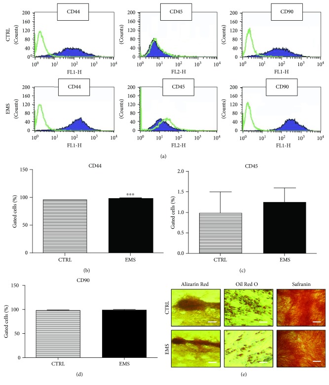
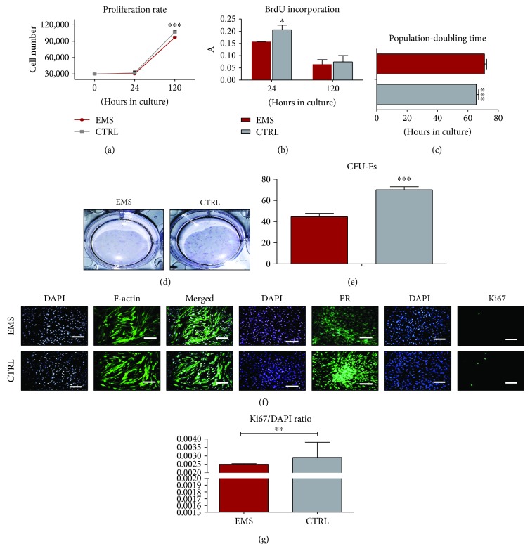
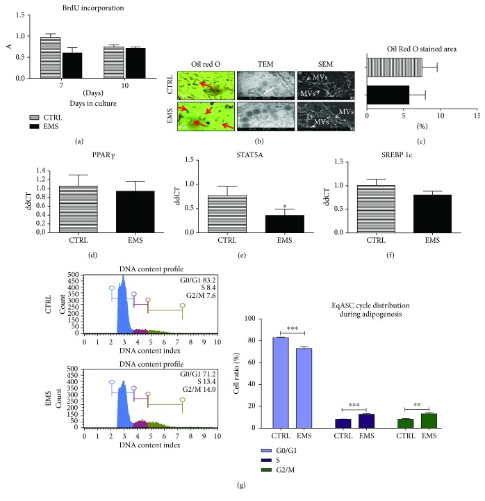
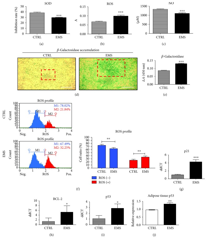
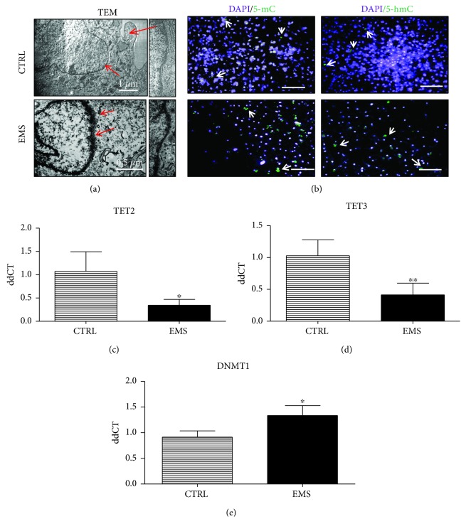
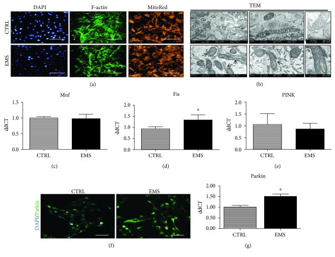
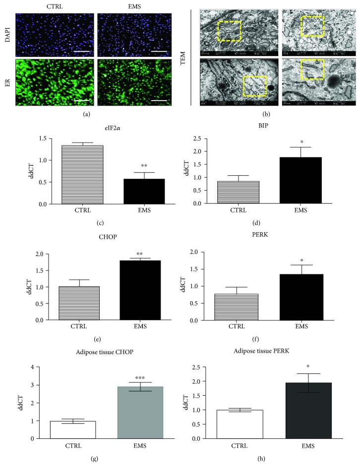
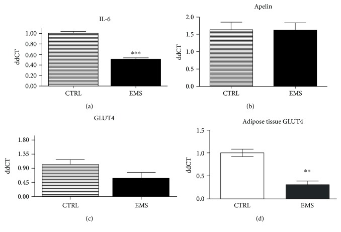
Mesenchymal stem cells (MSCs) are frequently used in both human and veterinary medicine because their unique properties, such as modulating the immune response and differentiating into multiple lineages, make them a valuable tool in cell-based therapies. However, many studies have indicated the age-, lifestyle-, and disease-related deterioration of MSC regenerative characteristics. However, it still needs to be elucidated how the patient's health status affects the effectiveness of MSC differentiation. In the present study, we isolated mesenchymal stem cells from adipose tissue (adipose-derived mesenchymal stem cells (ASCs)) from horses diagnosed with equine metabolic syndrome (EMS), a common metabolic disorder characterized by pathological obesity and insulin resistance. We investigated the metabolic status of isolated cells during adipogenic differentiation using multiple research methods, such as flow cytometry, PCR, immunofluorescence, or transmission and confocal microscopy. The results indicated the impaired differentiation potential of ASCEMS. Excessive ROS accumulation and ER stress are most likely the major factors limiting the multipotency of these cells. However, we observed autophagic flux during differentiation as a protective mechanism that allows cells to maintain homeostasis and remove dysfunctional mitochondria.


Figures

Figure 1
Figure 1
Immunophenotyping and multipotency assay. Representative histograms from flow cytometry analysis, green lines—isotope controls and purple lines—specific for surface antigen antibodies (a). Cells were analyzed for the expression of CD44 (b), CD45 (c), and CD90 (d). Moreover, to confirm the multipotency of isolated cells, they were induced into osteogenic, adipogenic, and chondrogenic lineages. Effectiveness of differentiation was confirmed by specific staining (e). Results were expressed as mean ± SD. Scale bar 100 μm. ∗∗∗p < 0.001.
Figure 2
Figure 2
Growth kinetics of ASCs cultured in control conditions. Cell number estimated using a resazurin-based assay (a) and incorporation of BrdU (b). Population-doubling time was calculated using the number of cells in each of the experimental time points (c). The ability of cells to form colonies originating from one cell was evaluated by CFU-F assay. Representative photographs showing colonies stained with pararosaniline (d) and quantitative data obtained by the application of a CFU-F algorithm (e). The morphology of cells was investigated using fluorescence staining for f-actin and endoplasmic reticulum (ER) (f). Moreover, proliferation of cells was estimated by immunofluorescence staining for the Ki67 antigen as presented on representative images. Data was quantified and the Ki67/DAPI ratio was calculated (g). Results are expressed as mean ± SD. Scale bar 100 μm. p < 0.05, ∗∗p < 0.01, and ∗∗∗p < 0.001.
Figure 3
Figure 3
Growth kinetics and morphology of ASCCTRL and ASCEMS cultured under adipogenic conditions. To evaluate the proliferation rate under adipogenic differentiation, incorporation of BrdU was established (a) in both of the investigated groups. To confirm adipogenesis and establish its effectiveness, cells were stained with Oil Red O. Moreover, the secretion of MVs was evaluated using SEM imaging while the ultrastructure of cells was visualized using a TEM microscope (b). The Oil Red O-stained area was quantified using Image J (c). Using RT-PCR, expression of PPARγ (d), STAT5A (e), and SREBP 1c (f) was established. Cell cycle (DNA content) was analyzed in both of the investigated cultures using a cell analyzer (g). Obtained results indicate a more effective differentiation in control cells as they stop to proliferate in order to differentiate into adipocytes. ASC isolated from EMS individuals seems to be more resistant to adipogenic stimuli. Results are expressed as mean ± SD. Scale bar 200 μm. p < 0.05, ∗∗p < 0.01, and ∗∗∗p < 0.001.
Figure 4
Figure 4
Assessment of oxidative stress factors and apoptosis under adipogenic conditions. To evaluate the levels of oxidative stress, the amount of SOD (a), ROS (b), and NO (c) in culture supernatants were assessed. Representative photographs showing the results of senescence-associated β-galactosidase staining. Boxed regions with red edges indicate the regions with excessive β-galactosidase accumulation. Accumulation of β-galactosidase was also quantified by spectrophotometric measurement of dye reduction (e). Intracellular accumulation of ROS was assessed with a cell analyzer (f). Moreover, the expression of the following transcripts was investigated using the RT-PCR method: p21 (g), BCL-2 (h), and p53 (i). mRNA levels of p53 was also analyzed in adipose tissue homogenates of healthy and EMS diagnosed horses (j). Results are expressed as mean ± SD. Scale bar 250 μm. p < 0.05, ∗∗p < 0.01, and ∗∗∗p < 0.001.
Figure 5
Figure 5
Epigenetic state of ASCCTRL and ASCEMS cultured under adipogenic conditions. TEM images of nuclei (a) and their higher magnification showing heterochromatin underneath the nuclear envelope. ASCEMS was characterized by a breakdown of heterochromatin associated with the inner nuclear membrane. Immunofluorescence staining for genomic distribution of 5-mC and 5-hmC (b) was performed in order to evaluate DNA methylation status. Using RT-PCR, expression of TET2 (c), TET3 (d), and DNMT1 (e) was evaluated. Obtained results indicate increased DNA methylation in ASCEMS. Results are expressed as mean ± SD. Scale bar 250 μm. p < 0.05 and ∗∗p < 0.01.
Figure 6
Figure 6
Assessment of autophagy in ASCCTRL and ASCEMS cultured under adipogenic conditions after the 10th day. Formation of autophagosomes and autolysosomes was visualized using TEM imaging. Circles with red edges indicate autophagosomes within the cell (a). Moreover, using immunofluorescence staining, LAMP2 localization was investigated under a confocal microscope (b). Using RT-PCR, the expression of autophagy-related genes including Beclin (c), LC3 (d), and LAMP 2 (e) was evaluated. Upregulation of LAMP2 indicates autolysosome formation which is required to complete the autophagic removal of damaged organelles. Interestingly, no differences in LAMP2 expression in adipose tissue (f) was observed. Results are expressed as mean ± SD. Scale bar 250 μm. p < 0.05.
Figure 7
Figure 7
Mitochondrial dynamics and clearance in ASCCTRL and ASCEMS cultured under adipogenic conditions. Using fluorescence staining, cells' cytoskeleton (f-actin) and mitochondrial net were visualized (a). Moreover, a TEM microscope allowed for a deeper assessment of mitochondrial morphology (b). Mitochondria from ASCCTRL presented typical, elongated morphology, while ASCEMS cells were characterized by mitochondrial aberrations including membrane and cristae raptures. Moreover, mitochondrial dynamics was assessed by RT-PCR as the expression of Mnf (c) and Fis (d) genes was investigated. However, no differences were observed between groups. Similarly, no differences in PINK expression were noted (e). To evaluate the amount of Parkin protein, immunofluorescence staining and RT-PCR were performed. Representative photographs obtained from a confocal microscope indicated increased Parkin accumulation in ASCEMS (f). Those data were also confirmed by the analysis of Parkin mRNA levels, as it was significantly upregulated in ASCEMS (g). Results are expressed as mean ± SD. Scale bar 250 μm. p < 0.05.
Figure 8
Figure 8
Endoplasmic reticulum stress in ASCCTRL and ASCEMS cultured under adipogenic conditions after the 10th day. ER condition and distribution were visualized under fluorescent and TEM microscopes. The more intense fluorescence ER signal was noted in ASCCTRL, where ER was robustly expanded throughout the cells (a). In the case of ASCEMS, ER was fragmented and disintegrated as observed under TEM (b). To further investigate ER stress in investigated cells, we establish the expression of eIF2α (c), BIP (d), CHOP (e), and PERK (f). Obtained results indicated an induction of unfolded protein response and ER stress in ASCEMS. Furthermore, we decided to evaluate ER stress in ASC niche-adipose tissue. Interestingly ER stress was also observed within adipocytes of EMS horses as increased expression of both CHOP (g) and PERK (h) transcripts was noted. Results are expressed as mean ± SD. Scale bar 250 μm. p < 0.05, ∗∗p < 0.01, and ∗∗∗p < 0.001.
Figure 9
Figure 9
Inflammation and insulin sensitivity. Using RT-PCR, the expression of IL-6 (a), apelin (b), and GLUT4 (c) was assessed in ASCs. Moreover, GLUT-4 levels were also established in adipose tissue of horses (d). Interestingly, no differences in GLUT4 expression were observed in ASC; however, it was significantly downregulated in adipose tissue of EMS horses. Results are expressed as mean ± SD. Scale bar 250 μm. ∗∗p < 0.01 and ∗∗∗p < 0.001.

Similar articles

Cited by

References

    1. Caplan A. I. Mesenchymal stem cells. Journal of Orthopaedic Research. 1991;9(5):641–650. doi: 10.1002/jor.1100090504. - DOI - PubMed
    1. Chamberlain G., Fox J., Ashton B., Middleton J. Concise review: mesenchymal stem cells: their phenotype, differentiation capacity, immunological features, and potential for homing. Stem Cells. 2007;25(11):2739–2749. doi: 10.1634/stemcells.2007-0197. - DOI - PubMed
    1. Grzesiak J., Marycz K., Wrzeszcz K., Czogała J. Isolation and morphological characterisation of ovine adipose-derived mesenchymal stem cells in culture. International Journal of Stem Cells. 2011;4(2):99–104. doi: 10.15283/ijsc.2011.4.2.99. - DOI - PMC - PubMed
    1. Marędziak M., Marycz K., Lewandowski D., Siudzińska A., Śmieszek A. Static magnetic field enhances synthesis and secretion of membrane-derived microvesicles (MVs) rich in VEGF and BMP-2 in equine adipose-derived stromal cells (EqASCs)—a new approach in veterinary regenerative medicine. In Vitro Cellular & Developmental Biology - Animal. 2015;51(3):230–240. doi: 10.1007/s11626-014-9828-0. - DOI - PMC - PubMed
    1. Hashemian S. J., Kouhnavard M., Nasli-Esfahani E. Mesenchymal stem cells: rising concerns over their application in treatment of type one diabetes mellitus. Journal of Diabetes Research. 2015;2015:19. doi: 10.1155/2015/675103.675103 - DOI - PMC - PubMed

LinkOut - more resources