. 2018 Jun 21:9:16.
doi: 10.1186/s13227-018-0104-3. eCollection 2018.

Formation of the initial kidney and mouth opening in larval amphioxus studied with serial blockface scanning electron microscopy (SBSEM)

Affiliations

Formation of the initial kidney and mouth opening in larval amphioxus studied with serial blockface scanning electron microscopy (SBSEM)

Nicholas D Holland. Evodevo. .

Abstract

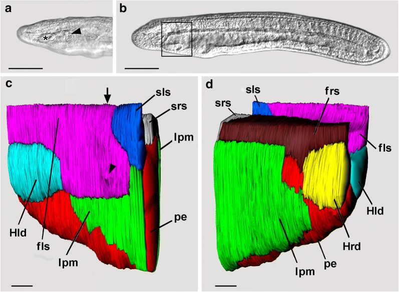
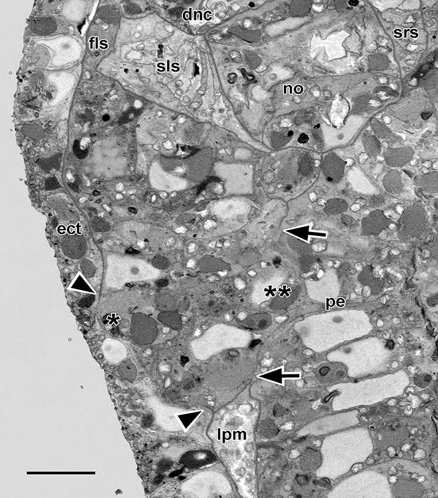
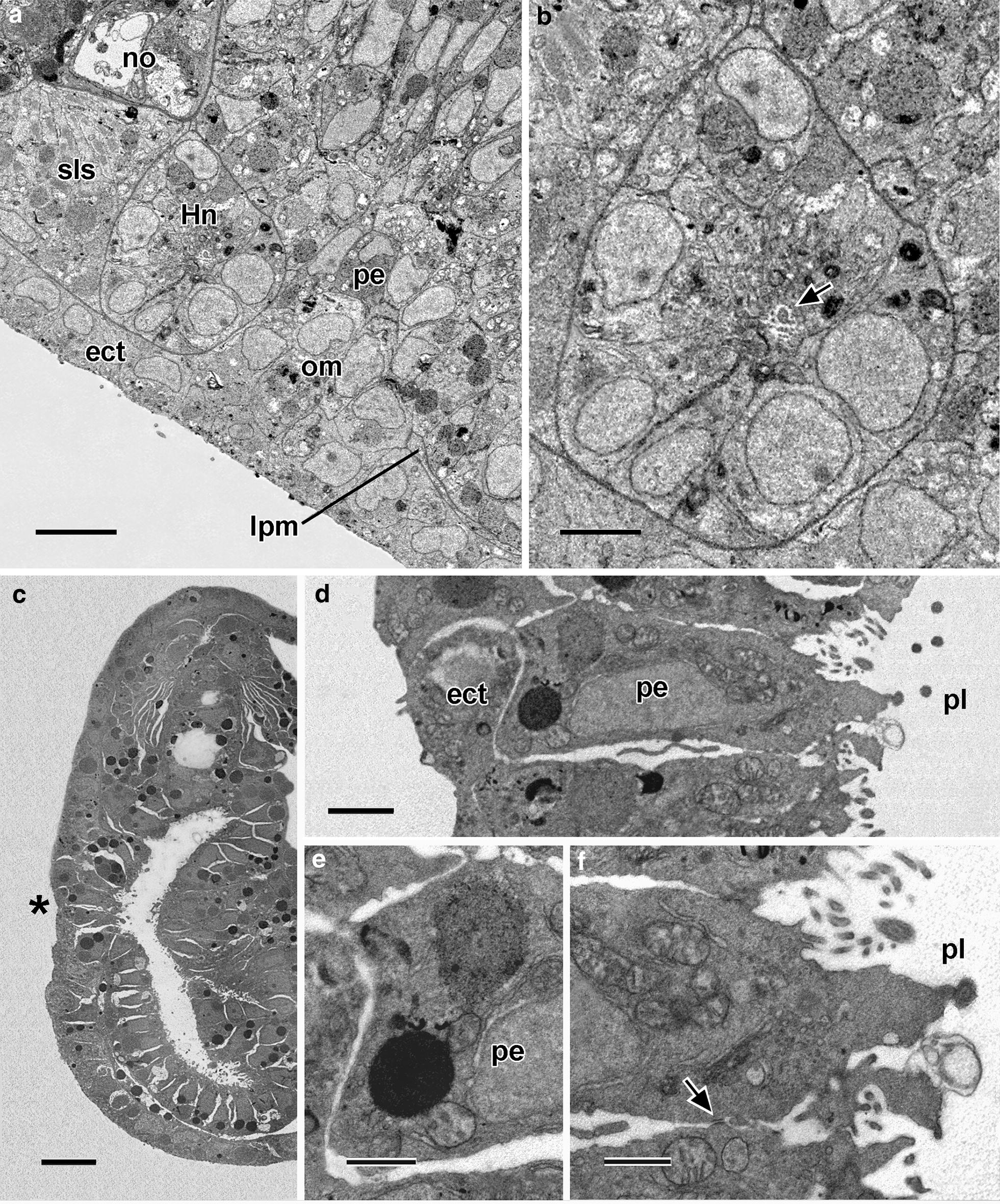
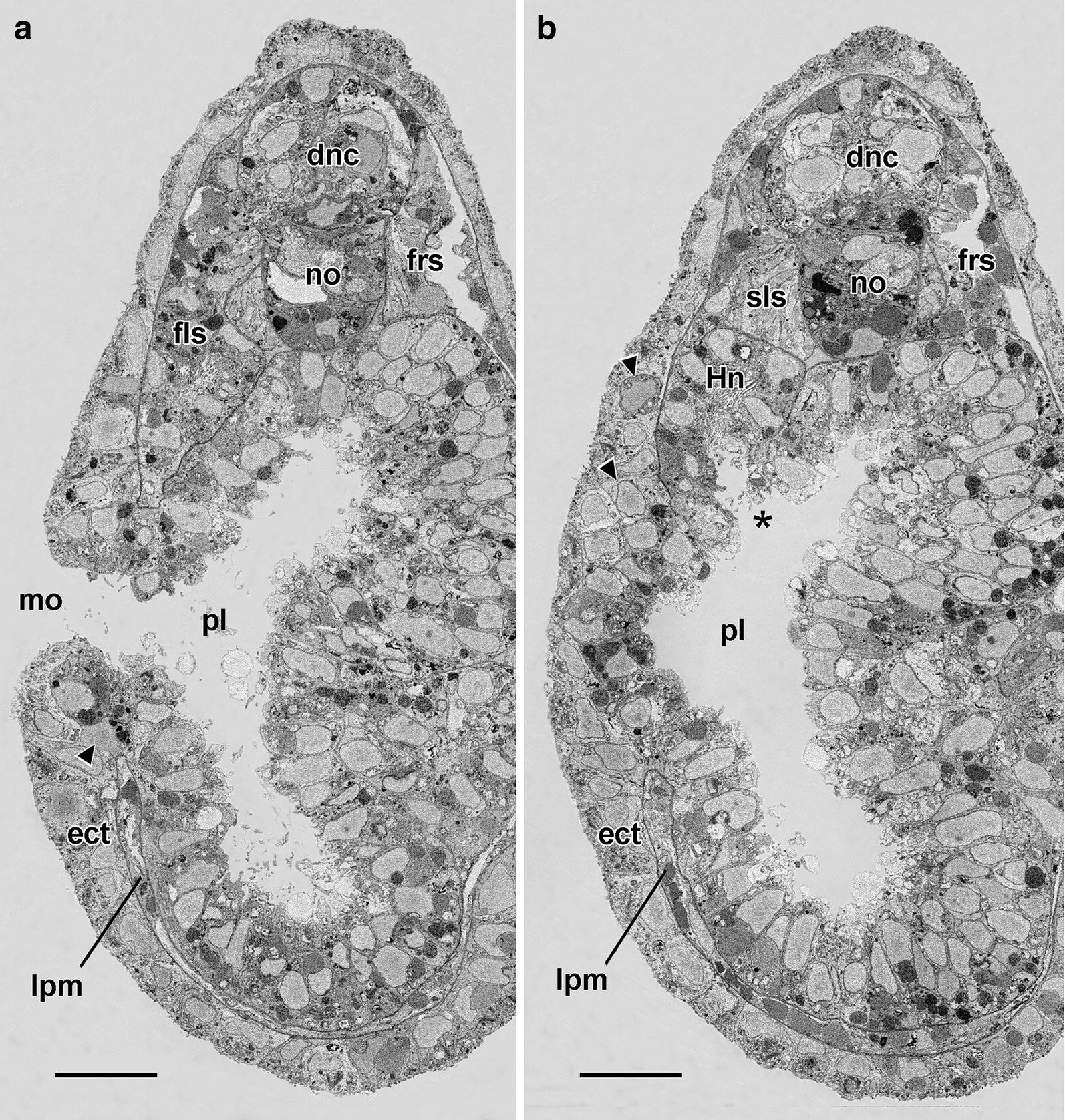
Background: For early larvae of amphioxus, Kaji et al. (Zool Lett 2:2, 2016) proposed that mesoderm cells are added to the rim of the forming mouth, giving it the quality of a coelomoduct without homology to the oral openings of other animals. They depended in part on non-serial transmission electron microscopic (TEM) sections and could not readily put fine structural details into a broader context. The present study of amphioxus larvae is based largely on serial blockface scanning electron microscopy (SBSEM), a technique revealing TEM-level details within an extensive anatomical volume that can be reconstructed in three dimensions.

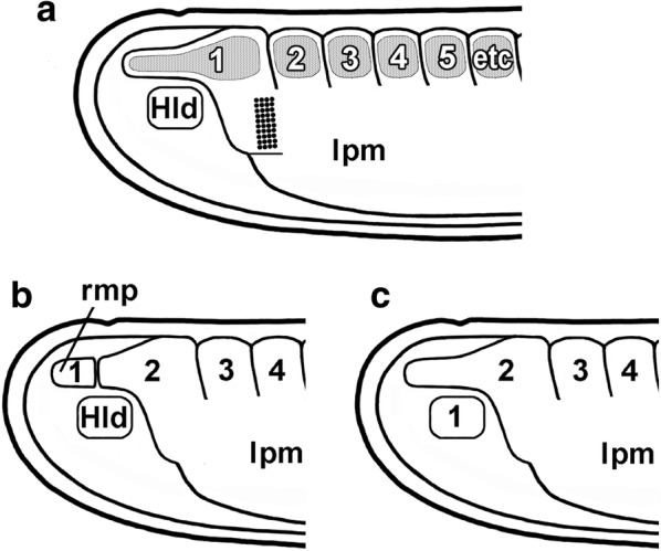
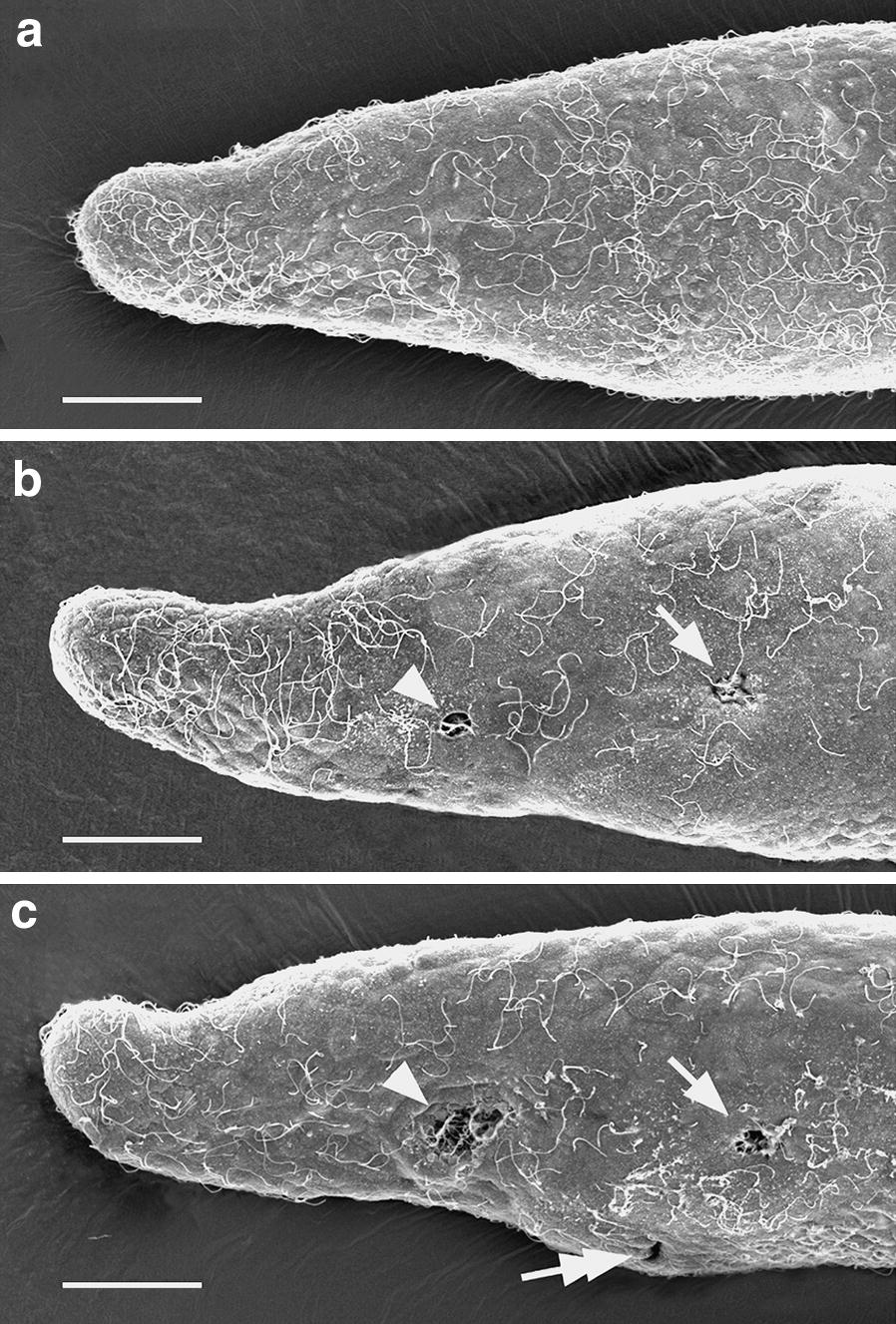
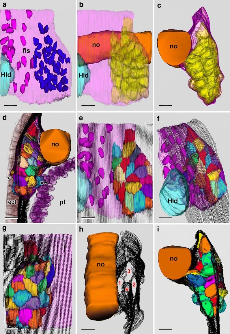
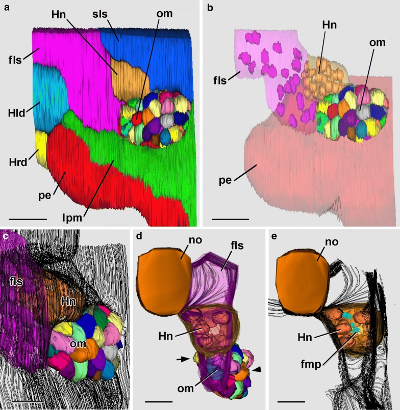
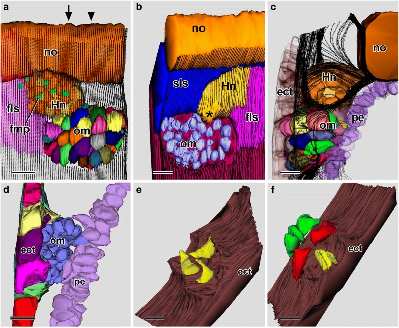
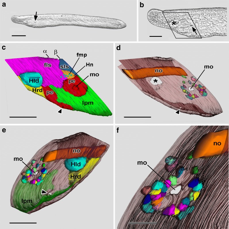
Results: In amphioxus larvae shortly before mouth formation, a population of compact mesoderm cells is present at the posterior extremity of the first left somite. As development continues, the more dorsal of these cells give rise to the initial kidney (Hatschek's nephridium), while the more ventral cells become interposed between the ectoderm and endoderm in a localized region where the mouth will soon penetrate. SBSEM reveals that, after the mouth has opened, a majority of these mesoderm cells can still be detected, sandwiched between the ectoderm and endoderm; they are probably myoblasts destined to develop into the perioral muscles.

Conclusions: SBSEM has provided the most accurate and detailed description to date of the tissues at the anterior end of amphioxus larvae. The present study supports the finding of Kaji et al. (2016) that the more dorsal of the cells in the posterior region of the first left somite give rise to the initial kidney. In contrast, the fate of the more ventral cells (called here the oral mesoderm) is less well understood. Although Kaji et al. (2016) implied that all of the oral mesoderm cells joined the rim of the forming mouth, SBSEM reveals that many of them are still present after mouth penetration. Even so, some of those cells go missing during mouth penetration and their fate is unknown. It cannot be ruled out that they were incorporated into the rim of the nascent mouth as proposed by Kaji et al. (2016). On the other hand, they might have degenerated or been shed from the larva during the morphogenetic interaction between the ectoderm and endoderm to form the mouth. The present SBSEM study, like Kaji et al. (2016), is based on static morphological data, and dynamic cell tracer experiments would be needed to decide among these possibilities.

Keywords: Amphioxus; Cephalochordata; Kidney; Lancelet; Mouth evolution; Serial blockface scanning electron microscopy (SBSEM).


Figures

Fig. 1
Fig. 1
The anterior region of amphioxus larvae before mouth opening. The somites (shaded) give rise to and are continuous with the lateral plate mesoderm (lpm). Historically, amphioxus somites have been numbered in three different ways. a The present study follows [43] in equating somites to myotomes (shaded). b Alternatively, Hatschek [44] counted a rostral muscular process (rmp) as the first myotome, but later abandoned this idea [45]. c In the third scheme, several authors [19, 26, 37, 46] defined Hatschek’s left (Hld) and right diverticula as the first somites. Coarse stippling in a indicates the zone of compact mesoderm cells in the first left somite
Fig. 2
Fig. 2
Conventional TEM of a cross section of a somite of an amphioxus larva shortly before mouth opening. The ectoderm (ect) is underlain by a basal lamina (arrowhead). The external cell layer (ecl) includes extremely thin cytoplasmic extensions (arrow) and is separated by an inconspicuous myocoel (black asterisk) from the myotome (mt). The myoblasts of the myotome contain myofilament bundles (white asterisks). Scale bar = 1 μm
Fig. 3
Fig. 3
Conventional SEM series to determine the time of mouth opening. a 23-h larva; none the ten sampled had a mouth. b 24-h larva; one of two larvae out of ten that had open mouth (arrow) and preoral ciliated pit (arrowhead). c 25-h larva; all of the ten sampled had a mouth (arrow), an opening of the club-shaped gland (tandem arrow), and a preoral ciliated pit (arrowhead). All scale bars = 25 μm
Fig. 4
Fig. 4
a DIC of anterior end of a living 22-h larva focused on the left surface, showing Hatschek’s left diverticulum (asterisk) and a population of compact mesoderm cells (arrowhead) comprising the posterior region of the first left somite. Scale bar = 100 μm. b DIC of a living 23-h larva with the midsagittal plane in focus. The boxed region indicates where SBSEM series were reconstructed in 3-D in c and d. Scale bar = 100 μm. c SBSEM of 23-h larva; left side view of a 3-D reconstruction; ectoderm is not shown. The arrowhead points to a small mesodermal protuberance showing where the mouth will later form. The arrow is explained in the following figure caption. Scale bar = 10 μm. d Right side view of a 3-D reconstruction. Scale bar = 10 μm. fls, first left somite; frs, first right somite; Hld, Hatschek’s left diverticulum; Hrd, Hatschek’s right diverticulum; lpm, lateral plate mesoderm; pe, pharyngeal endoderm; sls, second left somite; srs, second right somite
Fig. 5
Fig. 5
23-h larva. Single SBSEM scan through the arrowed level in Fig. 4c. The compact mesoderm cells of the first left somite bulge laterally (single asterisk) and medially (twin asterisk) toward the ectoderm and pharyngeal endoderm, respectively. For basal laminae, the arrowheads show the dorsoventral limits of the lateral window, and the arrowheads show the dorsoventral limits of the medial window. dnc, dorsal nerve cord; ect, ectoderm; fls, first left somite; lpm, lateral plate mesoderm; no, notochord; pe, pharyngeal endoderm; sls, second left somite; srs, second right somite. Scale bar = 5 μm
Fig. 6
Fig. 6
23-h larva. 3-D reconstructions based on SBSEM sequences. a Left side view of the posterior part of the first left somite (light pink) and part of Hatschek’s left diverticulum (light blue). Nuclei of the more anterior mesoderm cells are dark pink, and nuclei of the 48 compact mesoderm cells making up the posterior wall of the first left somite are dark blue. b Viewpoint of a with notochord orange and the first left somite light pink; the compact mesoderm cells of the posterior wall of the somite are shown in semitransparent yellow. c Similar to b, but turned to be viewed from the anterior. d View from posterior showing the compact mesoderm cells (in various colors) of the posterior wall of the first left somite; the notochord is orange, the nuclei of the endoderm cells are semitransparent purple, and the ectoderm cells (semitransparent brown) are locally compressed (asterisk) by the lateral protrusion of a few of the mesoderm cells; basal laminae are indicated by the parallel black traces. e View comparable to a, showing the nuclei of the more anterior mesoderm cells in dark pink, and the compact cells of the posterior wall in various colors; the basiepithelial basal lamina is indicated by the parallel black traces; a basal lamina-free area (the lateral window) is visible near the lower right. f The reconstruction in Fig. 5e turned to be viewed from the anterior left. g The compact mesoderm cells comprising the posterior wall of the first left somite viewed from the medial side; the subendodermal basal lamina (parallel black traces) is absent from an area (the medial window) near the bottom left. h The notochord and basal laminae (parallel black traces) of the left side from a dorso-anterior vantage point. The basal lamina ventral to the compact mesoderm cells of the first left somite is indicated by an asterisk; 1 and 2 are, respectively, the medial and lateral windows in the basal laminae, while 3 indicates the posterior communication of the first left somite with the lateral plate mesoderm. i Anterior view comparable to Fig. 5c; notochord is orange; compact mesoderm cells of the posterior wall of the first left somite in various colors, and basal lamina indicated by parallel black traces. All scale bars = 5 μm. ect, ectoderm; fls, first left somite, Hld, Hatschek’s left diverticulum, no, notochord; pl, pharyngeal lumen
Fig. 7
Fig. 7
24-h larva. 3-D reconstructions based on SBSEM sequences. a Left side view of a reconstruction of a region similar to Fig. 4c; individual cells are depicted for the oral mesoderm. Scale bar = 10 μm. b Same view as a, showing cells for the oral mesoderm; the nuclei of Hatschek’s nephridium and the more anterior mesoderm cells of the first left somite are show, respectively, in gold and pink. Scale bar = 10 μm. c Oblique view of the right anterior side of the larva showing nuclei for the first left somite and for Hatschek’s nephridium as well as the cells of the oral mesoderm, which tends to bulge outward through the lateral window in the subectodermal basal lamina (indicted by the parallel black traces). Scale bar = 5 μm. d Anterior view including nuclei of Hatschek’s nephridium (gold); the cells of the oral mesoderm (various colors) bulge laterally (arrowhead) and medially (arrow) in regions where the basal lamina (nor shown) has disappeared. Scale bar = 5 μm. e Anterior view comparable to d, but showing flagellar/microvillar processes (blue rectangles) and nuclei of the Hatschek’s nephridium (gold); basal laminae are indicated by the parallel black traces. Scale bar = 5 μm. fls, first left somite; fmp, flagellar/microvillar process; Hld, Hatschek’s left diverticulum; Hn, Hatschek’s nephridium; Hrd, Hatschek’s left diverticulum; lpm, lateral plate mesoderm; pe, pharyngeal endoderm; om, oral mesoderm; sls, second left somite
Fig. 8
Fig. 8
24-h larva. 3-D reconstructions based on SBSEM sequences. a Left-side structures from lateral viewpoint. The subectodermal basal lamina (indicated by parallel black traces) is interrupted (as a lateral window) over the oral mesoderm cells. Within Hatschek’s nephridium (rendered semitransparent) the cell nuclei (gold) are indicated as are several flagellar/microvillar processes (blue). The arrow and arrowhead are explained in the caption for Fig. 9. b Left-side structures from a medial viewpoint. The nuclei (blue) of the oral mesoderm cells are visible through a medial window in the subendodermal basal lamina. The asterisk marks an interruption in the basal lamina where Hatschek’s nephridium opens into the pharyngeal lumen. c View of left-side structures from the posterior showing that a basal lamina (parallel black traces) bounds Hatschek’s nephridium posteriorly but does not intervene between the oral mesoderm cells and the lateral plate mesoderm (toward viewer). d Similar view to c showing ectoderm cells (various colors), nuclei (blue) of the oral mesoderm, and nuclei (purple) of the pharyngeal endoderm. e Inner surface of the ectoderm (brown) viewed from anteriorly; dorsal and ventral are toward the upper right and lower left, respectively; three selected lateral cells of the oral mesoderm are shown in yellow. f Inner surface of the ectoderm (brown) viewed from posteriorly; dorsal and ventral are toward the upper right and lower left, respectively; for selected oral mesoderm cells, a lateral cell is yellow, a dorsal and a ventral cell are red, and three medial cells are green. ect, ectoderm; fls, first left somite; fmp, flagellar/microvillar process; Hld, Hatschek’s left diverticulum; Hn, Hatschek’s nephridium; no, notochord; pe, pharyngeal endoderm; om, oral mesoderm; sls, second left somite. All scale bars = 5 μm
Fig. 9
Fig. 9
24-h larva. a Single SBSEM scan through the level indicated by the arrow in Fig. 8a showing structures on the left side. Scale bar = 5 μm. b Enlargement of a showing Hatschek’s nephridium, including a flagellar/microvillar process (arrow). Scale bar = 2 μm. c 24.5-h larva. Conventional TEM cross section at the level of the arrowhead in Fig. 8a. The asterisk indicates the region of body wall where pharyngeal endoderm cells are contacting the ectoderm. Scale bar = 10 μm. d Enlargement of the cells nearest the asterisk in c; pharyngeal endoderm cell (toward left) extends basally to contact an ectoderm cell (toward left). Scale bar = 2 μm. e The basal region of pharyngeal endoderm cell in d associated with an ectoderm cell at the left. Scale bar = 1 μm. f The apical region of pharyngeal endoderm cell in d associated with an adjacent endoderm cell by an inconspicuous intermediate junction (arrow). Scale bar = 1 μm. ect, ectoderm; Hn, Hatschek’s nephridium; lpm, lateral plate mesoderm; no, notochord; om, oral mesoderm; pe, pharyngeal endoderm; om, oral mesoderm; sls, second left somite
Fig. 10
Fig. 10
25-h larva. a DIC of living 25-h larva with recently opened mouth (arrow). Scale bar = 100 μm. b Enlargement of anterior end of a; asterisk and arrow indicate, respectively, Hatschek’s left diverticulum and mouth. The rhombus indicates the rombohedral volume where SBSEM series were reconstructed in 3-D in ce. Scale bar = 50 μm. c Left side view of a 3-D reconstruction. The ectoderm is not shown. The arrowhead points to the external opening of the club-shaped gland; α and β indicate, respectively, the oblique planes of the SBSEM scans in Fig. 11a, b. Scale bar = 50 μm. d Same reconstruction as d, but showing the notochord and the semitransparent ectoderm, which is discontinuous over Hatschek’s left diverticulum (asterisk), opening of the club-shaped gland (arrowhead), and mouth (mo); the last is encircled by oral mesoderm cells (depicted in various colors). Scale bar = 50 μm. e Reconstruction like d, but turned to show left-side structures from a medio-posterior viewpoint. Scale bar = 50 μm. f enlargement of the oral region of e, with the subendodermal basal lamina indicated by parallel black traces. Scale bar = 20 μm. ect, ectoderm; fls, first left somite; fmp, flagellar/microvillar process; Hld, Hatschek’s left diverticulum; Hn, Hatschek’s nephridium; Hrd, Hatschek’s right diverticulum (often termed the rostral coelom by this stage of development); lpm, lateral plate mesoderm; mo, mouth; no, notochord; pe, pharyngeal endoderm; pl, pharyngeal lumen; om, oral mesoderm; sls, second left somite
Fig. 11
Fig. 11
25-h larva. a Single SBSEM scan through the oblique plane indicated by α in Fig. 10c. The arrowhead points to an oral mesoderm cell. Scale bar = 10 μm. b Single SBSEM scan through the oblique plane indicated by β in Fig. 10c. The arrowheads point to oral mesoderm cells, and the asterisk marks the opening of Hatschek’s nephridium into the pharyngeal lumen. Scale bar = 10 μm. dnc, dorsal nerve cord; ect, ectoderm; fls, first left somite; frs, first right somite; Hn, Hatschek’s nephridium; lpm, lateral plate mesoderm; mo, mouth; no, notochord; pl, pharyngeal lumen; sls, second left somite

Similar articles

Cited by

References

    1. Kaji T, Reimer JD, Morov AR, Kuratani S, Yasui K. Amphioxus mouth after dorso-ventral inversion. Zool Lett. 2016;2:2. doi: 10.1186/s40851-016-0038-3. - DOI - PMC - PubMed
    1. Peddie CJ, Collinson LM. Exploring the third dimension: volume electron microscopy comes of age. Micron. 2014;61:9–19. doi: 10.1016/j.micron.2014.01.009. - DOI - PubMed
    1. Titze B, Genoud C. Volume scanning electron microscopy for imaging biological ultrastructure. Biol Cell. 2016;108:307–323. doi: 10.1111/boc.201600024. - DOI - PubMed
    1. Holland ND. The long and winding path to understanding kidney structure in amphioxus—a review. Int J Dev Biol. 2017;61:683–688. doi: 10.1387/ijdb.170196nh. - DOI - PubMed
    1. Holland LZ, Yu JK. Cephalochordate (amphioxus) embryos: procurement, culture and basic methods. Methods Cell Biol. 2004;74:195–215. doi: 10.1016/S0091-679X(04)74009-1. - DOI - PubMed

LinkOut - more resources