

Association Between the Cilioretinal Artery and Choroidal Neovascularization in Age-Related Macular Degeneration: A Secondary Analysis From the Age-Related Eye Disease Study
- PMID: 29978186
- PMCID: PMC6142983
- DOI: 10.1001/jamaophthalmol.2018.2650
Association Between the Cilioretinal Artery and Choroidal Neovascularization in Age-Related Macular Degeneration: A Secondary Analysis From the Age-Related Eye Disease Study
Erratum in
-
Errors in Tables 2 and 3 and Results Section.JAMA Ophthalmol. 2019 Jul 1;137(7):856. doi: 10.1001/jamaophthalmol.2019.1384. JAMA Ophthalmol. 2019. PMID: 31046109 Free PMC article. No abstract available.
Abstract
Importance: A hemodynamic role in the pathogenesis of age-related macular degeneration (AMD) has been proposed, but to our knowledge, an association between retinal vasculature and late AMD has not been investigated.
Objective: To determine whether the presence and location of a cilioretinal artery may be associated with the risk of late AMD in the Age-Related Eye Disease Study (AREDS).
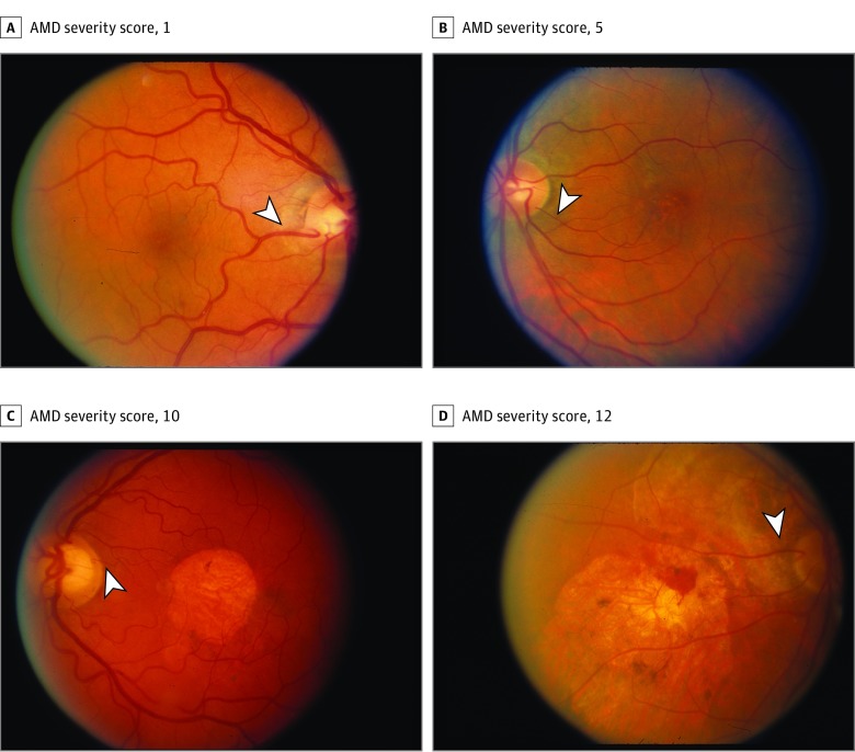
Design, setting, and participants: Retrospective analysis of prospective, randomized clinical trial data from 3647 AREDS participants. Fundus photographs of AREDS participants were reviewed by 2 masked graders for the presence or absence of a cilioretinal artery and whether any branch extended within 500 μm of the central macula. Multivariate regressions were used to determine the association of the cilioretinal artery and vessel location, adjusted for age, sex, and smoking status, with the prevalence of choroidal neovascularization (CNV) or central geographic atrophy (CGA) and AMD severity score for eyes at randomization and progression at 5 years.
Main outcomes and measures: Association of cilioretinal artery with prevalence and 5-year incidence of CNV or CGA.
Results: Among AREDS participants analyzed, mean (SD) age was 69.0 (5.0) years, with 56.3% female, 46.6% former smokers, and 6.9% current smokers. A total of 26.9% of patients had a cilioretinal artery in 1 eye, and 8.4% had the vessel bilaterally. At randomization, eyes with a cilioretinal artery had a lower prevalence of CNV (5.0% vs 7.6%; OR, 0.66; 95% CI, 0.51-0.85; P = .001) but no difference in CGA (1.1% vs 0.8%; OR, 1.33; 95% CI, 0.76-2.32; P = .31). In eyes without late AMD, those with a cilioretinal artery also had a lower mean (SD) AMD severity score (3.00 [2.35] vs 3.19 [2.40]; P = .02). At 5 years, eyes at risk with a cilioretinal artery had lower rates of progression to CNV (4.1% vs 5.5%; OR, 0.75; 95% CI, 0.56-1.00; P = .05) but no difference in developing CGA (2.2% vs 2.7%; OR, 0.83; 95% CI, 0.56-1.23; P = .35) or change in AMD severity score (0.65 [1.55] vs 0.73 [1.70]; P = .11). In patients with a unilateral cilioretinal artery, eyes with the vessel showed a lower prevalence of CNV than fellow eyes (4.7% vs 7.2%; P = .01).
Conclusions and relevance: The presence of a cilioretinal artery is associated with a lower risk of developing CNV, but not CGA, suggesting a possible retinal hemodynamic contribution to the pathogenesis of neovascular AMD.
Trial registration: ClinicalTrials.gov Identifier: NCT00000145.
Conflict of interest statement
Figures
Comment in
-
The Cilioretinal Artery-A Friend to Age-Related Macular Degeneration?JAMA Ophthalmol. 2018 Sep 1;136(9):1015-1016. doi: 10.1001/jamaophthalmol.2018.2644. JAMA Ophthalmol. 2018. PMID: 29978193 Free PMC article. No abstract available.
-
Statistical Issues on Evaluating Association Between the Cilioretinal Artery and Age-Related Macular Degeneration.JAMA Ophthalmol. 2019 Jul 1;137(7):855-856. doi: 10.1001/jamaophthalmol.2019.1112. JAMA Ophthalmol. 2019. PMID: 31046113 Free PMC article. No abstract available.
-
Statistical Issues on Evaluating Association Between the Cilioretinal Artery and Age-Related Macular Degeneration-Reply.JAMA Ophthalmol. 2019 Jul 1;137(7):856. doi: 10.1001/jamaophthalmol.2019.1115. JAMA Ophthalmol. 2019. PMID: 31046119 No abstract available.
References
Publication types
MeSH terms
Substances
Associated data
Grants and funding
LinkOut - more resources
Full Text Sources
Other Literature Sources
Medical
Research Materials
