Review
. 2018 Nov;45(12):2190-2200.
doi: 10.1007/s00259-018-4078-0. Epub 2018 Jul 6.

18F-NaF and 18F-FDG as molecular probes in the evaluation of atherosclerosis

Affiliations
Review

18F-NaF and 18F-FDG as molecular probes in the evaluation of atherosclerosis

Mikaela L McKenney-Drake et al. Eur J Nucl Med Mol Imaging. 2018 Nov.

Abstract

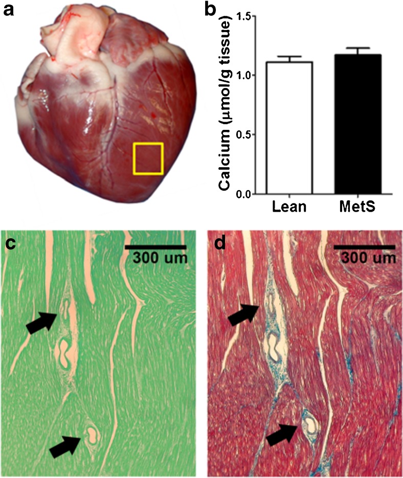
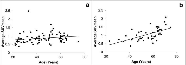
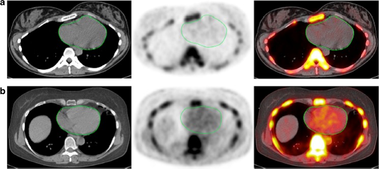
The early detection of atherosclerotic disease is vital to the effective prevention and management of life-threatening cardiovascular events such as myocardial infarctions and cerebrovascular accidents. Given the potential for positron emission tomography (PET) to visualize atherosclerosis earlier in the disease process than anatomic imaging modalities such as computed tomography (CT), this application of PET imaging has been the focus of intense scientific inquiry. Although 18F-FDG has historically been the most widely studied PET radiotracer in this domain, there is a growing body of evidence that 18F-NaF holds significant diagnostic and prognostic value as well. In this article, we review the existing literature on the application of 18F-FDG and 18F-NaF as PET probes in atherosclerosis and present the findings of original animal and human studies that have examined how well 18F-NaF uptake correlates with vascular calcification and cardiovascular risk.

Keywords: 18F-FDG; 18F-NaF; Atherosclerosis; Calcification; Cardiovascular disease quantification; Global assessment of cardiac disease.


Conflict of interest statement

None.

Figures

Fig. 1
Fig. 1
Progression from healthy arteries to complicated lesions. FDG and NaF uptake have long been known to precede vascular calcification evident on CT and intravascular ultrasonography (IVUS) [5, 6]. The paradigm shift is the stronger predictive power of NaF uptake and the occurrence of active calcification measured by 18F-NaF uptake in early coronary fatty streaks and preatheroma (CAC coronary artery calcium)
Fig. 2
Fig. 2
Coronary artery global molecular calcification score (GMCS) and percent injected dose per gram body weight 18F-NaF uptake. a A region of interest was drawn around the heart on each cardiac CT slice from which GMCS was calculated. b Pigs with metabolic syndrome (MetS; n = 11) had a GMCS almost 2.5-fold higher than lean pigs (n = 2; *p < 0.05)
Fig. 3
Fig. 3
IVUS images and quantification of early-stage CAD (type I, II, and III lesions). a, b, d Cross-sectional view of a coronary artery in Ossabaw pigs. a A lean pig (vascular wall traced in red). b A pig with metabolic syndrome and CAD (initial lumen traced in red, actual lumen traced in yellow); percent plaque burden = (initial − actual)/initial) × 100. c Pigs with metabolic syndrome (MetS; n = 7) had significantly more extensive CAD than lean pigs (n = 2) as shown by plaque burden quantification in the proximal 15 mm of the right coronary artery (p < 0.05). d One pig showed evidence of focal calcification (green arrow lesion, green lines acoustic shadowing). Distance between blue dots is 1 mm
Fig. 4
Fig. 4
Early stage CAD was quantified as percent wall coverage using IVUS. a Cross-sectional image of a coronary artery in an Ossabaw pig with metabolic syndrome with 100% wall coverage (concentric fatty streak, intimal thickening) and a plaque burden of about 13%, which would not be clinically significant. b Pigs with metabolic syndrome (MetS; n = 8) had a percent wall coverage about fivefold greater than lean pigs (n = 3; *p < 0.05). c RCA intimal wall coverage is significantly correlated with 18F-NaF uptake (p < 0.05). Distance between blue dots is 1 mm
Fig. 5
Fig. 5
Calcium content of the left ventricle. a Specimen was collected from the left ventricle away from any conduit artery (yellow box). b There is no difference in left ventricle calcium content between lean pigs (n = 6) and MetS pigs (n = 11). c, d Histopathology of the left ventricle: c von Kossa mineral staining on fast green background (showing mineral as black sediment)) showed no evidence of myocardial or microvasculature calcification in either lean pigs or MetS pigs; d Masson’s trichrome staining shows healthy nonfibrotic myocardium (arrows microvessels)
Fig. 6
Fig. 6
18F-NaF average SUVmean in the aortic arch wall in relation to age in healthy controls (a) and patients with cardiovascular risk factors (b). The average SUVmean (mean ± SD) in the aortic arch in healthy controls and patients were 0.87 ± 0.30 and 1.07 ± 0.37, respectively (p = 0.002). The Spearman correlation coefficients for the healthy controls and patients were 0.32 (p = 0.04) and 0.64 (p < 0.001), respectively
Fig. 7
Fig. 7
Transverse images (left CT, middle PET, right PET/CT) of the heart (green circles) in two clinically normal subjects (a 25 years old, b 61 years old). The global cardiac calcification scores were 12,492.44 in subject a and 18,424.70 in subject b. Normalizing the values to background NaF uptake increases the discrepancy between the subjects, resulting in 2.18 times the uptake in subject b than in subject a. Corresponding to the sites of NaF uptake in subject b, no structural calcification is seen on the corresponding CT scan and there is significant disparity between the PET and CT results. This is not an uncommon observation in this setting and clearly demonstrates the basis for assessment of cardiovascular calcification with these two different imaging modalities. While molecular imaging with NaF detects the earliest evidence for vascular calcification, evidence for calcification on CT largely reflects an end-stage disease process and therefore may be an irreversible pathologic state. Disparity between these two observations provides evidence for stage of calcification and has implications for the irreversibility of macrocalcification

References

    1. Lloyd-Jones D, Adams R, Carnethon M, De Simone G, Ferguson TB, Flegal K, et al. Heart disease and stroke statistics – 2009 update. Circulation. 2009;119:e21–e181. - PubMed
    1. Lozano R, Naghavi M, Foreman K, Lim S, Shibuya K, Aboyans V, et al. Global and regional mortality from 235 causes of death for 20 age groups in 1990 and 2010: a systematic analysis for the global burden of disease study 2010. Lancet. 2013;380:2095–2128. doi: 10.1016/S0140-6736(12)61728-0. - DOI - PMC - PubMed
    1. Fowkes FGR, Rudan D, Rudan I, Aboyans V, Denenberg JO, McDermott MM, et al. Comparison of global estimates of prevalence and risk factors for peripheral artery disease in 2000 and 2010: a systematic review and analysis. Lancet. 2013;382:1329–1340. doi: 10.1016/S0140-6736(13)61249-0. - DOI - PubMed
    1. Go AS, Mozaffarian D, Roger VL, Benjamin EJ, Berry JD, Blaha MJ, et al. Heart disease and stroke statistics – 2014 update: a report from the American Heart Association. Circulation. 2014;129:e28–e292. doi: 10.1161/CIRCULATIONAHA.113.003961. - DOI - PMC - PubMed
    1. McEvoy JW, Blaha MJ, DeFilippis AP, Budoff MJ, Nasir K, Blumenthal RS, et al. Coronary artery calcium progression: an important clinical measurement? A review of published reports. J Am Coll Cardiol. 2010;56:1613–1622. doi: 10.1016/j.jacc.2010.06.038. - DOI - PubMed

LinkOut - more resources