Review
. 2018 Oct;8(5):1483-1507.
doi: 10.1007/s13346-018-0551-3.

Metastatic and triple-negative breast cancer: challenges and treatment options

Affiliations
Review

Metastatic and triple-negative breast cancer: challenges and treatment options

Sumayah Al-Mahmood et al. Drug Deliv Transl Res. 2018 Oct.

Abstract

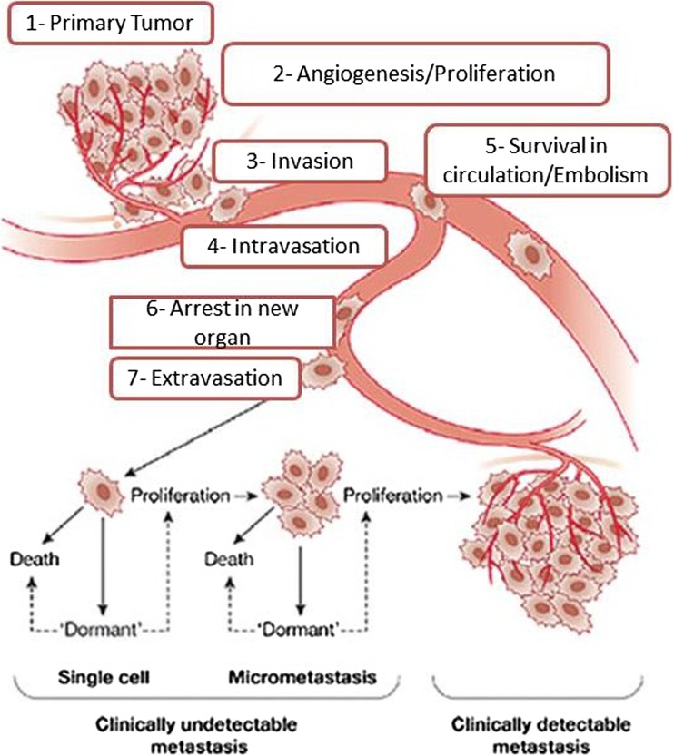
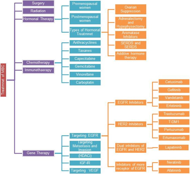
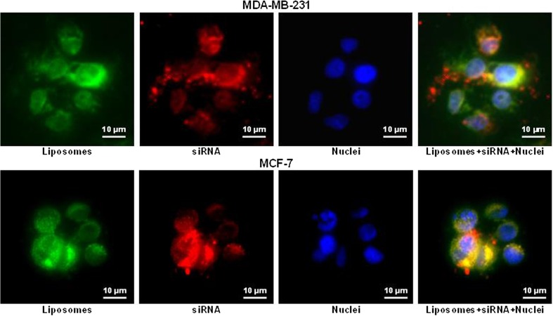
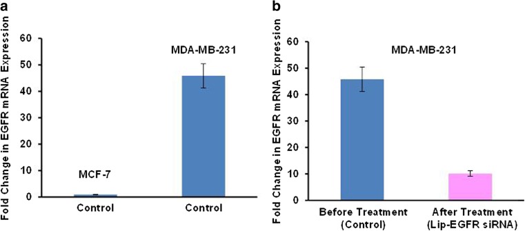
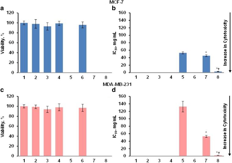
The major current conventional types of metastatic breast cancer (MBC) treatments include surgery, radiation, hormonal therapy, chemotherapy, or immunotherapy. Introducing biological drugs, targeted treatment and gene therapy can potentially reduce the mortality and improve the quality of life in patients with MBC. However, combination of several types of treatment is usually recommended. Triple negative breast cancer (TNBC) accounts for 10-20% of all cases of breast carcinoma and is characterized by the low expression of progesterone receptor (PR), estrogen receptor (ER), and human epidermal growth factor receptor 2 (HER2). Consequently, convenient treatments used for MBC that target these receptors are not effective for TNBC which therefore requires special treatment approaches. This review discusses the occurrence of MBC, the prognosis and predictive biomarkers of MBC, and focuses on the novel advanced tactics for treatment of MBC and TNBC. Nanotechnology-based combinatorial approach for the suppression of EGFR by siRNA and gifitinib is described.

Keywords: Combinatorial treatment of breast cancer; EGFR; Gefitinib; Liposomes; siRNA.


Conflict of interest statement

Conflict of interest

The authors declare that they have no conflict of interest.

Animal studies

All institutional and national guidelines for the care and use of laboratory animals were followed.

Figures

Fig. 1
Fig. 1
Major steps of breast cancer metastasis formation. Modified with permission from [39]
Fig. 2
Fig. 2
Role of CTCs in breast cancer in vitro and vivo. Modified and reproduced with permission from [55]
Fig. 3
Fig. 3
Models of MBC. a Tradition model of MBC. b Spontaneous metastasis assays. c Dynamic heterogeneity model. d Clonal dominance model. e Genometastasis hypothesis. f Gene expression profile. g Models of metastasis to lung, bone, and liver. h Parallel evolution model. i Breast cancer stem cells model. Pink represents non-metastasis breast tumor cells (good prognosis), red represents metastasis tumor cells (poor prognosis), yellow represents variant of tumor cells, green represents metastasis to bone, blue represents metastasis to liver, and purple represents metastasis to lung. Modified from [11]
Fig. 4
Fig. 4
The most common types of MBC treatment
Fig. 5
Fig. 5
The cellular internalization of siRNA delivered by liposomes. Representative images of human breast cancer (MDA-MB-231 and MCF-7) cells incubated within 24 h with liposomes (green fluorescence) containing siRNA (red fluorescence). Cell nuclei were stained with nuclear-specific dye (DAPI, blue fluorescence). Superimposition of red and green colors gives yellow color
Fig. 6
Fig. 6
The expression of EGFR mRNA. The relative quantity of EGFR gene expression in MCF-7 and MDA-MB-231 human breast cancer cells was calculated by the 2(DDCt) method using quantitative PCR. The levels of gene expression were represented as a fold change. Means ± SD are shown. a Expression of EGFR in MCF-7 and MDA-MB-231 cells incubated with media (control). b MDA-MB-231 cells before and after treatment. Cells were incubated within 24 h with liposomal siRNA targeted to EGFR mRNA (Lip-EGFR siRNA)
Fig. 7
Fig. 7
Viability of MCF-7 and MDA-MB-231 human breast cancer cells incubated 24 h with the indicated formulations. a, c Cytotoxicity of formulations that do not contain gefitinib in MCF-7 (a) and MDA-MB-231 (c) cells. b, d Cytotoxicity of formulations that contain gefitinib in MCF-7 (b) and MDA-MB-231 (d) cells. (1) Control (fresh media); (2) liposomes neutral; (3) liposomes cationic; (4) naked siRNA targeted to EGFR mRNA; (5) free gefitinib; (6) liposomal siRNA targeted to EGFR mRNA; (7) liposomal gefitinib; (8) liposomal siRNA targeted to EGFR mRNA + liposomal gefitinib. Means ± SD are shown. *P < 0.05 when compared with free gefitinib; P < 0.05 when compared with liposomal gefitinib
Fig. 8
Fig. 8
The role of MMPs in the progression and metastasis of cancer. Modified from [236]

References

    1. Lyden D, Welch DR, Psaila B. Cancer metastasis: biologic basis and therapeutics. Cambridge University Press; 2011.
    1. Gerratana L, Fanotto V, Bonotto M, Bolzonello S, Minisini AM, Fasola G, et al. Pattern of metastasis and outcome in patients with breast cancer. Clinical & Experimental Metastasis. 2015;32:125–133. - PubMed
    1. Kang Y, Siegel PM, Shu W, Drobnjak M, Kakonen SM, Cordón-Cardo C, et al. A multigenic program mediating breast cancer metastasis to bone. Cancer Cell. 2003;3:537–549. - PubMed
    1. Feys L, Descamps B, Vanhove C, Vral A, Veldeman L, Vermeulen S, et al. Radiation-induced lung damage promotes breast cancer lung-metastasis through CXCR4 signaling. Oncotarget. 2015;6:26615–26632. - PMC - PubMed
    1. Mansel WG, R. E., Fodstad, O. & Jiang. Metastasis of breast cancer. Netherland: Springer; 2007.

Publication types