

Newly characterized motile sperm domain-containing protein 2 promotes human breast cancer metastasis
- PMID: 29978511
- PMCID: PMC6588022
- DOI: 10.1002/ijc.31665
Newly characterized motile sperm domain-containing protein 2 promotes human breast cancer metastasis
Abstract
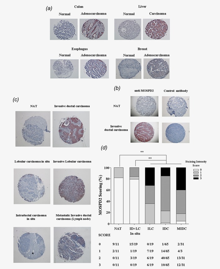
Breast cancer is the most frequently diagnosed cancer and the leading cause of cancer death among women worldwide. Breast cancer metastasis results in poor prognosis and increased mortality, but the mechanisms of breast cancer metastasis are yet to be fully resolved. Identifying distinctive proteins that regulate metastasis might be targeted to improve therapy in breast cancer. We previously described MOSPD2 as a surface membrane protein that regulates monocyte migration in vitro. In this study, we demonstrate for the first time that MOSPD2 has a major role in breast cancer cell migration and metastasis. MOSPD2 expression was highly elevated in invasive and metastatic breast cancer while it was absent or residual in normal tissue and in primary in situ tumors. In vitro experiments showed that silencing MOSPD2 in different breast cancer cell lines significantly inhibited cancer cell chemotaxis migration. Mechanistically, we found that silencing MOSPD2 profoundly abated phosphorylation events that are involved in breast tumor cell chemotaxis. In vivo, MOSPD2-silenced breast cancer cells exhibited marked impaired metastasis to the lungs. These results indicate that MOSPD2 plays a key role in the migration and metastasis of breast cancer cells and may be used to prevent the spreading of breast cancer cells and to mediate their death.
Keywords: MOSPD2; breast cancer; metastasis; migration.
© 2018 The Authors. International Journal of Cancer published by John Wiley & Sons Ltd on behalf of UICC.
Figures


Similar articles
-
MOSPD2 is a receptor mediating the LEAP-2 effect on monocytes/macrophages in a teleost, Boleophthalmus pectinirostris.Zool Res. 2020 Nov 18;41(6):644-655. doi: 10.24272/j.issn.2095-8137.2020.211. Zool Res. 2020. PMID: 33124217 Free PMC article.
-
Identification of Motile Sperm Domain-Containing Protein 2 as Regulator of Human Monocyte Migration.J Immunol. 2017 Mar 1;198(5):2125-2132. doi: 10.4049/jimmunol.1601662. Epub 2017 Jan 30. J Immunol. 2017. PMID: 28137892 Free PMC article.
-
MOSPD2 is a therapeutic target for the treatment of CNS inflammation.Clin Exp Immunol. 2020 Aug;201(2):105-120. doi: 10.1111/cei.13448. Epub 2020 May 18. Clin Exp Immunol. 2020. PMID: 32353176 Free PMC article.
-
CXCL16/CXCR6 chemokine signaling mediates breast cancer progression by pERK1/2-dependent mechanisms.Oncotarget. 2015 Jun 10;6(16):14165-78. doi: 10.18632/oncotarget.3690. Oncotarget. 2015. PMID: 25909173 Free PMC article.
-
Cullin3 promotes breast cancer cells metastasis and epithelial-mesenchymal transition by targeting BRMS1 for degradation.Oncotarget. 2015 Dec 8;6(39):41959-75. doi: 10.18632/oncotarget.5999. Oncotarget. 2015. PMID: 26544623 Free PMC article.
Cited by
-
Cell-free DNA analysis reveals POLR1D-mediated resistance to bevacizumab in colorectal cancer.Genome Med. 2020 Feb 22;12(1):20. doi: 10.1186/s13073-020-0719-6. Genome Med. 2020. PMID: 32087735 Free PMC article.
-
MOSPD2 is a receptor mediating the LEAP-2 effect on monocytes/macrophages in a teleost, Boleophthalmus pectinirostris.Zool Res. 2020 Nov 18;41(6):644-655. doi: 10.24272/j.issn.2095-8137.2020.211. Zool Res. 2020. PMID: 33124217 Free PMC article.
-
Deciphering MOSPD1's impact on breast cancer progression and therapeutic response.Biol Direct. 2024 Oct 5;19(1):88. doi: 10.1186/s13062-024-00531-9. Biol Direct. 2024. PMID: 39369222 Free PMC article.
-
Addressing the mean-variance relationship in spatially resolved transcriptomics data with spoon.bioRxiv [Preprint]. 2024 Nov 8:2024.11.04.621867. doi: 10.1101/2024.11.04.621867. bioRxiv. 2024. Update in: Biostatistics. 2024 Dec 31;26(1):kxaf012. doi: 10.1093/biostatistics/kxaf012. PMID: 39574747 Free PMC article. Updated. Preprint.
-
Screening of differentially expressed genes and identification of NUF2 as a prognostic marker in breast cancer.Int J Mol Med. 2019 Aug;44(2):390-404. doi: 10.3892/ijmm.2019.4239. Epub 2019 Jun 11. Int J Mol Med. 2019. PMID: 31198978 Free PMC article.
References
MeSH terms
Substances
LinkOut - more resources
Full Text Sources
Other Literature Sources
Medical
Molecular Biology Databases
