

Molecular regulation of MCU: Implications in physiology and disease
- PMID: 29980025
- PMCID: PMC6119482
- DOI: 10.1016/j.ceca.2018.06.006
Molecular regulation of MCU: Implications in physiology and disease
Abstract
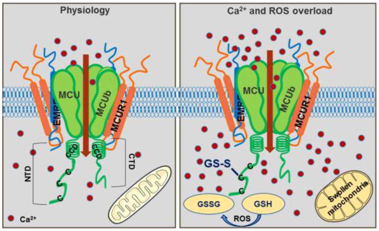
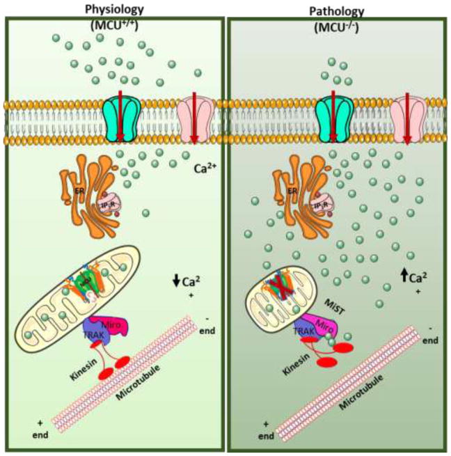
Ca2+ flux across the inner mitochondrial membrane (IMM) regulates cellular bioenergetics, intra-cellular cytoplasmic Ca2+ signals, and various cell death pathways. Ca2+ entry into the mitochondria occurs due to the highly negative membrane potential (ΔΨm) through a selective inward rectifying MCU channel. In addition to being regulated by various mitochondrial matrix resident proteins such as MICUs, MCUb, MCUR1 and EMRE, the channel is transcriptionally regulated by upstream Ca2+ cascade, post transnational modification and by divalent cations. The mode of regulation either inhibits or enhances MCU channel activity and thus regulates mitochondrial metabolism and cell fate.
Keywords: Calcium; Channel; EMRE; MCU; MCUR1; MCUb; MICU1; Magnesium; MiST; Miro1; Mitochondria; Oxidants; SLC25A23.
Copyright © 2018 Elsevier Ltd. All rights reserved.
Figures
References
-
- Berridge MJ, Lipp P, Bootman MD. The versatility and universality of calcium signalling. Nature Reviews Molecular Cell Biology. 2000;1:11. - PubMed
-
- Berridge MJ, Bootman MD, Roderick HL. Calcium: Calcium signalling: dynamics, homeostasis and remodelling. Nature Reviews Molecular Cell Biology. 2003;4:517–529. - PubMed
-
- Penner R, Fasolato C, Hoth M. Calcium influx and its control by calcium release. Curr Opin Neurobiol. 1993;3(3):368–74. - PubMed
-
- Putney JW, McKay RR. Capacitative calcium entry channels. BioEssays. 1999;21(1):38–46. - PubMed
-
- Gunter TE, Pfeiffer DR. Mechanisms by which mitochondria transport calcium. Am J Physiol. 1990;258(5 Pt 1):C755–86. - PubMed
Publication types
MeSH terms
Substances
Grants and funding
LinkOut - more resources
Full Text Sources
Other Literature Sources
Miscellaneous
