

Prosodic and phonetic subtypes of primary progressive apraxia of speech
- PMID: 29980072
- PMCID: PMC6171111
- DOI: 10.1016/j.bandl.2018.06.004
Prosodic and phonetic subtypes of primary progressive apraxia of speech
Erratum in
-
Corrigendum to "Prosodic and phonetic subtypes of primary progressive apraxia of speech" [Brain Lang. 184 (2018) 54-65].Brain Lang. 2020 Jun;205:104792. doi: 10.1016/j.bandl.2020.104792. Epub 2020 Apr 2. Brain Lang. 2020. PMID: 32248963 Free PMC article. No abstract available.
Abstract
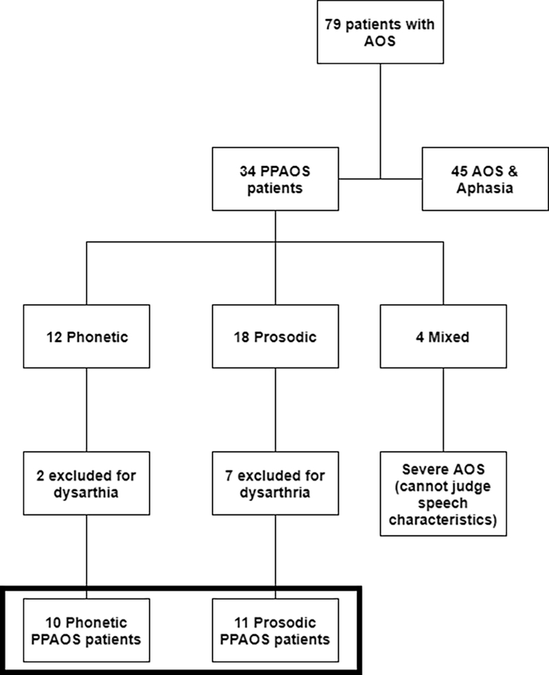
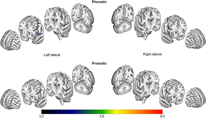
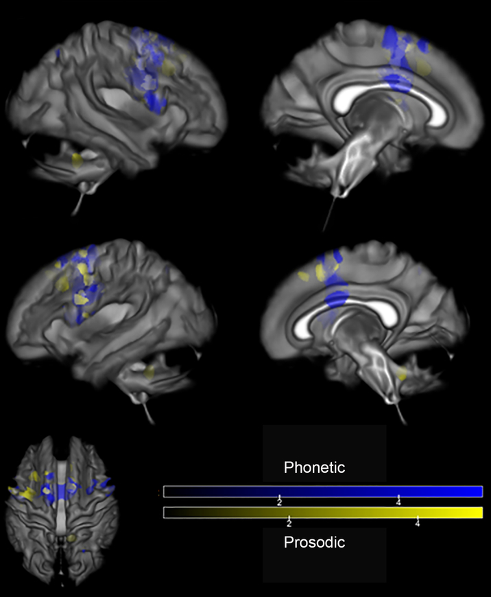
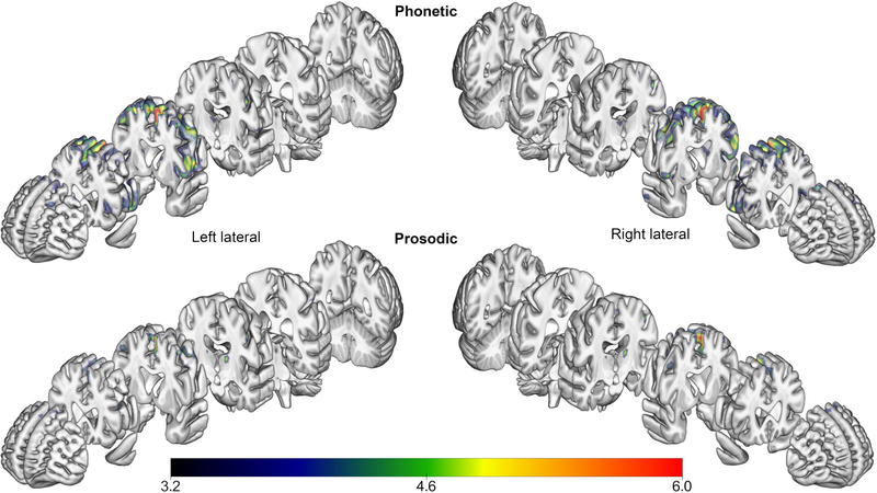
Primary progressive apraxia of speech (PPAOS) is a clinical syndrome in which apraxia of speech is the initial indication of neurodegenerative disease. Prior studies of PPAOS have identified hypometabolism, grey matter atrophy, and white matter tract degeneration in the frontal gyri, precentral cortex, and supplementary motor area (SMA). Recent clinical observations suggest two distinct subtypes of PPAOS may exist. Phonetic PPAOS is characterized predominantly by distorted sound substitutions. Prosodic PPAOS is characterized predominantly by slow, segmented speech. Demographic, clinical, and neuroimaging data (MRI, DTI, and FDG-PET) were analyzed to validate these subtypes and explore anatomic correlates. The Phonetic subtype demonstrated bilateral involvement of the SMA, precentral gyrus, and cerebellar crus. The Prosodic subtype demonstrated more focal involvement in the SMA and right superior cerebellar peduncle. The findings provide converging evidence that differences in the reliably determined predominant clinical characteristics of AOS are associated with distinct imaging patterns, independent of severity.
Keywords: Diffusion tensor imaging; Magnetic resonance imaging; Positron-emission tomography; Primary progressive aphasia; Primary progressive apraxia of speech.
Copyright © 2018 Elsevier Inc. All rights reserved.
Figures
References
-
- Alario FX, Chainay H, Lehericy S, & Cohen L (2006). The role of the supplementary motor area (SMA) in word production. Brain Research, 1076(1), 129–143. - PubMed
-
- Albert MS, DeKosky ST, Dickson D, Dubois B, Feldman HH, Fox NC, &al e. (2011). The diagnosis of mild cognitive impairment due to Alzheimer’s disease: Recommendations from the National Institute on Aging-Alzheimer’s Association workgroups on diagnostic guidelines for Alzheimer’s disease. Alzheimer’s and Dementia, 7, 270–279. - PMC - PubMed
-
- Ashburner J, & Friston KJ (2000). Voxel-Based Morphometry—The Methods. Neuroimage, 11(6), 805–821. - PubMed
-
- Ashburner J, & Friston KJ (2005). Unified segmentation. Neuroimage, 26(3), 839–851. - PubMed
Publication types
MeSH terms
Grants and funding
LinkOut - more resources
Full Text Sources
Other Literature Sources
Medical
