Vulvar Hemangioma: Case Report
- PMID: 29980161
- PMCID: PMC10316934
- DOI: 10.1055/s-0038-1657786
Vulvar Hemangioma: Case Report
Abstract
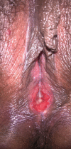
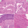
Hemangioma is a benign neoplasm that may affect the vulva, and it can cause functional or emotional disability. This article reports the case of a 52-year-old female patient with a history of a genital ulcer for the past 3 years and who had undergone various treatments with creams and ointments. The patient was biopsied and diagnosed with vulvar hemangioma and was subsequently submitted to surgical excision of the lesion. We emphasize the importance of following the steps of the differential diagnosis and proceeding with a surgical approach only if necessary.
O hemangioma é uma neoplasia benigna que pode afetar a vulva e pode causar incapacidade funcional ou emocional. Este artigo relata o caso de uma paciente de 52 anos com história de úlcera genital nos últimos 3 anos, submetida a diversos tratamentos com cremes e pomadas. A paciente foi biopsiada e diagnosticada com hemangioma vulvar e subsequentemente submetida a excisão cirúrgica da lesão. Ressaltamos a importância de seguir as etapas do diagnóstico diferencial e proceder a uma abordagem cirúrgica somente se necessário.
Thieme Revinter Publicações Ltda Rio de Janeiro, Brazil.
Conflict of interest statement
The authors have no conflicts of interest to declare.
Figures


References
-
- Gontijo B, Silva C MR, Pereira L B.Hemangioma da infância An Bras Dermatol 200378651–673.. Doi: 10.1590/S0365-05962003000600002
-
- Gampper T J, Morgan R F.Vascular anomalies: hemangiomas Plast Reconstr Surg 200211002572–585., quiz 586, discussion 587–588 - PubMed
-
- Pethe V V, Chitale S V, Godbole R N, Bidaye S V. Hemangioma of the ovary--a case report and review of literature. Indian J Pathol Microbiol. 1991;34(04):290–292. - PubMed
-
- Bava G L, Dalmonte P, Oddone M, Rossi U.Life-threatening hemorrhage from a vulvar hemangioma J Pediatr Surg 20023704E6. Doi: 10.1053/jpsu.2002.31645 - PubMed
-
- Cebesoy F B, Kutlar I, Aydin A.A rare mass formation of the vulva: giant cavernous hemangioma J Low Genit Tract Dis 2008120135–37.. Doi: 10.1097/LGT.0b013e3181255e85 - PubMed
Publication types
MeSH terms
LinkOut - more resources
Full Text Sources
Other Literature Sources
Medical
Miscellaneous
