

The Ashitaba (Angelica keiskei) Chalcones 4-hydroxyderricin and Xanthoangelol Suppress Melanomagenesis By Targeting BRAF and PI3K
- PMID: 29980517
- PMCID: PMC6317334
- DOI: 10.1158/1940-6207.CAPR-18-0092
The Ashitaba (Angelica keiskei) Chalcones 4-hydroxyderricin and Xanthoangelol Suppress Melanomagenesis By Targeting BRAF and PI3K
Abstract
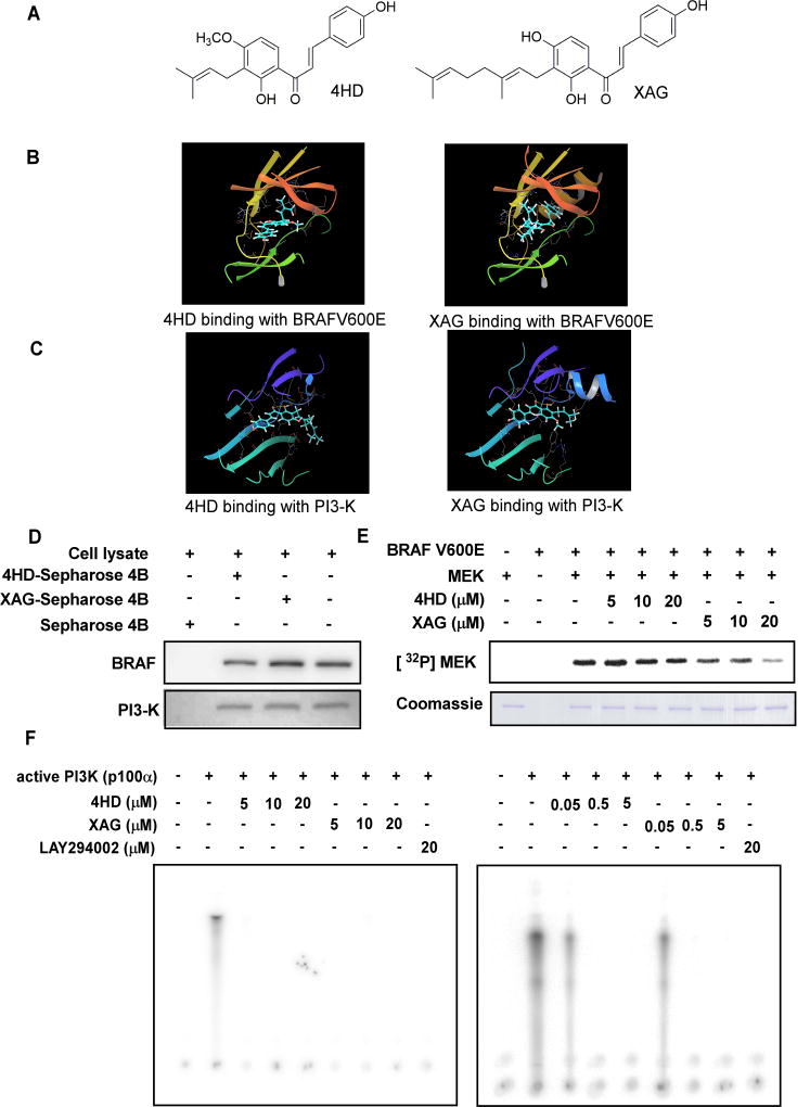
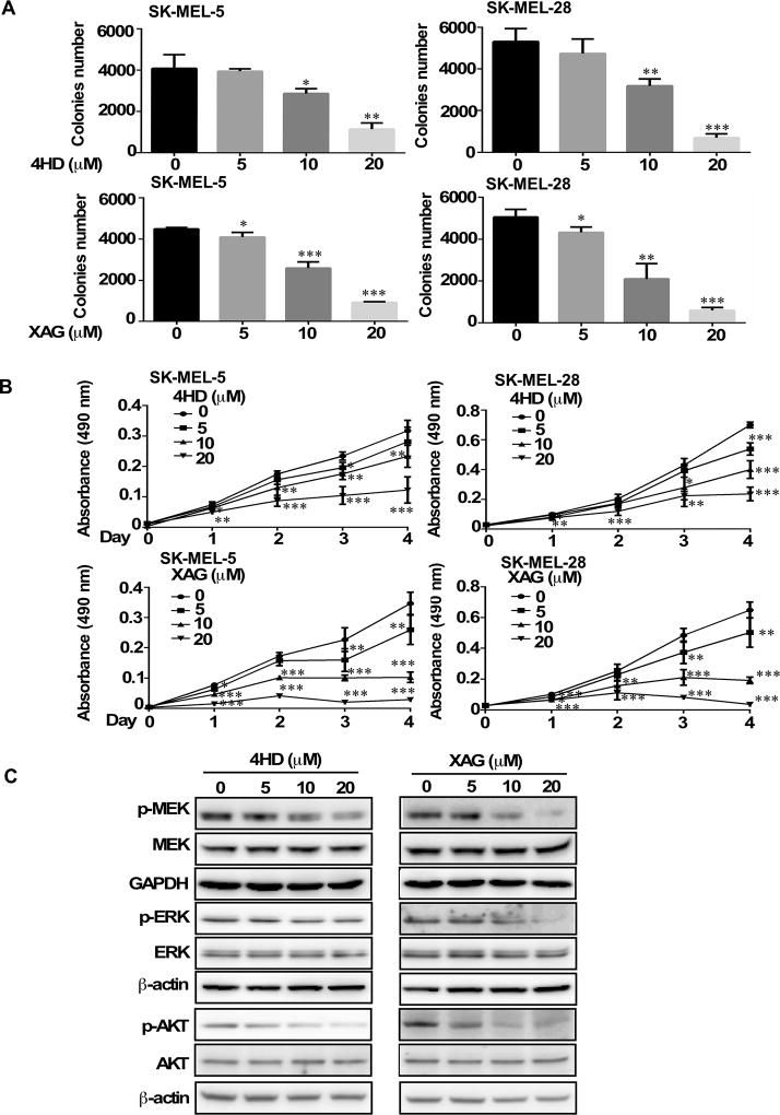
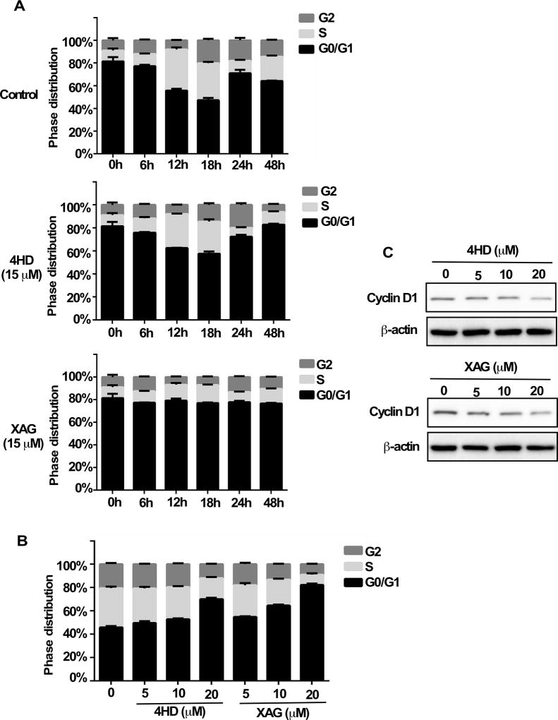
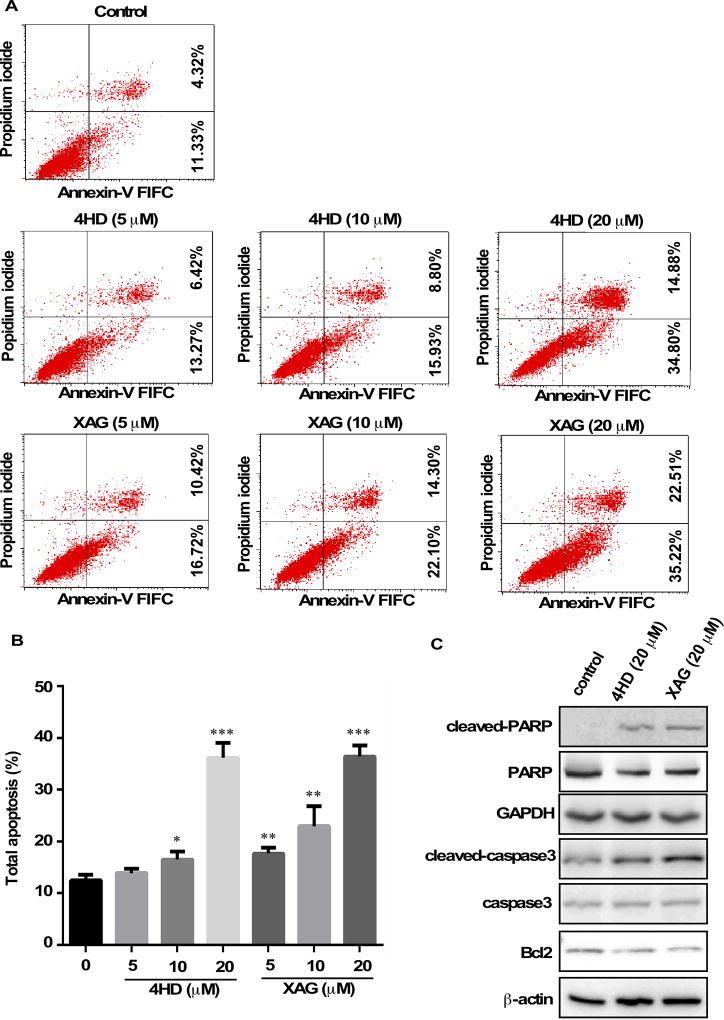
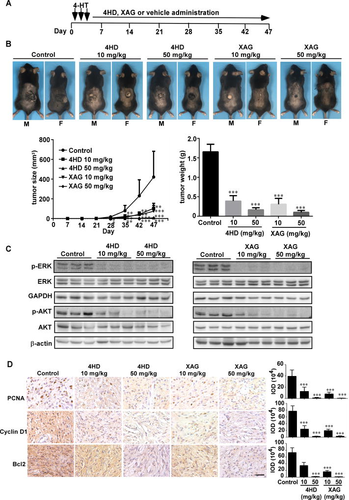
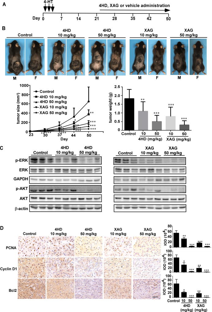
Malignant melanoma is an aggressive tumor of the skin and still lacks effective preventive and therapeutic treatments. In melanoma, both the BRAF/MEK/ERK and PI3-K/AKT signaling pathways are constitutively activated through multiple mechanisms, which result in cell-cycle progression and prevention of apoptosis. Therefore, the development of novel strategies for targeting BRAF and PI3K are of utmost importance. In this study, we found that Ashitaba (Angelica keiskei) chalcones, 4-hydroxyderricin (4HD) and xanthoangelol (XAG), suppressed melanoma development by directly targeting both BRAFV600E and PI3K, which blocked the activation of downstream signaling. This led to the induction of G1 phase cell-cycle arrest and apoptosis in melanoma cells. Importantly, 4HD or XAG dramatically attenuated tumor incidence and volume in the BRAF-activated Pten-deficient melanoma mouse model. Our findings suggest that 4HD and XAG are promising chemopreventive or potential therapeutic agents against melanomagenesis that act by targeting both BRAF and PI3K, providing hope for rapid clinical translation. Cancer Prev Res; 11(10); 607-20. ©2018 AACR.
©2018 American Association for Cancer Research.
Conflict of interest statement
Figures
Similar articles
-
4-Hydroxyderricin and xanthoangelol from Ashitaba (Angelica keiskei) suppress differentiation of preadiopocytes to adipocytes via AMPK and MAPK pathways.Mol Nutr Food Res. 2013 Oct;57(10):1729-40. doi: 10.1002/mnfr.201300020. Epub 2013 May 16. Mol Nutr Food Res. 2013. PMID: 23681764
-
Two chalcones, 4-hydroxyderricin and xanthoangelol, stimulate GLUT4-dependent glucose uptake through the LKB1/AMP-activated protein kinase signaling pathway in 3T3-L1 adipocytes.Nutr Res. 2015 Jul;35(7):618-25. doi: 10.1016/j.nutres.2015.05.010. Epub 2015 May 30. Nutr Res. 2015. PMID: 26077869
-
Two Prenylated Chalcones, 4-Hydroxyderricin, and Xanthoangelol Prevent Postprandial Hyperglycemia by Promoting GLUT4 Translocation via the LKB1/AMPK Signaling Pathway in Skeletal Muscle Cells.Mol Nutr Food Res. 2024 Mar;68(5):e2300538. doi: 10.1002/mnfr.202300538. Epub 2024 Jan 24. Mol Nutr Food Res. 2024. PMID: 38267744
-
Toxicological assessment of Ashitaba Chalcone.Food Chem Toxicol. 2015 Mar;77:111-9. doi: 10.1016/j.fct.2014.12.021. Epub 2015 Jan 7. Food Chem Toxicol. 2015. PMID: 25576957 Review.
-
Human relevance of NRAS/BRAF mouse melanoma models.Eur J Cell Biol. 2014 Jan-Feb;93(1-2):82-6. doi: 10.1016/j.ejcb.2013.10.010. Epub 2013 Nov 4. Eur J Cell Biol. 2014. PMID: 24342721 Review.
Cited by
-
4-Hydroxyderricin Promotes Apoptosis and Cell Cycle Arrest through Regulating PI3K/AKT/mTOR Pathway in Hepatocellular Cells.Foods. 2021 Aug 29;10(9):2036. doi: 10.3390/foods10092036. Foods. 2021. PMID: 34574146 Free PMC article.
-
Molecular Mechanisms of Antiproliferative Effects of Natural Chalcones.Cancers (Basel). 2021 May 31;13(11):2730. doi: 10.3390/cancers13112730. Cancers (Basel). 2021. PMID: 34073042 Free PMC article. Review.
-
Angelica keiskei Impacts the Lifespan and Healthspan of Drosophila melanogaster in a Sex and Strain-Dependent Manner.Pharmaceuticals (Basel). 2023 May 12;16(5):738. doi: 10.3390/ph16050738. Pharmaceuticals (Basel). 2023. PMID: 37242522 Free PMC article.
-
Effects of Microbial Transformation on the Biological Activities of Prenylated Chalcones from Angelica keiskei.Foods. 2022 Feb 14;11(4):543. doi: 10.3390/foods11040543. Foods. 2022. PMID: 35206019 Free PMC article.
-
Plants-Derived Biomolecules as Potent Antiviral Phytomedicines: New Insights on Ethnobotanical Evidences against Coronaviruses.Plants (Basel). 2020 Sep 21;9(9):1244. doi: 10.3390/plants9091244. Plants (Basel). 2020. PMID: 32967179 Free PMC article. Review.
References
-
- Owens B. Melanoma. Nature. 2014;515:S109. - PubMed
-
- Miller AJ, Mihm MC. Melanoma. The New England Journal of Medicine. 2006;355:51–65. - PubMed
-
- Davies H, Bignell GR, Cox C, Stephens P, Edkins S, Clegg S, et al. Mutations of the BRAF gene in human cancer. Nature. 2002;417:949–54. - PubMed
-
- Garnett MJ, Marais R. Guilty as charged: B-RAF is a human oncogene. Cancer Cell. 2004;6:313–9. - PubMed
-
- Dhomen N, Marais R. BRAF signaling and targeted therapies in melanoma. Hematol Oncol Clin North Am. 2009;23:529–45. ix. - PubMed
Publication types
MeSH terms
Substances
Grants and funding
LinkOut - more resources
Full Text Sources
Other Literature Sources
Medical
Research Materials
Miscellaneous
