. 2018 Aug 10;293(32):12429-12439.
doi: 10.1074/jbc.RA118.004096. Epub 2018 Jul 6.

Mechanism-based inhibition of human persulfide dioxygenase by γ-glutamyl-homocysteinyl-glycine

Affiliations

Mechanism-based inhibition of human persulfide dioxygenase by γ-glutamyl-homocysteinyl-glycine

Omer Kabil et al. J Biol Chem. .

Abstract

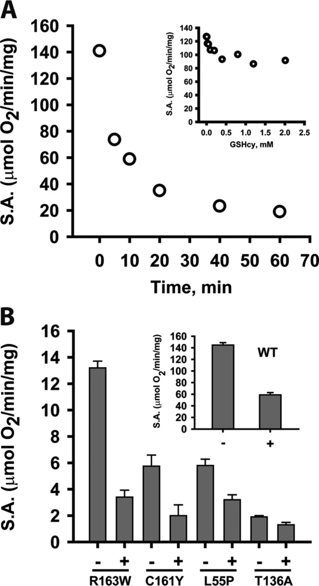
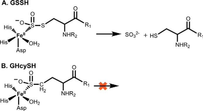
Hydrogen sulfide (H2S) is a signaling molecule with many beneficial effects. However, its cellular concentration is strictly regulated to avoid toxicity. Persulfide dioxygenase (PDO or ETHE1) is a mononuclear non-heme iron-containing protein in the sulfide oxidation pathway catalyzing the conversion of GSH persulfide (GSSH) to sulfite and GSH. PDO mutations result in the autosomal-recessive disorder ethylmalonic encephalopathy (EE). Here, we developed γ-glutamyl-homocysteinyl-glycine (GHcySH), in which the cysteinyl moiety in GSH is substituted with homocysteine, as a mechanism-based PDO inhibitor. Human PDO used GHcySH as an alternative substrate and converted it to GHcy-SO2H, mimicking GS-SO2H, the putative oxygenated intermediate formed with the natural substrate. Because GHcy-SO2H contains a C-S bond rather than an S-S bond in GS-SO2H, it failed to undergo the final hydrolysis step in the catalytic cycle, leading to PDO inhibition. We also characterized the biochemical penalties incurred by the L55P, T136A, C161Y, and R163W mutations reported in EE patients. The variants displayed lower iron content (1.4-11-fold) and lower thermal stability (1.2-1.7-fold) than WT PDO. They also exhibited varying degrees of catalytic impairment; the kcat/Km values for R163W, L55P, and C161Y PDOs were 18-, 42-, and 65-fold lower, respectively, and the T136A variant was most affected, with a 200-fold lower kcat/Km Like WT enzyme, these variants were inhibited by GHcySH. This study provides the first characterization of an intermediate in the PDO-catalyzed reaction and reports on deficits associated with EE-linked mutations that are distal from the active site.

Keywords: dioxygenase; enzyme kinetics; enzyme mechanism; ethylmalonic acid; ethylmalonic encephalopathy; hydrogen sulfide; iron; persulfide dioxygenase; sulfide oxidation.


Conflict of interest statement

The authors declare that they have no conflicts of interest with the contents of this article

Figures

Figure 1.
Figure 1.
Structural analysis of PDO mutations. The structure of human PDO (Protein Data Bank entry 4CHL) was used to map the locations of PDO mutations characterized in this study. A, iron (yellow sphere) ligands (His-79, His-135, and Asp-154) that form the 2-His-1-Asp facial triad are shown in stick representation. Locations of the six residues mutated in patients that were characterized previously and/or herein, Leu-55, Thr-136, Thr-152, Cys-161, Arg-163, and Asp-196, are shown by magenta spheres. B, close-up showing the side-chain interactions of Thr-136, which would be lost in the T136A mutation. C, close-up of Leu-55 displays interactions that would potentially be lost due to the L55P mutation. D, close-up of the interaction involving Cys-161, which would be perturbed in the C161Y mutant. E, close-up of Arg-163 highlights the interactions that might be lost in the R163W mutant.
Figure 2.
Figure 2.
Proposed reaction mechanism for PDO. A, in the resting state, Fe(II) is coordinated by a 2-His-1-Asp facial triad, and the remaining three coordination sites are occupied by waters (1). Binding of GSSH displaces a water molecule as the sulfane sulfur coordinates to the iron (2). Oxygen binding displaces a second water molecule and gives rise to the Fe(III)-superoxo complex (3), which exists in resonance with a structure (4) in which the coordinated sulfur has a radical cation character. Recombination of the iron-coordinated sulfur leads to formation of a cyclic-peroxo intermediate (5). Homolytic cleavage of the O–O bond results in a sulfoxy-cation and an Fe(II)-activated oxygen (6). Recombination (7) followed by hydrolysis yields the products, sulfite and GSH (8), and restores the enzyme to its resting state (1). B, structures of GSSH, GHcySH, and GSH.
Figure 3.
Figure 3.
Kinetic analysis of PDO mutants. A–D, the dependence of PDO activity on GSSH concentration for L55P (A), T136A (B), C161Y (C), and R163W (D) PDO. Oxygen consumption by PDO in 100 mm sodium phosphate buffer, pH 7.4, was monitored at 22 °C in the presence of varying concentrations of GSSH. The data are representative of three independent experiments. Data were fitted with the Hill equation as described under “Experimental procedures.” S.A., specific activity.
Figure 4.
Figure 4.
Spectral analysis of GSH and GHcySH binding to ferric-PDO. UV-visible spectra of ferric PDO solutions (150 μm) in 100 mm HEPES, pH 8, were recorded in the presence of increasing concentrations of GSH (A) and GHcySH (B) at 22 °C under aerobic conditions. The initial and final spectra are shown in solid lines in black and red, respectively. The change in absorbance at 600 nm was used to estimate the Kd for GSH (C) and GHcySH (D). Data are representative of three or four independent experiments and were fit to the Michaelis–Menten equation as described under “Experimental procedures.”
Figure 5.
Figure 5.
HPLC analysis of the PDO reaction mixture with GHcyS. A, HPLC analysis of a reaction mixture containing PDO (500 μm) and GHcySH (1 mm). The peaks with retention times of 7.5 min and 21 min represent DNB-GHcy-SO2H and DNB-GHcyS-CAA, respectively. B, a control sample was prepared as in A, but lacking PDO, and it showed DNB-GHcyS-CAA. In C and D, standards for DNB-GHcySH (C) and DNB-GHcyS-CAA (D) are shown. The peak at 4–5 min is associated with DNFB, which was present in excess.
Figure 6.
Figure 6.
MS analysis of the reaction of PDO with GHcySH. LC/MS spectra were extracted for the reaction mixture at 4.8 min and the control (minus PDO) sample at 20.3 min. A, for the reaction sample, peaks corresponding to DNB-labeled GHcySH (m/z = 488) and DNB-labeled GHcy-SO2H (m/z = 520) were observed. B, for the control sample, a peak corresponding to DNB-GHcyS-CAA (m/z = 546) was seen. C, MS/MS spectra were extracted at 4.8 min for DNB-GHcy-SO2H. Fragmentation produced peaks corresponding to the loss of DNB, glycine, and CH(NH2)COOH moiety of glutamate (m/z = 177). The CO2HCH2CH2NH2CHCO2H fragment of glutamate was also observed (m/z = 133). D, the MS/MS spectra were extracted at 21 min for DNB-GHcyS-CAA. Fragmentation produced peaks corresponding to the loss of glycine (m/z = 471). The fragment corresponding to protonated glutamate was also observed (m/z = 148).
Figure 7.
Figure 7.
Inhibition of WT and mutant PDOs by GHcySH. A, time-dependent inhibition of PDO by GHcySH. PDO activity was measured in the presence of GHcySH using the standard oxygen consumption assay as described under “Experimental procedures.” Solutions of 2 μg of PDO in 100 mm sodium phosphate buffer, pH 7.4, were pre-incubated aerobically with 1 mm GHcySH for various times (0–60 min) before sealing the reaction chamber and initiating the reaction by injecting GSSH (3 mm final concentration). The data are representative of three independent experiments. Inset, dependence of PDO inhibition on GHcySH concentration. PDO activity was measured in the presence of GHcySH as described under “Experimental procedures.” B, inhibition of WT and mutant PDOs with GHcySH. Reactions were performed under Vmax conditions for WT (inset) and mutant PDOs as described under “Experimental procedures” with 0.5–170 μg of enzyme pre-incubated with 1 mm GHcySH for 10 min before initiating the reaction with 3 mm GSSH. Reactions were performed at 22 °C. The data are representative of three independent experiments. S.A., specific activity.
Figure 8.
Figure 8.
Dioxygenation of GSSH versus GHcySH. A, with GSSH, the sulfinic acid intermediate undergoes hydrolysis, presumably by the iron-bound water, to give sulfite and GSH. B, with GHcySH, the corresponding sulfinic acid species represents a dead end product.

References

    1. Burlina A., Zacchello F., Dionisi-Vici C., Bertini E., Sabetta G., Bennet M. J., Hale D. E., Schmidt-Sommerfeld E., and Rinaldo P. (1991) New clinical phenotype of branched-chain acyl-CoA oxidation defect. Lancet 338, 1522–1523 10.1016/0140-6736(91)92338-3 - DOI - PubMed
    1. García-Silva M. T., Campos Y., Ribes A., Briones P., Cabello A., Santos Borbujo J., Arenas J., and Garavaglia B. (1994) Encephalopathy, petechiae, and acrocyanosis with ethylmalonic aciduria associated with muscle cytochrome c oxidase deficiency. J. Pediatr. 125, 843–844 10.1016/S0022-3476(06)80197-6 - DOI - PubMed
    1. Tiranti V., Briem E., Lamantea E., Mineri R., Papaleo E., De Gioia L., Forlani F., Rinaldo P., Dickson P., Abu-Libdeh B., Cindro-Heberle L., Owaidha M., Jack R. M., Christensen E., Burlina A., and Zeviani M. (2006) ETHE1 mutations are specific to ethylmalonic encephalopathy. J. Med. Genet. 43, 340–346 - PMC - PubMed
    1. Tiranti V., D'Adamo P., Briem E., Ferrari G., Mineri R., Lamantea E., Mandel H., Balestri P., Garcia-Silva M. T., Vollmer B., Rinaldo P., Hahn S. H., Leonard J., Rahman S., Dionisi-Vici C., et al. (2004) Ethylmalonic encephalopathy is caused by mutations in ETHE1, a gene encoding a mitochondrial matrix protein. Am. J. Hum. Genet. 74, 239–252 10.1086/381653 - DOI - PMC - PubMed
    1. Tiranti V., Viscomi C., Hildebrandt T., Di Meo I., Mineri R., Tiveron C., Levitt M. D., Prelle A., Fagiolari G., Rimoldi M., and Zeviani M. (2009) Loss of ETHE1, a mitochondrial dioxygenase, causes fatal sulfide toxicity in ethylmalonic encephalopathy. Nat. Med. 15, 200–205 10.1038/nm.1907 - DOI - PubMed

Publication types

MeSH terms

Associated data

LinkOut - more resources