

Lactate potentiates angiogenesis and neurogenesis in experimental intracerebral hemorrhage
- PMID: 29980670
- PMCID: PMC6035243
- DOI: 10.1038/s12276-018-0113-2
Lactate potentiates angiogenesis and neurogenesis in experimental intracerebral hemorrhage
Abstract
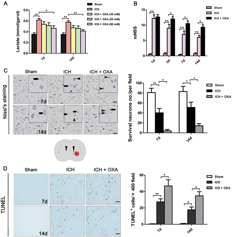
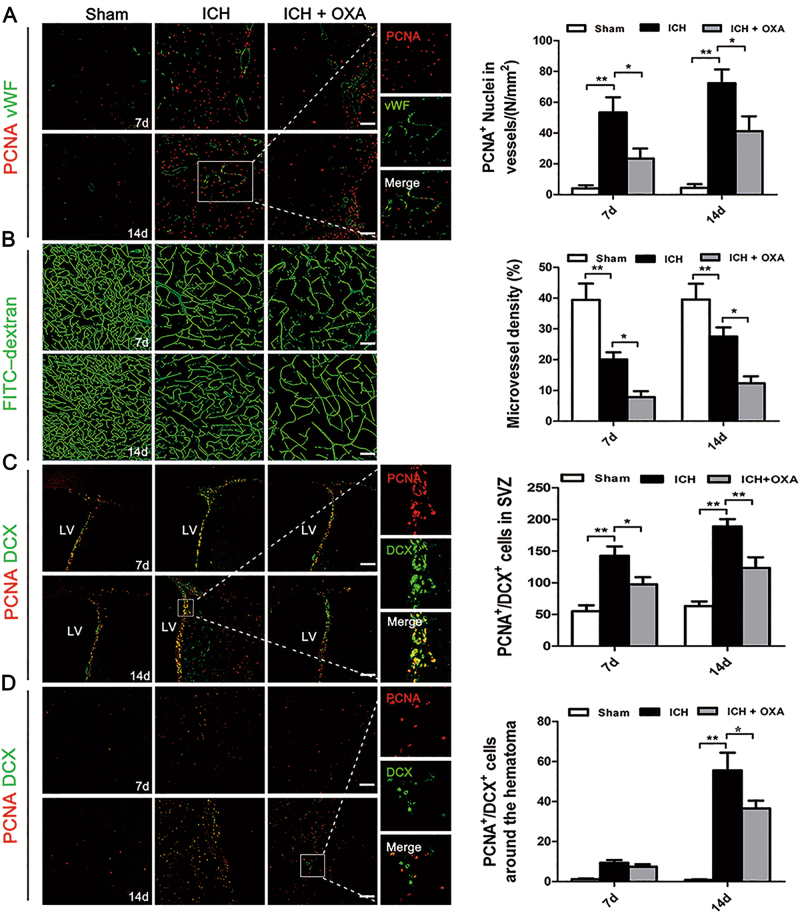
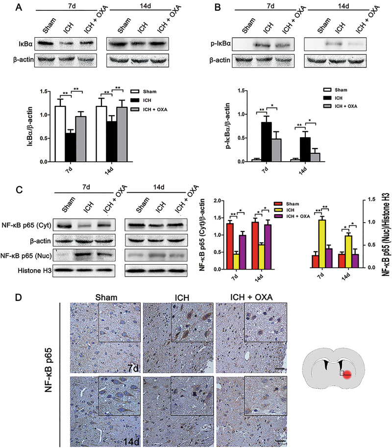
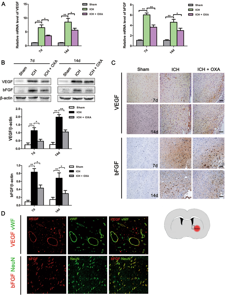
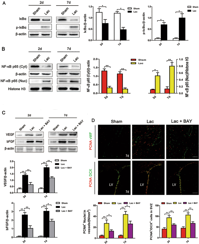
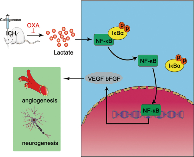
Lactate accumulation has been observed in the brain with intracerebral hemorrhage (ICH). However, the outcome of lactate accumulation has not been well characterized. Here, we report that lactate accumulation contributes to angiogenesis and neurogenesis in ICH. In the first set of the experiment, a rat model of ICH was induced by injecting collagenase into the brain. The effects of lactate accumulation on the neurological function, apoptosis, and numbers of newborn endothelial cells and neurons, as well as the proliferation-associated signaling pathway, were evaluated in the rat brain. In the second set, exogenous L-lactate was infused into intact rat brains so that its effects could be further assessed. Following ICH, lactate accumulated around the hematoma; the numbers of PCNA+/vWF+ nuclei and PCNA+/DCX+ cells were significantly increased compared with the numbers in the Sham group. Moreover, ICH induced translocation of nuclear factor-kappa B (NF-κB) p65 into the nucleus, resulting in a notable upregulation of VEGF and bFGF mRNAs and proteins compared with the levels in the Sham controls. Administration of a lactate dehydrogenase inhibitor dramatically inhibited these effects, decreased the vascular density, and aggravated neurological severity scores and apoptosis after ICH. After exogenous L-lactate infusion, the numbers of PCNA+/vWF+ nuclei and PCNA+/DCX+ cells were strikingly increased compared with the numbers in the Sham controls. In addition, lactate facilitated NF-κB translocation to induce increased transcription of VEGF and bFGF. Co-infusion with an NF-κB inhibitor significantly inhibited these effects. These data suggest that lactate potentiates angiogenesis and neurogenesis by activating the NF-κB signaling pathway following ICH.
Conflict of interest statement
The authors declare that they have no conflict of interest.
Figures
Similar articles
-
Leukemia Inhibitory Factor Decreases Neurogenesis and Angiogenesis in a Rat Model of Intracerebral Hemorrhage.Curr Med Sci. 2019 Apr;39(2):298-304. doi: 10.1007/s11596-019-2034-2. Epub 2019 Apr 23. Curr Med Sci. 2019. PMID: 31016525
-
Cerebral angiogenesis after collagenase-induced intracerebral hemorrhage in rats.Brain Res. 2007 Oct 17;1175:134-42. doi: 10.1016/j.brainres.2007.08.028. Epub 2007 Aug 19. Brain Res. 2007. PMID: 17888890
-
Effects of high-mobility group box1 on cerebral angiogenesis and neurogenesis after intracerebral hemorrhage.Neuroscience. 2013 Jan 15;229:12-9. doi: 10.1016/j.neuroscience.2012.10.054. Epub 2012 Nov 5. Neuroscience. 2013. PMID: 23137544
-
Buyang huanwu decoction promotes angiogenesis via vascular endothelial growth factor receptor-2 activation through the PI3K/Akt pathway in a mouse model of intracerebral hemorrhage.BMC Complement Altern Med. 2015 Mar 28;15:91. doi: 10.1186/s12906-015-0605-8. BMC Complement Altern Med. 2015. PMID: 25886469 Free PMC article.
-
Stem Cell Therapy: A Promising Therapeutic Method for Intracerebral Hemorrhage.Cell Transplant. 2018 Dec;27(12):1809-1824. doi: 10.1177/0963689718773363. Epub 2018 Jun 5. Cell Transplant. 2018. PMID: 29871521 Free PMC article. Review.
Cited by
-
Astrocytes, reactive astrogliosis, and glial scar formation in traumatic brain injury.Neural Regen Res. 2025 Apr 1;20(4):973-989. doi: 10.4103/NRR.NRR-D-23-02091. Epub 2024 May 17. Neural Regen Res. 2025. PMID: 38989932 Free PMC article.
-
Hypoxia induced lactate acidosis modulates tumor microenvironment and lipid reprogramming to sustain the cancer cell survival.Front Oncol. 2023 Jan 25;13:1034205. doi: 10.3389/fonc.2023.1034205. eCollection 2023. Front Oncol. 2023. PMID: 36761981 Free PMC article. Review.
-
Effects of lactate and carbon monoxide interactions on neuroprotection and neuropreservation.Med Gas Res. 2021 Oct-Dec;11(4):158-173. doi: 10.4103/2045-9912.318862. Med Gas Res. 2021. PMID: 34213499 Free PMC article.
-
The Key Role of Mitochondrial Function in Health and Disease.Antioxidants (Basel). 2023 Mar 23;12(4):782. doi: 10.3390/antiox12040782. Antioxidants (Basel). 2023. PMID: 37107158 Free PMC article. Review.
-
MCT4-driven CAF-mediated metabolic reprogramming in breast cancer microenvironment is a vulnerability targetable by miR-425-5p.Cell Death Discov. 2024 Mar 14;10(1):140. doi: 10.1038/s41420-024-01910-x. Cell Death Discov. 2024. PMID: 38485929 Free PMC article.
References
Publication types
MeSH terms
Substances
LinkOut - more resources
Full Text Sources
Other Literature Sources
Miscellaneous
