. 2018 Jul 6;9(1):2644.
doi: 10.1038/s41467-018-05062-2.

Intermedin protects against sepsis by concurrently re-establishing the endothelial barrier and alleviating inflammatory responses

Affiliations

Intermedin protects against sepsis by concurrently re-establishing the endothelial barrier and alleviating inflammatory responses

Fei Xiao et al. Nat Commun. .

Abstract

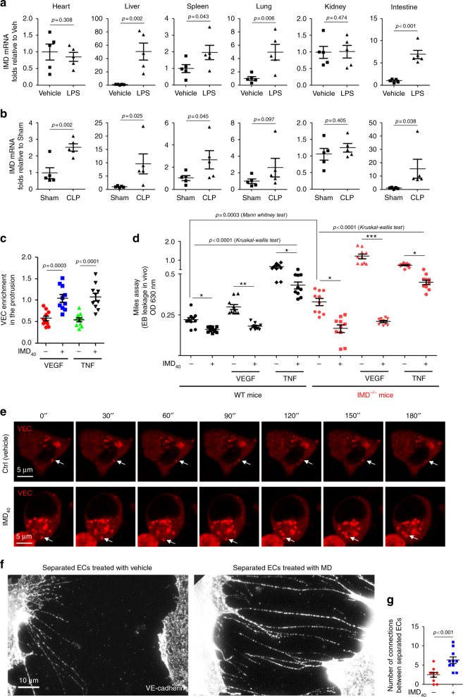
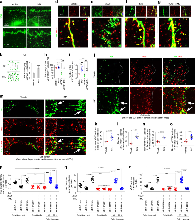
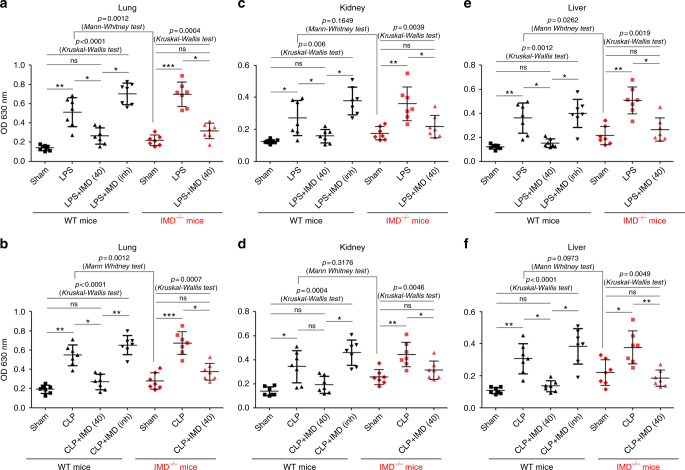
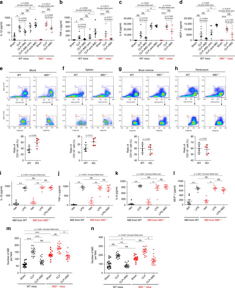
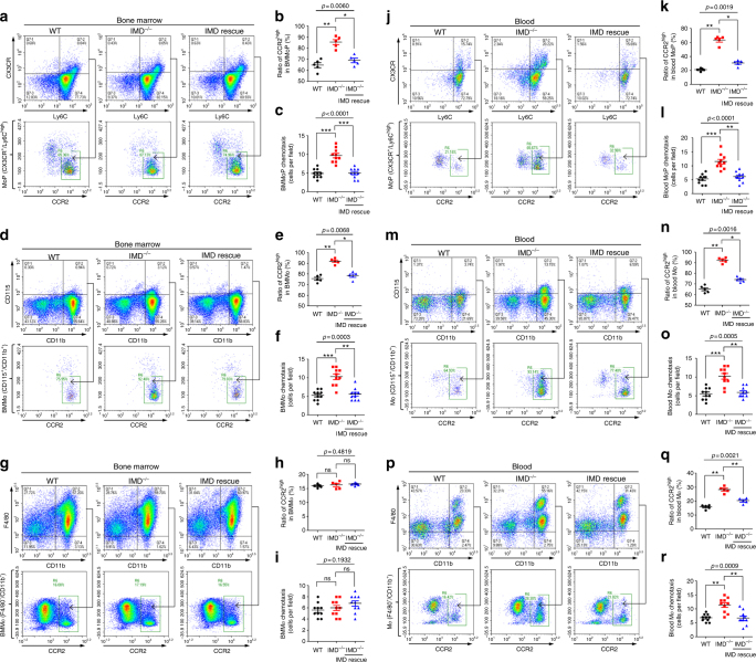
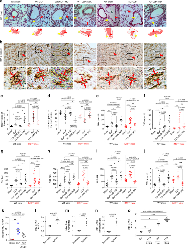
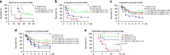
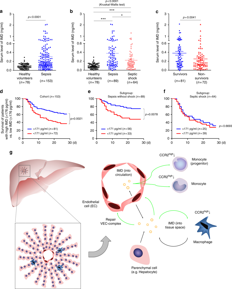
Sepsis is a life-threatening condition caused by dysregulated host responses to infection. Widespread vascular hyperpermeability and a "cytokine storm" are two pathophysiological hallmarks of sepsis. Here, we show that intermedin (IMD), a member of the calcitonin family, alleviates organ injury and decreases mortality in septic mice by concurrently alleviating vascular leakage and inflammatory responses. IMD promotes the relocation of vascular endothelial cadherin through a Rab11-dependent pathway to dynamically repair the disrupted endothelial junction. Additionally, IMD decreases inflammatory responses by reducing macrophage infiltration via downregulating CCR2 expression. IMD peptide administration ameliorates organ injuries and significantly improves the survival of septic mice, and the experimental results correlate with the clinical data. Patients with high IMD levels exhibit a lower risk of shock, lower severity scores, and greatly improved survival outcomes than those with low IMD levels. Based on our data, IMD may be an important self-protective factor in response to sepsis.


Conflict of interest statement

The authors declare no competing interests.

Figures

Fig. 1
Fig. 1
IMD was significantly up-regulated in septic mice, repairing the endothelial junction by promoting VEC relocation. a, b The Balb/c mice received intraperitoneally (i.p.) injection of LPS (24 mg/kg) or vehicle (saline), or had sham or CLP surgery. After 9 h, mice were sacrificed, and level of IMD mRNA in major organs was measured by real-time RT-PCR. c The HUVEC monolayer was treated with vehicle (PBS), IMD40 (2 μM), VEGF (50 ng/ml), TNF-α (20 ng/ml) alone, or treated with VEGF or TNF-α for 2 h followed by treatment of IMD40. The density of VEC signal referred to F-actin at the cell–cell contact was quantified using 10 randomly chosen fields from two experiments. d The Miles assay was performed as described in Methods. The Evans-Blue (EB) leakage (OD 630 nm) of WT or IMD−/− mice was quantified (n = 10 mice). e HUVECs were transfected with the lentivirus that expresses mCherry-tagged VEC (red), and a time-lapse photography was performed with a 30-s interval. The arrows indicate the location of a single VEC endosome at each time point. f The completely separated ECs were re-connected by the extended filopodia with the presence of IMD40. g The number of anastomosed filopodia connecting two completely separated HUVECs was quantified using 10 randomly chosen fields from two experiments. The data of (a–d, g) were presented as scatter plots with mean ± SEM. Significance was assessed by Mann–Whitney test (a–c,g) and one-way ANOVA (Kruskal–Wallis test) followed by non-parametric Dunn’s post-hoc analysis (d)
Fig. 2
Fig. 2
Rab11 facilitates the IMD-induced VEC re-localization. a The HUVECs treated with vehicle (PBS) or IMD40 (2 μM) were stained with VEC (green). b The schematic of the VEC endosome exchange at the cell–cell contact. c The VEC exchange was quantified using 10 randomly chosen fields from two experiments and expressed relative to the vehicle group. d–g The Antibody feeding assay was performed as described in Methods. The HUVEC monolayer was treated with PBS, IMD, VEGF, or VEGF plus IMD. The internalized VEC endosomes were detected by pre-incubation of anti-VEC (green), and double-stained with anti-Rab11 (red). The yellow arrows indicate the VEC+/Rab11+ double-positive vesicles, the yellow arrowheads indicate the Rab11+ vesicles fused to the VEC-complexes, and the green arrows indicate the VEC+ endosomes that did not co-localized with Rab11. h, i The percentage of VEC+/Rab11+ vesicles in total VEC+ vesicles and the absolute number of them were quantified using 10 randomly chosen fields. j, m The Antibody feeding assay showed the VEC+ and Rab11+ vesicles at the cell border of HUVECs. k, l, n, o The number of VEC+ or VEC+/Rab11+ double-positive vesicles in the area of cell border where cells did not contact with adjacent ones (k,l), or the area from where the filopodia extended to connect the separated ECs (n, o) were quantified using 10 randomly chosen fields. p, q, r The HUVECs were transfected with the shRNAs of Rab11 (shR-377860, shR-377861, shR-411101), followed by the treatment of VEGF with or without IMD40. shR-411101 that targets the 3′-UTR of Rab11 was rescued by transfecting the vector that expresses wild type or mutant Rab11 lacking 3′-UTR. The VEC staining intensity at the cell–cell contact, and the VEC+ vesicles at the cell border and in the filopodia were quantified using 10 randomly chosen field. All data were presented as scatter plots with mean ± SEM. Significance was assessed by Mann–Whitney test (c, k, l, n, o) and Kruskal–Wallis test followed by non-parametric Dunn’s post-hoc analysis (p–r)
Fig. 3
Fig. 3
IMD alleviated the vessel leakage in lung, kidney, and liver. C57/BL6 female (8–10 weeks, 20–25 g, housed in SPF conditions, n = 7) WT or IMD−/− mice were injected IMD40 (0.5 mg/kg) or IMDinh (1 mg/kg) subcutaneously 1 h before LPS administration (24 mg/kg) or CLP surgery. The EB leakage of lung, kidney, and liver from LPS-treated mice (a–c) or CLP-treated mice (d–f) was determined by optical density (OD 630 nm). Data was presented as scatter plots with mean ± SD (n = 7 per group). Significance was assessed by Kruskal–Wallis test followed by non-parametric Dunn’s post-hoc analysis. The control mice (WT or IMD−/−) was compared by Mann–Whitney test separately
Fig. 4
Fig. 4
IMD ameliorated the inflammatory response. a–d IMD40 (0.5 mg/kg), or IMDinh (1 mg/kg), or anti-IMD mAb (2.5 mg/kg) was injected into WT or IMD−/− mice 1 h before CLP or sham surgery. The blood samples were harvested 9 h after surgery. The concentration of IL-1β (a), IL-6 (b), TNF-α (c), and MCP (d) was determined by ELISA. e–h The macrophage profile on steady state of blood, spleen, bone marrow, and peritoneum. Gating strategy: using CD11b and F4/80 to identify the macrophage. The lowest panels: the macrophage profile (%) was presented as scatter plots with mean ± SEM (n = 5 mice). i–l The peritoneal macrophages were isolated from WT or IMD−/− mice, and tested for cytokine productions after the stimulation of LPS. m, n The macrophage infiltration in liver and lung 9 h after surgery was quantified. Number of macrophages per field was presented as scatter plots with mean ± SEM using 15 randomly chosen fields from three mice. Significance was assessed by Mann–Whitney test (e-h) or Kruskal–Wallis test followed by non-parametric Dunn’s post-hoc analysis (a–d, m, n)
Fig. 5
Fig. 5
IMD suppressed the recruitment of macrophages from bone marrow to the periphery via decreasing CCR2high cells. The bone marrow samples (a, d, g) and blood samples (j, m, p) from WT, IMD−/−, or IMD−/− mice rescued by IMD40 injection were analyzed using flow cytometry. Gating strategy: monocyte progenitors (MoP, identified as Ly6Chigh/CX3CR+); monocytes (Mo, identified as CD115+/CD11b+); macrophages (Mϕ, identified as F4-80+/CD11b+). Ratio of CCR2high (%) in BMMoP (b), BMMo (e), BMMϕ (h), blood MoP (k), blood Mo (n), and blood Mϕ (q) were quantified. The chemotaxis toward MCP-1 (CCL2) of the BMMoP (c), BMMo (f), BMMϕ (i), blood MoP (l), blood Mo (o), and blood Mϕ (r) were tested and quantified. Significance was assessed by Kruskal–Wallis test followed by non-parametric Dunn’s post-hoc analysis
Fig. 6
Fig. 6
IMD alleviated the lung oedema and acute kidney injuries in sepsis. a Representative images show the HE-stained lungs. Lower panels: diagrams of the peri-vascular cuffs. The red arrows indicate the vessel area, and the yellow arrows indicate the cuff area. The ratio of peri-vascular cuff area/vessel area in a randomly chosen field was calculated using ImagePro Plus, and each data (the dot that was shown in the image) represents a mean value of five randomly chosen fields from one mouse, and the statistical data were calculated using six mice (n = 6). b The PHA-E-stained kidney samples. The red arrows point the brush borders, which indicate the thick microvilli-covered surface of proximal tubules. Lower panels: the measurement of the brush border thickness. c The ratio of lung cuffing area referred to the vessel area per lung (d) and the thickness of the brush border (pixels) per kidney were measured using Image-pro Plus and presented as plots ± SD. Each dot represents one mean level of five randomly chosen fields from one mouse, and the statistical data were calculated using six mice (n = 6). e–j After 9 h of CLP surgery, the blood concentration of BUN, CREA, ALT, AST, ALP, and TBIL was measured. Data were presented as scatter plots with mean ± SEM (n = 12). k The mRNA level of IMD in the liver relative to the control (Sham) were quantified. l–n The mRNA level of IMD in HUVEC (l), THP-1 (m), and peritoneal macrophage (n) were incubated in normoxia and hypoxia (1% O2). o The mRNA level of IMD in HUVECs treated with different doses of LPS. Significance was assessed by Kruskal–Wallis test followed by non-parametric Dunn’s post-hoc analysis (c–k, o) and t test (l–n). The control mice (WT or IMD−/−) was compared by Mann–Whitney test (c, d) or t test (e–j) particularly
Fig. 7
Fig. 7
IMD significantly improved the survival of septic mice. a–e Kaplan–Meier survival analysis of the septic mice. a, b The mice were treated with saline, IMD40 (0.5 mg/kg) or IMDinh (1 mg/kg) 1 h before CLP surgery (short-term procedure, n = 10; or long-term procedure, n = 16) and every day for 3 days. c, d The mice (n = 20) received long-term CLP procedure treated with IMD40 at different dose (c) or timing (d). e The extended long-term CLP model using WT, IMD−/− mice, and IMD−/− mice (n = 12) rescued by IMD40 (subcutaneous injection, 0.25 mg/kg/day, 1 h before surgery and every day for 3 days). Each group was compared with the CLP-alone group, and significance was assessed by log rank test
Fig. 8
Fig. 8
IMD level indicates survival outcome in septic patients. a The IMD levels in healthy volunteers and in patients with sepsis were presented as scatter plots with mean ± SEM. b The IMD levels in healthy volunteers and in septic patients with or without shock. c The IMD levels in the survived and non-survived sepsis patients. d–f Kaplan–Meier survival analysis of all patients with sepsis (d), sepsis without shock (e), and septic shock (f) with IMD levels ≥ 171 pg/ml (the median) versus those <171 pg/ml. Significance was assessed by Mann–Whitney test (a, c), Kruskal–Wallis test followed by non-parametric Dunn’s post-hoc analysis (b), and log rank test (d–f). g The sources of IMD and its induction in response to sepsis: the circulating IMD in the blood is mainly from the endothelial cells and functions like an endocrine factor. Meanwhile, the parenchymal cells (such as hepatocytes) and infiltrated monocytes/macrophages are the two main sources of IMD in organ tissues, functioning in a paracrine/autocrine manner. During sepsis, the bacterial endotoxin or the widespread hypoxia triggers the transcription of IMD, which in turn repairs the VEC complex through a Rab11-dependent pathway and reduces the secretion of inflammatory factors by downregulating the CCR2high monocytes and macrophages, resulting in self-protective effect. Notably, the IMD peptides released from macrophage themselves can inhibit further infiltration of macrophages into the local tissue by downregulating CCR2, which may represent a feedback loop that maintains the immune balance that was previously disrupted in response to the severe infection

References

    1. Riedemann NC, Guo RF, Ward PA. Novel strategies for the treatment of sepsis. Nat. Med. 2003;9:517–524. doi: 10.1038/nm0503-517. - DOI - PubMed
    1. Russell JA. Management of sepsis. N. Engl. J. Med. 2006;355:1699–1713. doi: 10.1056/NEJMra043632. - DOI - PubMed
    1. Deutschman CS, Tracey KJ. Sepsis: current dogma and new perspectives. Immunity. 2014;40:463–475. doi: 10.1016/j.immuni.2014.04.001. - DOI - PubMed
    1. Spicer A, Calfee CS. Fixing the leak: targeting the vascular endothelium in sepsis. Crit. Care. 2012;16:177. doi: 10.1186/cc11829. - DOI - PMC - PubMed
    1. Lee WL, Slutsky AS. Sepsis and endothelial permeability. N. Engl. J. Med. 2010;363:689–691. doi: 10.1056/NEJMcibr1007320. - DOI - PubMed

Publication types

MeSH terms