. 2018 Jul 6;9(1):2642.
doi: 10.1038/s41467-018-05024-8.

Structures of DNA-bound human ligase IV catalytic core reveal insights into substrate binding and catalysis

Affiliations

Structures of DNA-bound human ligase IV catalytic core reveal insights into substrate binding and catalysis

Andrea M Kaminski et al. Nat Commun. .

Abstract

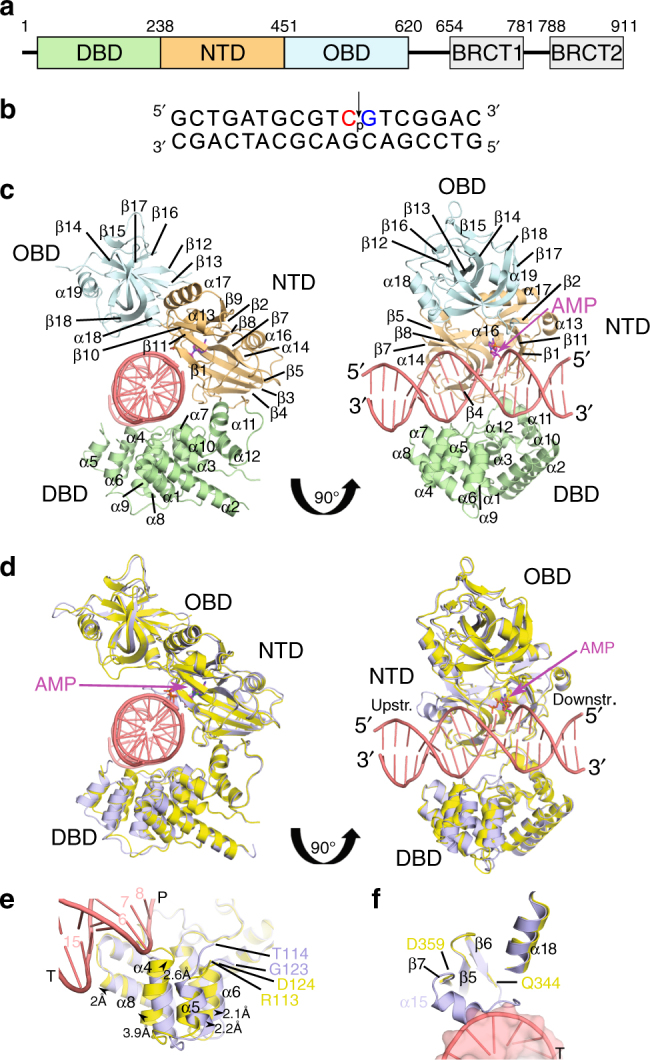
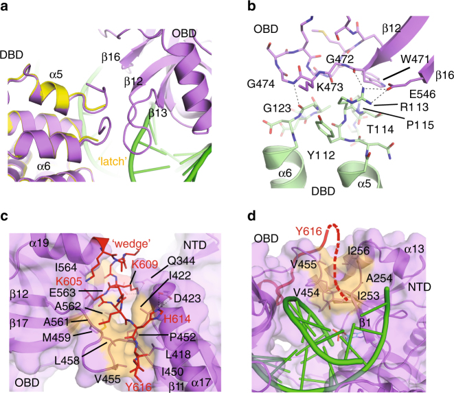
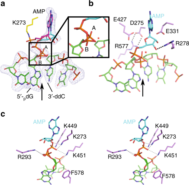
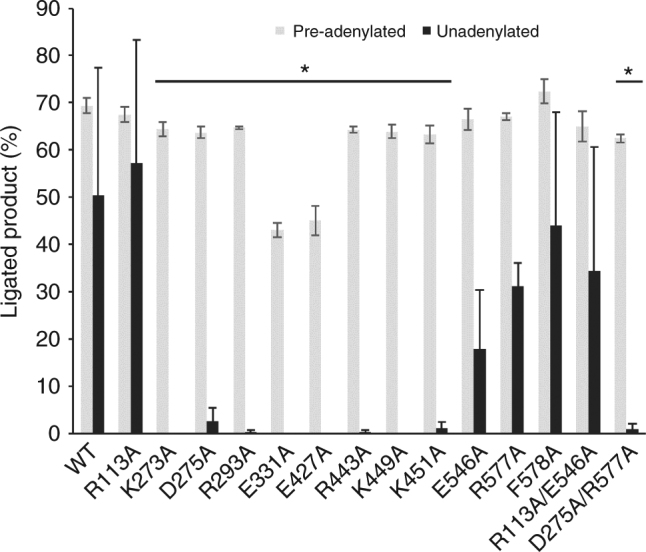
DNA ligase IV (LigIV) performs the final DNA nick-sealing step of classical nonhomologous end-joining, which is critical for immunoglobulin gene maturation and efficient repair of genotoxic DNA double-strand breaks. Hypomorphic LigIV mutations cause extreme radiation sensitivity and immunodeficiency in humans. To better understand the unique features of LigIV function, here we report the crystal structure of the catalytic core of human LigIV in complex with a nicked nucleic acid substrate in two distinct states-an open lysyl-AMP intermediate, and a closed DNA-adenylate form. Results from structural and mutagenesis experiments unveil a dynamic LigIV DNA encirclement mechanism characterized by extensive interdomain interactions and active site phosphoanhydride coordination, all of which are required for efficient DNA nick sealing. These studies provide a scaffold for defining impacts of LigIV catalytic core mutations and deficiencies in human LIG4 syndrome.


Conflict of interest statement

The authors declare no competing interests.

Figures

Fig. 1
Fig. 1
Structural characterization of the lysyl-adenylate form of the human DNA LigIV catalytic domain. a Schematic of human LigIV subdomain architecture. b Nicked DNA substrate co-crystallized with LigIV, with the backbone discontinuity indicated (arrow). The primers upstream and downstream of the nick were dideoxy-terminated (red) or phosphorylated (blue), respectively. c Ribbon diagram of the lysyl-adenylate (magenta) form of LigIV (DBD in green; NTD in orange; OBD in blue) bound to the nicked DNA substrate (pink), as viewed down the axis of the DNA helix (left). On the right, the complex has been rotated 90° around the y-axis, to view the DNA. In this orientation, the nicked DNA substrate is observed in the same orientation as in b, with the upstream duplex on the left, and the downstream duplex on the right. The relative positions of the protein subdomains, along with the secondary structural elements, are labeled as in Supplementary Fig. 3. d Comparison of the DNA-bound, lysyl-adenylate form of LigIV (protein in yellow; DNA in pink; AMP in magenta), with that of the apoprotein (protein in light blue; ATP in green; PDB ID code 3W5O). Orientations are the same as in c. e Structural differences observed in the DBD of the apoprotein (light blue, PDB ID code 3W5O) vs. the DNA-bound lysyl-adenylate complex (protein in yellow, DNA in pink). Direction and measured distances for movement of paired atoms are shown as black arrows. f Binding of a nicked DNA substrate (pink) to the catalytic domain of LigIV (yellow) results in unraveling of α-helix 15 observed in the apoprotein (light blue)
Fig. 2
Fig. 2
Detailed analysis of the lysyl-adenylate complex. a Disposition of the LigIV lysyl-adenylate (magenta) covalently linked to Lys273, relative to the nick in the DNA backbone (pink). The interatomic distance between the oxygens on the 5′-phosphate on the downstream primer and the phosphorous atom of the AMP is indicated by the black dashed line (~5.4 Å). A simulated annealing omit Fo − Fc difference electron density map (gray) is shown, contoured at 2.5σ. b Interactions with the lysyl-adenylate (magenta) and the protein residues (yellow) lining the nucleotide-binding site. Putative hydrogen-bonding interactions with the AMP hydroxyls are indicated by black dashed lines, and are listed in detail in Supplementary Table 2. The LigIV apoprotein structure (PDB ID code 3W5O) was superimposed onto the DNA-bound structure, and the position of its ATP molecule is shown in green. c Nick sensing by LigIV involves multiple putative interactions (black dashed lines) between protein sidechains (yellow), the adenylate (magenta), and the 5′-phosphate on the DNA (pink). The nick junction is indicated by a black arrow
Fig. 3
Fig. 3
The DNA–adenylate complex adopts a catalytically competent closed conformation. a Ribbon diagram of the LigIV catalytic domain (purple) bound to a nicked DNA substrate (green), where the adenylate group (cyan) has been covalently transferred from Lys273 to the 5′-phosphate on the downstream strand. The complex is viewed down the axis of the DNA helix. b Superposition of the open lysyl-adenylate (protein in yellow, DNA in pink) and closed DNA–adenylate (protein in purple, DNA in green) complexes of LigIV. The hinge region is marked with a black dashed box. During the open to closed transition, the OBD domain undergoes a complex “swivel-and-close” motion to encircle the DNA, the direction of which is indicated by the spiral arrow. Secondary structural elements in ribbon diagrams are labeled as in Supplementary Fig. 3. The ordered region of the C-terminal tail in the closed DNA–adenylate complex is shown in red and the ‘latch’ is indicated in orange. c Rendering of the electrostatic surface of the OBD subdomain in the closed conformation, with the bound nicked DNA–adenylate substrate (DNA in green, adenylate in cyan). The electrostatic surface potential was calculated using the Adaptive Poisson–Boltzmann Solver tool in PyMOL, which ranges from −6 kT e−1 (electronegative, red) to 6 kT e−1 (electropositive, blue). Regions of neutral charge are shown in white. The nicked DNA substrate (green) with the 5′-adenylate (cyan) is drawn in stick. The nick junction is indicated by a black arrow. The trajectories of the upstream and downstream duplex are indicated by a dashed black line. d, e Detailed aspects of conformational differences between the open (yellow) and closed (protein in purple, DNA in green) conformations in the OBD subdomain (d, linker between NTD and OBD delineated by a black dashed box, and Insert 2 by a magenta oval) and the loop between β6 and β7 (e). α15 observed in the apoprotein structure (PDB ID 3W5O) is drawn transparently in gray. f Detailed view of the interactions of the β6-β7 connecting loop with the DNA substrate. Putative hydrogen-bonding interactions with the DNA substrate are shown as black dashed lines
Fig. 4
Fig. 4
Striking structural features of the LigIV catalytic domain. a Comparison of the “latch” (orange) in the open (yellow) and closed DNA–adenylate complex (protein in purple, DNA in green). b Details of “latch” interactions between the DBD (green) and OBD (purple) subdomains, with protein sidechains drawn in stick. c Disposition of the “wedge” motif. The C-terminal tail (red) becomes ordered in the closed conformation, and covers a hydrophobic channel (orange) between the OBD and NTD subdomains, the potential trajectory of which continues along the protein surface toward the downstream DNA (green) duplex (d). Putative hydrogen-bonding interactions are indicated by black dashed lines. A detailed list of “latch” interactions is given in Supplementary Table 3
Fig. 5
Fig. 5
Analysis of interactions involving the DNA–adenylate prior to nick sealing. a The lysyl-adenylate from the open complex (K273 in yellow, AMP in magenta) was superimposed with the DNA–adenylate (AMP in cyan, DNA in green). Alternate conformations of the primer terminal 5′-phosphate are shown and indicated as “A” or “B” (magnified view in inset), and the putative position of the 3′-OH is marked with a red asterisk. A simulated annealing omit Fo − Fc difference electron density map (gray) is shown for residues in the closed DNA–adenylate complex, contoured at 2.5σ. b Candidates for divalent metal binding within the LigIV active site. The nick junction is indicated by a black arrow. c Stereo diagram of potential interactions involved in charge neutralization and/or substrate stabilization. A detailed list of the hydrogen-bonding interactions is given in Supplementary Table 2
Fig. 6
Fig. 6
Analysis of nick ligation by the wild-type and mutant LigIV catalytic domains. Levels of ligation activity for wild type and mutant LigIV catalytic domain constructs were examined using either unadenylated (black bars) or pre-adenylated (gray bars) substrates. 1 mM ATP was included in reactions with the unadenylated substrate, to aid in enzyme adenylation, and was not included in reactions with the pre-adenylated substrate. Extent of ligation product formation for each LigIV variant is indicated, as mean ± standard deviation. (*, p < 0.05, WT vs. LigIV variant)
Fig. 7
Fig. 7
Structural comparison of the major mammalian DNA ligases. a Global superposition of the closed DNA-bound conformations of LigIV (purple, DNA in green), LigI (gray, PDB ID code 1X9N), and LigIII (khaki, PDB ID code 3L2P). Structural differences in the NTD (b), OBD (c), and DBD (d) subdomains of the mammalian ligases (DNA binding path in green). e Regions of interest in LigIV that contribute to its unique biological behaviors. Labels for secondary structural elements are representative of LigIV (numbered as in Supplementary Fig 3). Structurally disparate motifs are marked by asterisks, as follows: blue asterisk, α-helix that is conserved in LigI and LigIII structures, but is disordered (α15) in the DNA-bound structure of LigIV; gray, short α-helix present only in LigI (Asp647-Ile651); red asterisk, α11 is considerably longer in LigI than in LigIV; magenta asterisk, short helix present only in LigI (Pro341-Gly344); yellow asterisk, α5-α6 and Insert 1 regions in LigIV are dissimilar to the equivalent region in LigI and LigIII
Fig. 8
Fig. 8
Structural insights into LIG4 syndrome mutations and polymorphisms. a Known LIG4 syndrome mutations (residues in space-filling representation, hot pink) mapped onto the LigIV closed DNA–adenylate structure (protein in purple, DNA in green). b Relative positions of mutations near the AMP (cyan) binding site of human LigIV that have been associated with LigIV deficiency. c Conformation of the LigIV N-terminus (orange), and its putative role in protein folding and stability. Thr9 makes a putative hydrogen bond with Gln146, which could be disrupted when mutated to isoleucine. d Disposition of residues in Motif Va (magenta, with Gly469 in red), in relation to the bound DNA substrate (green)

Similar articles

Cited by

References

    1. Pascal JM. DNA and RNA ligases: structural variations and shared mechanisms. Curr. Opin. Struct. Biol. 2008;18:96–105. doi: 10.1016/j.sbi.2007.12.008. - DOI - PubMed
    1. Prigent C, Satoh MS, Daly G, Barnes DE, Lindahl T. Aberrant DNA repair and DNA replication due to an inherited enzymatic defect in human DNA ligase I. Mol. Cell. Biol. 1994;14:310–317. doi: 10.1128/MCB.14.1.310. - DOI - PMC - PubMed
    1. Akbari M, et al. Overexpression of DNA ligase III in mitochondria protects cells against oxidative stress and improves mitochondrial DNA base excision repair. DNA Repair. 2014;16:44–53. doi: 10.1016/j.dnarep.2014.01.015. - DOI - PMC - PubMed
    1. Shokolenko IN, et al. Mitochondrial DNA ligase is dispensable for the viability of cultured cells but essential for mtDNA maintenance. J. Biol. Chem. 2013;288:26594–26605. doi: 10.1074/jbc.M113.472977. - DOI - PMC - PubMed
    1. Tomkinson AE, et al. Completion of base excision repair by mammalian DNA ligases. Prog. Nucleic Acid. Res. Mol. Biol. 2001;68:151–164. doi: 10.1016/S0079-6603(01)68097-8. - DOI - PubMed

Publication types