

Short-term Dynamics after Single- and Three-piece Acrylic Intraocular Lens Implantation: A Swept-source Anterior Segment Optical Coherence Tomography Study
- PMID: 29980770
- PMCID: PMC6035277
- DOI: 10.1038/s41598-018-28609-1
Short-term Dynamics after Single- and Three-piece Acrylic Intraocular Lens Implantation: A Swept-source Anterior Segment Optical Coherence Tomography Study
Abstract
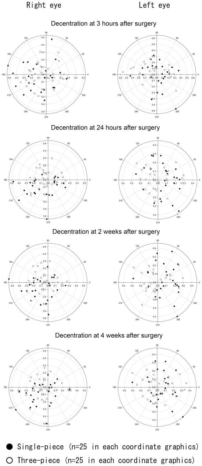
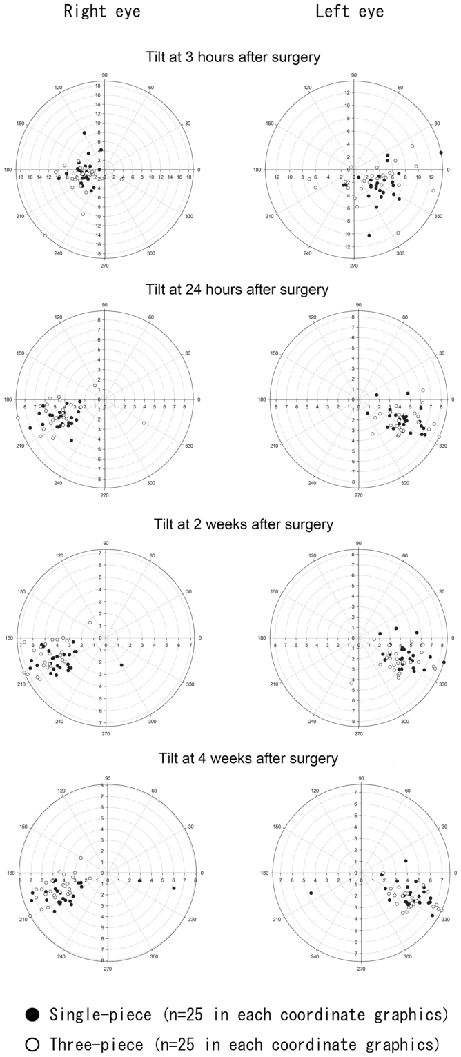
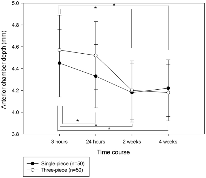
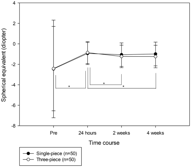
Accurate alignment of an intraocular lens (IOL) is indispensable for achieving accurate postoperative refractive outcomes. Thus, we evaluated decentration and tilt of single- and three-piece IOLs, as well as anterior chamber depth (ACD), at 3 hours, 24 hours, 2 weeks, and 4 weeks after cataract surgery, using swept-source anterior segment optical coherence tomography. There was no significant difference in postoperative visual acuity between eyes with single- or three-piece IOLs. Absolute values of IOL decentration at 24 hours and 2 weeks after surgery were significantly larger (P = 0.008 and 0.046, respectively) in eyes with the single-piece IOL than in those with the three-piece IOL. Both single- and three-piece IOLs tended to tilt toward the inferotemporal direction; however, there was no significant difference in the absolute values of IOL tilt at any postoperative time point. ACD at 24 hours after surgery was significantly deeper (P = 0.009) in eyes with the three-piece IOL, compared with eyes with the single-piece IOL. Therefore, although both single- and three-piece IOL locations varied transiently after surgery, IOL locations were similar between both IOLs at 4 weeks after surgery and were not associated with any statistical difference in visual function.
Conflict of interest statement
The authors declare no competing interests.
Figures
Similar articles
-
Effect of Intraocular Lens Tilt and Decentration on Visual Acuity, Dysphotopsia and Wavefront Aberrations.Vision (Basel). 2020 Sep 14;4(3):41. doi: 10.3390/vision4030041. Vision (Basel). 2020. PMID: 32937750 Free PMC article. Review.
-
[Comparison of stability of acrylic intraocular lens and transparency of lens capsule using Pentacam Scheimpflug System].Zhonghua Yan Ke Za Zhi. 2011 Apr;47(4):298-302. Zhonghua Yan Ke Za Zhi. 2011. PMID: 21612677 Clinical Trial. Chinese.
-
Optical coherence tomography assessment of capsule closure after cataract surgery.J Cataract Refract Surg. 2005 Feb;31(2):330-6. doi: 10.1016/j.jcrs.2004.04.057. J Cataract Refract Surg. 2005. PMID: 15767154
-
Prospective intrapatient comparison of 6.0-millimeter optic single-piece and 3-piece hydrophobic acrylic foldable intraocular lenses.Ophthalmology. 2006 Apr;113(4):585-90. doi: 10.1016/j.ophtha.2005.10.064. Ophthalmology. 2006. PMID: 16581420 Clinical Trial.
-
Tilt and decentration with various intraocular lenses: A narrative review.World J Clin Cases. 2022 Apr 26;10(12):3639-3646. doi: 10.12998/wjcc.v10.i12.3639. World J Clin Cases. 2022. PMID: 35647149 Free PMC article. Review.
Cited by
-
Predicting Residual Astigmatism in Cataract Surgery.Vision (Basel). 2022 Nov 24;6(4):70. doi: 10.3390/vision6040070. Vision (Basel). 2022. PMID: 36548932 Free PMC article. Review.
-
Rotational stability of modified toric intraocular lens.PLoS One. 2021 Mar 1;16(3):e0247844. doi: 10.1371/journal.pone.0247844. eCollection 2021. PLoS One. 2021. PMID: 33647069 Free PMC article.
-
Effect of Intraocular Lens Tilt and Decentration on Visual Acuity, Dysphotopsia and Wavefront Aberrations.Vision (Basel). 2020 Sep 14;4(3):41. doi: 10.3390/vision4030041. Vision (Basel). 2020. PMID: 32937750 Free PMC article. Review.
-
Analysis of intraocular lens tilt and decentration after cataract surgery in eyes with high myopia using the anterior segment optical coherence tomography.Sci Rep. 2024 Nov 14;14(1):27987. doi: 10.1038/s41598-024-78759-8. Sci Rep. 2024. PMID: 39543310 Free PMC article.
-
Risk Factor for Transient Hyperopic Refractive Outcome at Acute Postoperative Period After Panoptix Intraocular Lens Implantation.Clin Ophthalmol. 2021 Jun 16;15:2499-2503. doi: 10.2147/OPTH.S318286. eCollection 2021. Clin Ophthalmol. 2021. PMID: 34163134 Free PMC article.
References
Publication types
MeSH terms
Substances
LinkOut - more resources
Full Text Sources
Other Literature Sources
Medical
