. 2019 Feb;10(2):104-119.
doi: 10.1007/s13238-018-0563-2. Epub 2018 Jul 6.

TMEM43-S358L mutation enhances NF-κB-TGFβ signal cascade in arrhythmogenic right ventricular dysplasia/cardiomyopathy

Affiliations

TMEM43-S358L mutation enhances NF-κB-TGFβ signal cascade in arrhythmogenic right ventricular dysplasia/cardiomyopathy

Guoxing Zheng et al. Protein Cell. 2019 Feb.

Abstract

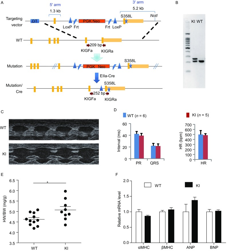
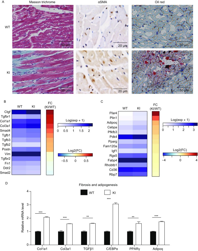
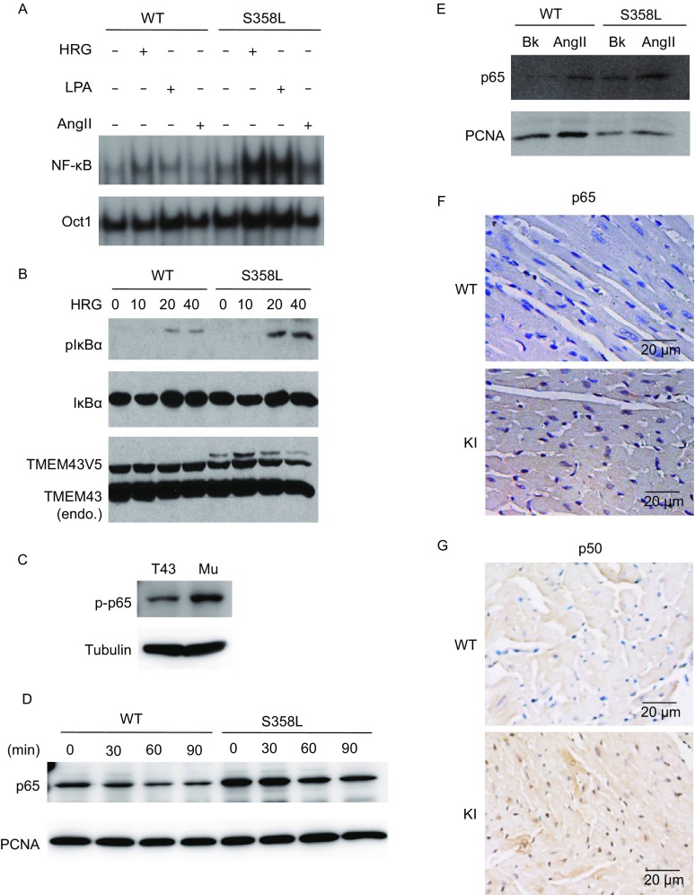
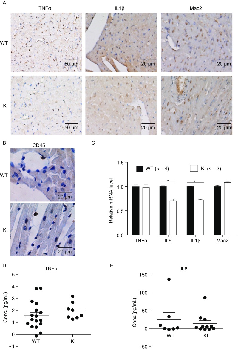
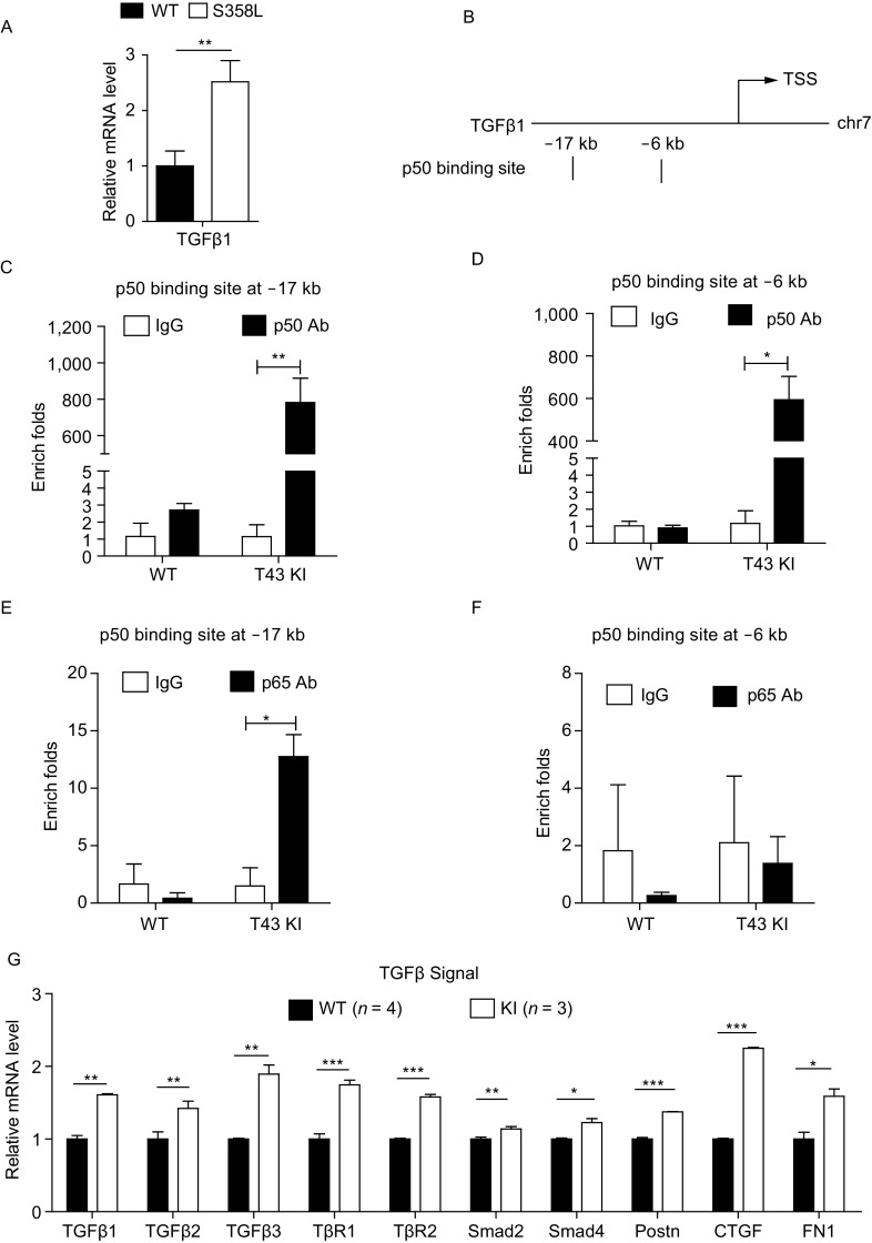
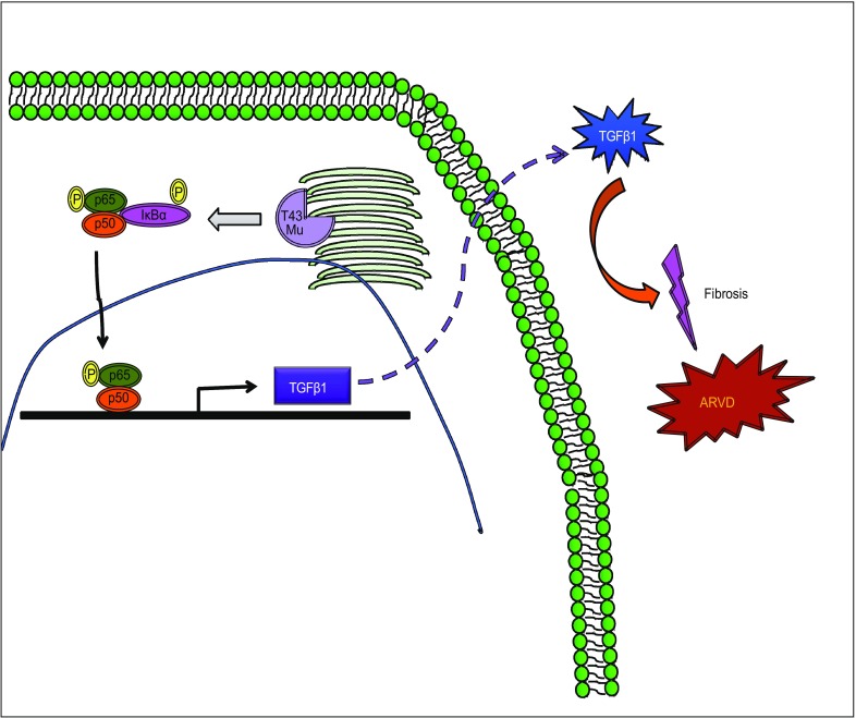
Arrhythmogenic right ventricular dysplasia/cardiomyopathy (ARVD/C) is a genetic cardiac muscle disease that accounts for approximately 30% sudden cardiac death in young adults. The Ser358Leu mutation of transmembrane protein 43 (TMEM43) was commonly identified in the patients of highly lethal and fully penetrant ARVD subtype, ARVD5. Here, we generated TMEM43 S358L mouse to explore the underlying mechanism. This mouse strain showed the classic pathologies of ARVD patients, including structural abnormalities and cardiac fibrofatty. TMEM43 S358L mutation led to hyper-activated nuclear factor κB (NF-κB) activation in heart tissues and primary cardiomyocyte cells. Importantly, this hyper activation of NF-κB directly drove the expression of pro-fibrotic gene, transforming growth factor beta (TGFβ1), and enhanced downstream signal, indicating that TMEM43 S358L mutation up-regulates NF-κB-TGFβ signal cascade during ARVD cardiac fibrosis. Our study partially reveals the regulatory mechanism of ARVD development.

Keywords: ARVD; NF-κB; TGFβ; TMEM43; fibrosis; knock-in mouse.


Figures

Figure 1
Figure 1
Generation and characterization of ARVD TMEM43 mutation mice. (A) The strategy of the construction of TMEM43 S358L mutation mouse. DT gene provided negative selection, while Neo gene provided positive selection of homo-recombination ES clones. Targeting vector carrying S358L point mutation was homo-recombinated with wild type genome through 1.3 kb 5′ arm and 5.2 kb 3′ arm. The obtained mutation mice were mated with whole body expression EIIα-Cre mice to remove the Neo selection cassette in the intron of Tmem43 gene. (B) Genotyping of TMEM43 S358L knockin (KI) heterozygous mice had a 252 bp and 209 bp PCR product with primer sets of KIGFa and KIGRa, while wild type (WT) mice only had 209 bp product. DNA sequencing of the 252 bp PCR products verified the genomic mutation. (C) The represented figures in ultrasound analysis of littermate WT and TMEM43 mutation KI mice, indicating enlarged heart chambers in KI mice. (D) Electrocardiograph of littermate WT (n = 6) and KI (n = 5) mice. The heart rate (HR), PR interval and QRS interval were compared between the groups of WT and KI mice. Student’s t-test was used for statics analysis. (E) The heart weights and body weights of 8-week old male WT (n = 11) and KI (n = 9) mice were detected and the ratios of heart weights verse body weights were calculated. (F) Quantity PCR assay showed the mRNA level of the markers of cardiac hypertrophy in the hearts of WT (n = 5) and KI (n = 7) mice. Student’s t-test was used for statics analysis
Figure 2
Figure 2
TMEM43 mutation mice reproduced ARVD fibrofatty features. (A) The hearts of 8-week old male WT (n = 17) and KI (n = 8) mice were fixed and sectioned in paraffin. The sections were subjected to masson’s trichrome staining (left panel). The blue staining indicated cardiac fibrosis region. The paraffin sections of the hearts of 8-week old male WT and KI mice were subjected to immunohistological staining via αSMA antibody (middle panel).The frozen sections of the hearts of 8-week old male WT (n = 3) and KI (n = 3) mice were stained with oil red (right panel). (B) The heat map showed the mRNA transcriptional level of the genes of cardiac fibrosis in RNA-seq assay of the hearts of WT (n = 4) and KI (n = 3) mice. (C) The heat map showed the mRNA level of the genes of cardiac adipogenesis in RNA-seq assay of the hearts of WT (n = 4) and KI (n = 3) mice. (D) Quantity PCR assay showed the mRNA level of the markers of cardiac fibrosis and adipogeneis in the hearts of WT (n = 4) and KI (n = 3) mice. Gapdh served as endogenous control. Student’s t-test was used for statics analysis. ** indicated P-value < 0.01, *** indicated P-value < 0.001
Figure 3
Figure 3
TMEM43 ARVD mutant enhanced NF-κB activity. (A) Primary human cardiomyocytes stably expressing V5-tagged TMEM43 WT or S358L mutant were serum starved for overnight and stimulated with HRG (50 ng/mL), LPA (10 μmol/L) or Ang II (1 μmol/L) for 60 min. Nuclear extracts were isolated and subjected to EMSA analysis with P32 labelled nucleotide probe of p65 binding substrate. OCT-1 served as a loading control. (B) Cardiomyocytes stably expressing vector or V5-tagged TMEM43 WT or S358L mutant were serum starved for overnight and stimulated with 50 ng/mL of HRG for the indicated time. The cell lysates were subjected to immunoblotting of the indicated antibodies. (C) The total protein of A549 cells stably expressing TMEM43 WT or S358L mutant were collected and lysed. The cell lysates were subjected to immunoblotting of the indicated antibodies. (D) A549 cells stably expressing TMEM43 WT or S358L mutant were serum starved for overnight and stimulated with or without AngII (4 μmol/L) for the indicated time. The nuclear extracts were subjected to immunoblotting of p65 antibody and PCNA antibody. (E) The MEF cells derived from WT and KI mice were serum starved for overnight and stimulated with or without AngII (10 μmol/L) for 60 min. The nuclear extracts were collected and subjected to immunoblotting of p65 antibody and control PCNA antibody. (F) The paraffin sections of the hearts of 8-week old male WT and KI mice were performed immunohistological staining via p65 antibody. (G) The paraffin sections of the hearts of 8-week old male WT and KI mice were performed immunohistological staining via p50 antibody
Figure 4
Figure 4
The inflammation did not increase in TMEM43 mutation mice. (A) The paraffin sections of the hearts of 8-week old male WT and KI mice were performed immunohistological staining via pro-inflammatory cytokine TNFα, IL1β antibody and macrophage marker MAC2 antibody. (B) The paraffin sections of the hearts of 8-week old male WT and KI mice were performed immunohistological staining via CD45 antibody. (C) Quantity PCR assay showed the mRNA level of pro-inflammatory cytokines in the hearts of WT (n = 4) and KI (n = 3) mice. Student’s t-test was used for statics analysis. The star * indicated P-value < 0.05 in Student’s t-test. (D) The level of TNFα cytokine in the serum of 8-week old male WT (n = 17) and KI (n = 8) mice were analyzed via enzyme linked immunosorbent assay (ELISA). (E) The level of IL6 cytokine in the serum of 8-week old male WT (n = 7) and KI (n = 11) mice was analyzed via ELISA
Figure 5
Figure 5
NF-κB1 synergized with p65 to directly control Tgfβ1 expression, thus enhanced TGFβ signaling activity. (A) mRNA isolated from cardiomyocytes stably expressing V5-tagged TMEM43 WT or S358L mutant were measured by quantitative real-time PCR. Student’s t-test was used for statics analysis. Two stars ** indicated P-value < 0.01 in Student’s t-test. (B) Qiagen website predicted the NF-κB1 (p50) binding sites on the promoter of Tgfβ1 at mouse genome. (C) The hearts of 8-week old male WT and KI mice were subjected to Chromatin Immunoprecipitation (ChIP) assay via IgG and p50 antibodies. The enriched folds of antibody binding were assayed by qPCR at −17 kb binding site. (D) The enriched folds of antibody binding were assayed by qPCR on −6 kb binding site. (E) The hearts of 8-week old male WT and KI mice were subjected to Chromatin Immunoprecipitation (ChIP) assay via IgG and p65 antibodies. The enriched folds of antibody binding were assayed by qPCR on −17 kb binding site. (F) The enriched folds of antibody binding were assayed by qPCR on −6 kb binding site. Student’s t-test was used for statics analysis. One star * indicated P-value < 0.05, two stars ** indicated P-value < 0.01 in Student’s t-test. (G) Quantity PCR assay showed the mRNA level of genes of TGFβ signal in the hearts of WT (n = 4) and KI mice (n = 3). Gapdh served as endogenous control. Student’s t-test was used for statics analysis. * indicated P-value < 0.05, ** indicated P-value < 0.01, *** indicated P-value < 0.001
Figure 6
Figure 6
TMEM43 S358L mutant enhanced cardiac fibrosis during ARVD development. TMEM43 S358L mutation hyper-activates NF-κB signal, which further induces Tgfβ1 expression and enhances TGFβ signaling pathway. Increasing activation of TGFβ signaling promotes fibrosis during ARVD development

Similar articles

Cited by

References

    1. Asimaki A, Tandri H, Duffy ER, Winterfield JR, Mackey-Bojack S, Picken MM, Cooper LT, Wilber DJ, Marcus FI, Basso C, et al. Altered desmosomal proteins in granulomatous myocarditis and potential pathogenic links to arrhythmogenic right ventricular cardiomyopathy. Circ Arrhythm Electrophysiol. 2011;4:743–752. doi: 10.1161/CIRCEP.111.964890. - DOI - PMC - PubMed
    1. Baskin B, Skinner JR, Sanatani S, Terespolsky D, Krahn AD, Ray PN, Scherer SW, Hamilton RM. TMEM43 mutations associated with arrhythmogenic right ventricular cardiomyopathy in non-Newfoundland populations. Hum Genet. 2013;132:1245–1252. doi: 10.1007/s00439-013-1323-2. - DOI - PubMed
    1. Bassaganya-Riera J, Song R, Roberts PC, Hontecillas R. PPAR-gamma activation as an anti-inflammatory therapy for respiratory virus infections. Viral Immunol. 2010;23:343–352. doi: 10.1089/vim.2010.0016. - DOI - PubMed
    1. Bengtsson L, Otto H. LUMA interacts with emerin and influences its distribution at the inner nuclear membrane. J Cell Sci. 2008;121:536–548. doi: 10.1242/jcs.019281. - DOI - PubMed
    1. Campian ME, Verberne HJ, Hardziyenka M, de Groot EA, van Moerkerken AF, van Eck-Smit BL, Tan HL. Assessment of inflammation in patients with arrhythmogenic right ventricular cardiomyopathy/dysplasia. Eur J Nucl Med Mol Imaging. 2010;37:2079–2085. doi: 10.1007/s00259-010-1525-y. - DOI - PMC - PubMed

Publication types