. 2018 Sep 6;46(15):7716-7730.
doi: 10.1093/nar/gky568.

Histone H1 acetylation at lysine 85 regulates chromatin condensation and genome stability upon DNA damage

Affiliations

Histone H1 acetylation at lysine 85 regulates chromatin condensation and genome stability upon DNA damage

Yinglu Li et al. Nucleic Acids Res. .

Abstract

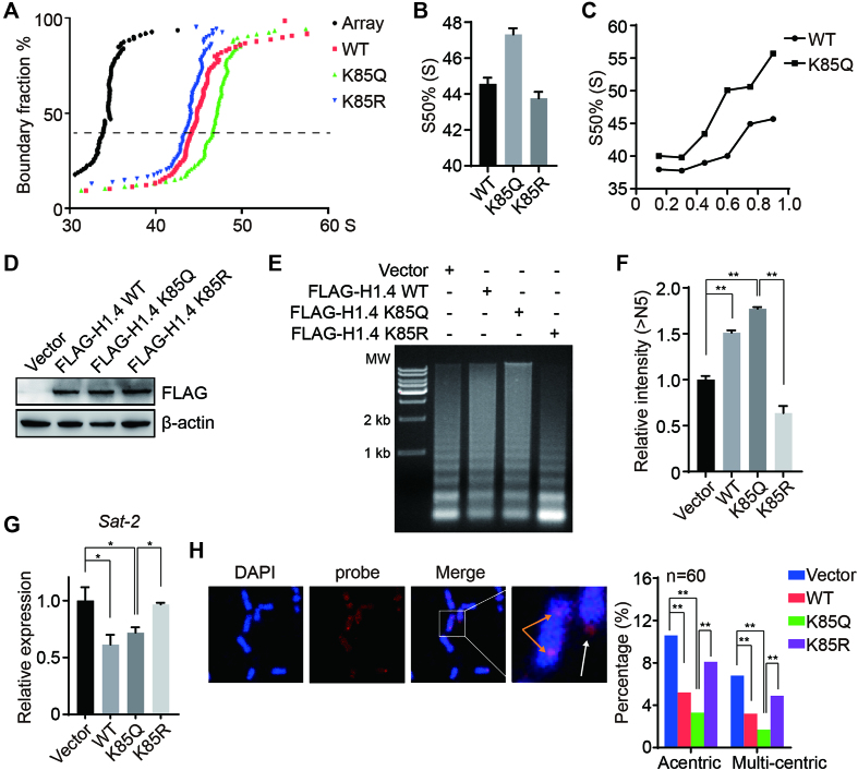
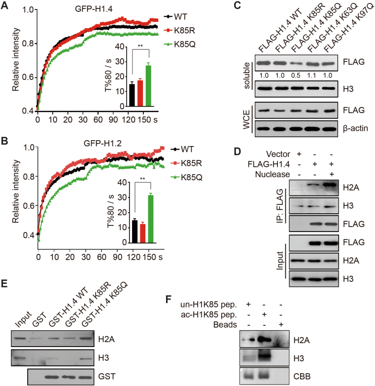
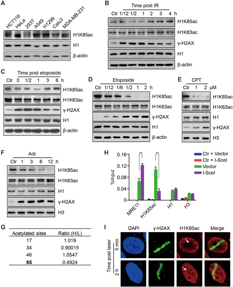
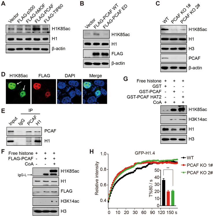
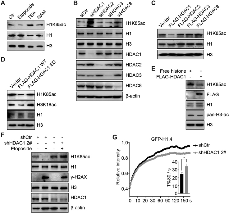
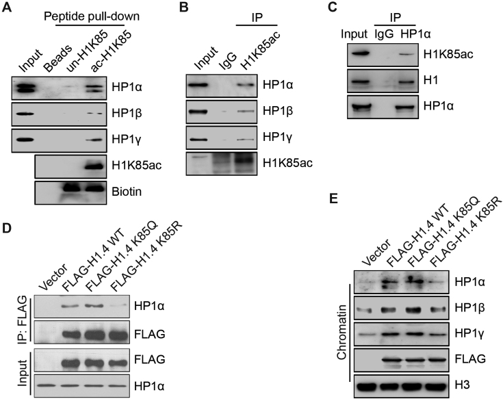
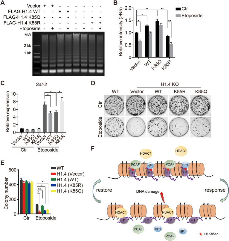
Linker histone H1 has a key role in maintaining higher order chromatin structure and genome stability, but how H1 functions in these processes is elusive. Here, we report that acetylation of lysine 85 (K85) within the H1 globular domain is a critical post-translational modification that regulates chromatin organization. H1K85 is dynamically acetylated by the acetyltransferase PCAF in response to DNA damage, and this effect is counterbalanced by the histone deacetylase HDAC1. Notably, an acetylation-mimic mutation of H1K85 (H1K85Q) alters H1 binding to the nucleosome and leads to condensed chromatin as a result of increased H1 binding to core histones. In addition, H1K85 acetylation promotes heterochromatin protein 1 (HP1) recruitment to facilitate chromatin compaction. Consequently, H1K85 mutation leads to genomic instability and decreased cell survival upon DNA damage. Together, our data suggest a novel model whereby H1K85 acetylation regulates chromatin structure and preserves chromosome integrity upon DNA damage.


Figures

Figure 1.
Figure 1.
Acetylation-mimic H1K85 mutations cause chromatin condensation. (A) Recombinant WT, K85Q or K85R HIS-tagged H1.4 was added to the reconstituted nucleosome array and subjected to in vitro analytical ultracentrifugation assay. A nucleosome array without H1.4 was used as a control. (B) Values from (A) at a boundary fraction of 50%. Data represent the means ± SD. (C) Sedimentation coefficient of the nucleosome array containing an increased ratio of H1 to nucleosome at a 50% boundary fraction. (D) H1.4 KO HeLa cells were stably-transfected with WT, K85Q and K85R H1.4 plasmids and whole cell lysates were analyzed by immunoblotting. (E) H1.4 KO HeLa cells were transfected with the indicated plasmids and chromatin fractions were extracted and analyzed by micrococcal nuclease (MNase) sensitivity assay. (F) Quantifications of lane signal intensities (upper bands, >N5) in (D). All data represent the means ± SD. (G) Sat-2 relative expression was measured by real-time PCR in H1.4 KO HeLa cells stably-transfected with WT, K85Q or K85R H1.4 plasmids. (H) H1.4 KO HeLa cells were stably transfected with the indicated plasmids and subjected to FISH. Metaphase chromosomes were labeled using a pan-centromere probe (red). Chromosomes were counterstained with DAPI (blue). The white arrow indicates acentric chromosomes, and the orange arrow indicates multiple-centric chromosomes. The centromere aberrances were statistically analyzed (n = 50).
Figure 2.
Figure 2.
Acetylation-mimic H1K85 mutation alters H1 dynamics and promotes H1 binding to core histones. (A and B) HCT116 cells were transfected with WT, K85R and K85Q GFP-H1.4 or GFP-H1.2 and subjected to FRAP. T%80 indicates the time when the fluorescent signals recovered to 80% of the original intensity after bleaching. All data represent the means ± SD. (C) HCT116 cells were transfected with the indicated plasmids and chromatin-bound proteins were extracted by salt extraction. Whole cell extracts and soluble proteins were analyzed by immunoblotting. The relative band intensity from three independent immunoblots are shown, relative to FLAG-H1.4 WT which was normalized to 1. (D) HCT116 cells were transfected with FLAG-H1.4 or an empty-vector. Cell extracts were immunoprecipitated using FLAG-conjugated M2 beads with or without nuclease and then analyzed by immunoblotting. (E) WT, K85R and K85Q GST-H1.4 or GST alone were incubated with recombinant H3 or H2A and analyzed by in vitro binding assay. (F) Biotin-tagged unmodified K85 or acetylated K85 peptides were incubated with recombinant H3 or H2A and analyzed by in vitro binding assay.
Figure 3.
Figure 3.
Acetylation of H1K85 is dynamically regulated in response to DNA damage. (A) Protein from different cancer cell lines was extracted and whole cell lysates were analyzed by immunoblotting. (B and C) HCT116 cells were treated with 10 Gy IR or etoposide (40 μM) for 1 h and released for the indicated times and whole cell lysates were analyzed by immunoblotting. (D) HCT116 cells were treated with etoposide (40 μM) for the indicated times and whole cell lysates were analyzed by immunoblotting. (E and F) HCT116 cells were treated with CPT for 12 h at the indicated concentrations or adriamycin (Adr, 1 μM) for the indicated times and histones were analyzed by immunoblotting. (G) H1 acetylation sites identified by SILAC. The relative ratio of the adriamycin treated group (H) to the control group (L) is shown. (H) DR-GFP U2OS cells were transfected with I-SceI endonuclease or an empty vector and subjected to ChIP with the indicated antibodies. Ctr indicates a specific genomic locus distal from the I-SceI cut site and was used as a negative control. MRE11 was used as a positive control. All data represent the means ± SD. (I) HCT116 cells were micro-irradiated and analyzed by immunofluorescence at 5 min or 2 h post-irradiation. The white arrow indicates the irradiation path.
Figure 4.
Figure 4.
PCAF acetylates H1K85 acetylation in vivo and in vitro. (A and B) HCT116 cells were transfected with the indicated plasmids and whole cell lysates were analyzed by immunoblotting. (C) WT and PCAF KO (1# and 2#) HCT116 cells were analyzed by immunoblotting. (D) PCAF KO (1#) cells were transfected with FLAG-PCAF and analyzed by immunofluorescence. The arrows indicate PCAF KO cells with overexpressed FLAG-PCAF plasmids. (E) HCT116 cells lysates were immunoprecipitated using an anti-IgG, anti-PCAF or anti-H1 antibody and analyzed by immunoblotting. (F) FLAG-PCAF was purified from HEK293T cells and subjected to in vitro acetylation assay with or without acetyl-CoA using free histones as substrates. IgG-L indicates the light chain of IgG. (G) GST, GST-PCAF or GST-PCAF HAT2 was purified from Escherichia coli and subjected to in vitro acetylation assay using free histones as substrates. (H) PCAF KO (1#, 2#) and WT cells were transfected with GFP-H1.4 and subjected to FRAP analysis. All data represent the means ± SD.
Figure 5.
Figure 5.
HDAC1 deacetylates H1K85 in vivo and in vitro. (A) HCT116 cells were treated with etoposide (40 μM) for 2 h, TSA (1 μM) for 12 h or NAM (5 mM) for 12 h. Histone was extracted and analyzed by immunoblotting. (B) HCT116 cells were transfected with the indicated siRNAs and whole cell extracts were analyzed by immunoblotting. (C and D) HCT116 cells were transfected with the indicated plasmids and histone was analyzed by immunoblotting. (E) FLAG-HDAC1 was purified from HEK293T cells and subjected to in vitro deacetylation assay using free histones as substrates. The reaction was analyzed by immunoblotting. (F) Stable HDAC1 knockdown (shHDAC1, 2#) or control cells (shCtr) were treated with etoposide and whole cell extracts were analyzed by immunoblotting. (G) Stable HDAC1 knockdown (shHDAC1, 2#) or control cells (shCtr) were transfected with GFP-H1.4 and analyzed by FRAP. All data represent the means ± SD.
Figure 6.
Figure 6.
H1K85 acetylation promotes the recruitment of heterochromatin protein 1 (HP1). (A) Chromatin-bound proteins of HCT116 cells were extracted and incubated with biotin-tagged unmodified K85, acetylated K85 peptide or beads alone and analyzed by in vitro peptide pull-down assay. (B and C) Chromatin-bound proteins of HCT116 cells were extracted and then immunoprecipitated using the indicated antibodies for co-IP and then analyzed by immunoblotting. (D) WT, K85Q and K85R FLAG-H1.4 or empty-vector alone were transfected into HeLa cells. Cell extracts were immunoprecipitated using FLAG-conjugated M2 beads and then analyzed by immunoblotting. (E) WT, K85Q and K85R FLAG-H1.4 or empty-vector were transfected into HeLa cells. Chromatin-bound proteins were extracted and analyzed by immunoblotting.
Figure 7.
Figure 7.
H1K85 acetylation regulates genome stability and cell survival in response to DNA damage. (A) HCT116 cells were transfected with the indicated plasmids and treated with etoposide (40 μM) for 2 h. Cells were extracted and analyzed by MNase sensitivity assay. (B) Quantifications of lane signal intensity (upper bands, >N5) in (A). All data represent the means ± SD. (C) Relative expression of Sat-2 in H1.4 KO cells stably-transfected with vector, WT, K85Q or K85R H1.4 after etoposide treatment (20 μM) for 4 h, determined by real-time PCR. (D and E) HCT116 cells were stably transfected with the indicated plasmids and subjected to colony formation assay after etoposide treatment (10 μM) for 2 h. All data represent the means ± SD. (F) Model of H1K85ac-mediated regulation of chromatin structure in the context of DNA damage. H1K85ac is balanced by PCAF and HDAC1. H1K85ac compacts chromatin by tethering H1 to core histones and recruiting HP1. In response to DNA damage, H1K85ac rapidly decreases, leading to reduced H1 binding affinity to chromatin and reduced enrichment of HP1, resulting in chromatin decondensation. Upon completion of DNA repair, H1K85ac levels increase and permit the restoration of chromatin structure.

References

    1. Fyodorov D.V., Zhou B.R., Skoultchi A.I., Bai Y.. Emerging roles of linker histones in regulating chromatin structure and function. Nat. Rev. Mol. Cell Biol. 2018; 19:192–206. - PMC - PubMed
    1. Hergeth S.P., Schneider R.. The H1 linker histones: multifunctional proteins beyond the nucleosomal core particle. EMBO Rep. 2015; 16:1439–1453. - PMC - PubMed
    1. Fan Y., Nikitina T., Zhao J., Fleury T.J., Bhattacharyya R., Bouhassira E.E., Stein A., Woodcock C.L., Skoultchi A.I.. Histone H1 depletion in mammals alters global chromatin structure but causes specific changes in gene regulation. Cell. 2005; 123:1199–1212. - PubMed
    1. Bayona-Feliu A., Casas-Lamesa A., Reina O., Bernues J., Azorin F.. Linker histone H1 prevents R-loop accumulation and genome instability in heterochromatin. Nat. Commun. 2017; 8:283. - PMC - PubMed
    1. Song F., Chen P., Sun D., Wang M., Dong L., Liang D., Xu R.M., Zhu P., Li G.. Cryo-EM study of the chromatin fiber reveals a double helix twisted by tetranucleosomal units. Science. 2014; 344:376–380. - PubMed

Publication types

MeSH terms