. 2018 Sep 20;25(9):1157-1167.e4.
doi: 10.1016/j.chembiol.2018.05.017. Epub 2018 Jul 5.

A Quantitative Chemoproteomic Platform to Monitor Selenocysteine Reactivity within a Complex Proteome

Affiliations

A Quantitative Chemoproteomic Platform to Monitor Selenocysteine Reactivity within a Complex Proteome

Daniel W Bak et al. Cell Chem Biol. .

Abstract

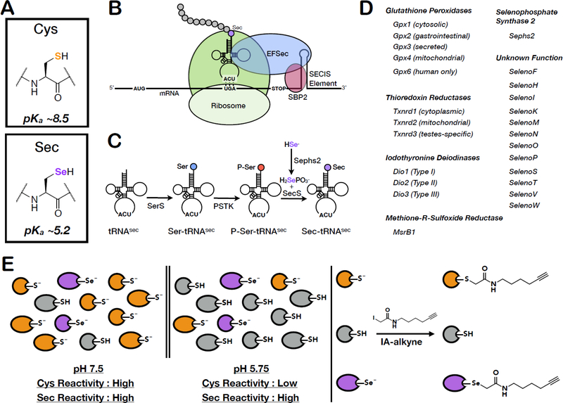
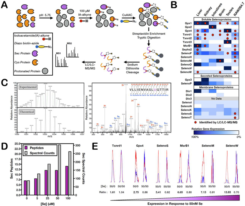
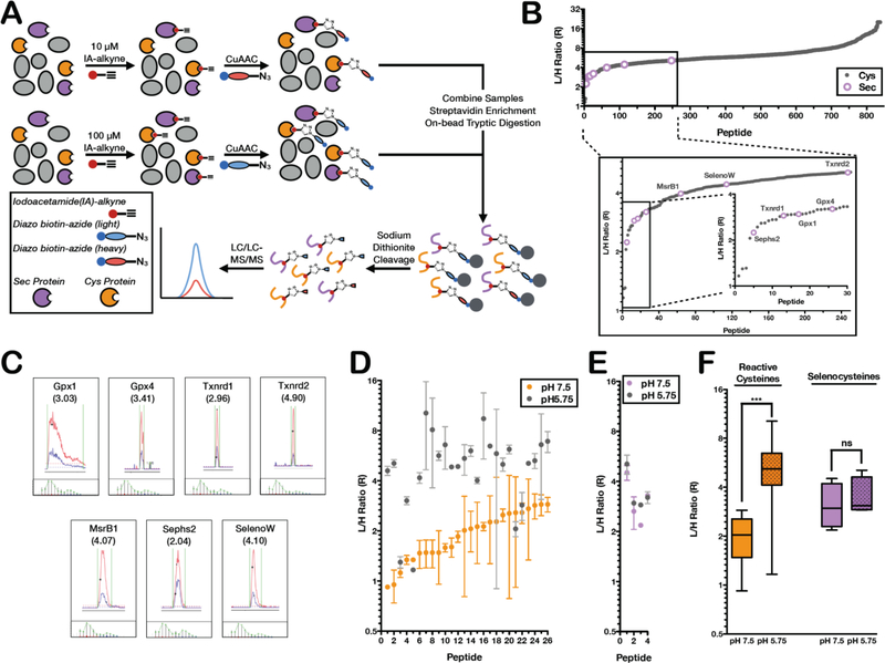
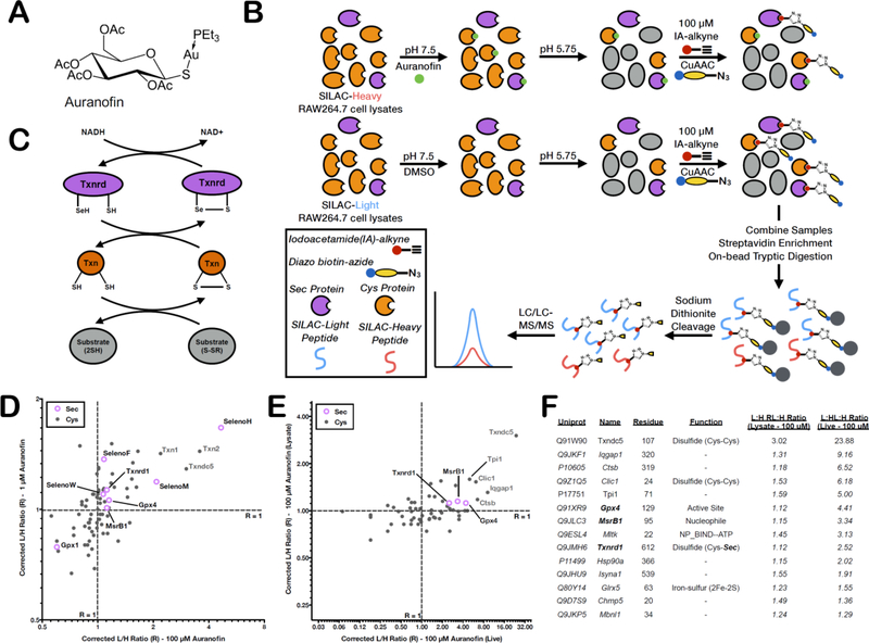
Mammalian selenocysteine (Sec)-containing proteins, selenoproteins, are important to (patho)physiological processes, including redox homeostasis. Sec residues have been recalcitrant to mass spectrometry-based chemoproteomic methods that enrich for reactive cysteine (Cys) residues with electrophilic chemical probes, despite confirmed reactivity of Sec with these electrophiles. Highly abundant Cys peptides likely suppress low-abundant Sec peptides. By exploiting the decreased pKa of Sec relative to Cys, we have developed a chemoproteomic platform that relies on low pH (pH 5.75) electrophile labeling, reducing Cys reactivity and enhancing identification of Sec-containing peptides across mouse tissues and cell lines. The utility of this Sec-profiling platform is underscored by evaluation of the selectivity of auranofin, an inhibitor of the selenoprotein, thioredoxin reductase, against both reactive Cys- and Sec-containing proteins. Platform limitations pertain to the non-physiological low-pH conditions that could perturb protein structure and function. Future work necessitates the discovery of Sec-selective electrophiles that function at physiological pH.

Keywords: chemoproteomics; cysteine; isoTOP-ABPP; selenocysteine; selenoprotein.


Figures

Figure 1.
Figure 1.. Mechanism of Sec incorporation, the mammalian selenoproteins and pH-dependent reactivity of Sec and Cys.
(A) Chemical structures and side-chain pKa values of Cys and Sec. (B) Sec-incorporation into proteins via a Sec-specific tRNA (Sec-tRNA[Ser]Sec) that recognizes an internal UGA (stop) codon within the gene. UGA-codon recognition is facilitated by trans-acting proteins, eEFSec (Sec-specific translation elongation factor) and SBP2 (SECIS-binding protein 2), which recognize and bind to a cis-acting SECIS (Sec insertion sequence) 3’UTR hairpin element. (C) The generation of Sec-charged tRNASec is accomplished in multiple steps; 1) SerS (Seryl-tRNA synthase) initially aminoacylates tRNA[Ser]Sec with serine to generate seryl-tRNA[Ser]Sec, 2) serine is phosphorylated by PSTK (phosphoseryl-tRNA[Ser]Sec kinase) to generate phosphoseryl-tRNA[Ser]Sec, and 3) selenophosphate, generated by Sephs2 (selenophosphate synthase 2), is incorporated into the amino acid backbone by SecS (Sec Synthase) to form Sec-tRNA[Ser]Sec. (D) The complete 25-member mammalian selenoproteome (24 proteins in mice) categorized by homology and function. (E) Due to differences in Cys and Sec pKa values, low-pH conditions result in protonation and loss in reactivity of the majority of Cys residues, whilst maintaining Sec deprotonation and reactivity. IA-alkyne labeling at low pH will therefore enrich for Sec-containing proteins.
Figure 2.
Figure 2.. Identification of Sec peptides by low-pH isoTOP-ABPP
(A) Workflow for low-pH isoTOP-ABPP where IA-alkyne labeling is performed at pH 5.75, followed by CuAAC-mediated incorporation of a diazo biotin-azide linker, streptavidin enrichment, on-bead trypsin digestion and elution of IA-labeled peptides for MS analysis. (B) IA-labeled Sec residues identified by low-pH isoTOP-ABPP (red dots) across five mouse tissues and a mouse macrophage cell line (RAW264.7). Sec-peptide identification by isoTOP-ABPP correlated well with known gene expression data (blue shading). (C) The theoretical and experimental MS1 isotopic envelope and representative annotated MS2 spectra for the Gpx1 Sec peptide. MS1/MS2 spectra for all identified Sec peptides are presented in Table S2. (D) Quantification of the total number of Sec peptides (purple bars) and Sec peptide spectral counts (gray bars) identified from cultured RAW264.7 cells upon selenium supplementation of the cell-culture media at varying concentrations. (E) Extracted light and heavy ion chromatographs demonstrating the varying dependence in Sec reactivity of selenoproteins in response to selenium limitation. L:H ratios are shown for each Sec residue and represent the change in reactivity (a presumed measure of selenoprotein abundance) when IA-alkyne labeling was performed in lysates from cells grown in media supplemented with 50 nM selenium (light) or 0 nM selenium (heavy) or as a control 50 nM selenium (light and heavy). L:H ratio ~ 1 indicates no change in Sec reactivity (or no increase in selenoprotein expression), while L:H ratios > 1 indicate a significant increase in Sec reactivity upon selenium supplementation (or an increase in selenoprotein expression).
Figure 3.
Figure 3.. Reactivity profiling of (seleno)cysteine residues by low-pH isoTOP-ABPP.
(A) Workflow for reactivity profiling of (seleno)cysteine residues at low pH. The low-pH proteome is labeled with either 100 μM or 10 μM IA-alkyne and appended to either an isotopically light or heavy diazo biotin-azide tag, respectively. The light and heavy samples analyzed with the general isoTOP-ABPP workflow. Reactivity is determined as the ratio of light to heavy (R) peptide intensities by LC/LC-MS/MS. (B) The low-pH reactivity of Sec (purple open circles) and Cys (gray closed circles) residues. Inset shows the top ~250 most reactive Sec and Cys residues. (C) Representative light and heavy MS1 chromatographs and isotopic envelopes for the seven identified Sec peptides. (D) The reactivity of the 26 hyperreactive Cys residues (R<3 at pH 7.5) at neutral pH (orange circles) and low pH (gray circles). (E) A comparison of the reactivity of 4 selenocysteine residues at neutral pH (purple circles) and low pH (gray circles). (F) Box plot comparing the global loss in Cys reactivity at low pH versus the minimal reactivity change of Sec residues. Significance is calculated as *** < 0.005, paired Student’s t-test (two-tailed), with n = 26 for cysteine and n = 4 for selenocysteine.
Figure 4.
Figure 4.. Screening the TrxR inhibitor, auranofin against the (seleno)cysteinome.
(A) Chemical structure of the Txnrd inhibitor, auranofin. (B) The Txnrd/Txn antioxidant cycle, which utilizes NADPH as an electron acceptor. (C) Workflow for using low-pH isoTOP-ABPP to screen the selectivity of Auranofin. SILAC light and heavy RAW264.7 lysates are treated with vehicle or auranofin at pH 7.5 then buffer exchanged to pH 5.75 for low-pH IA-alkyne labeling, followed by the standard isoTOP-ABPP workflow. (D) Correlation Plot of the L:H ratios (R) for Cys (gray closed circles) and Sec (open purple circles) residues at two different auranofin concentrations (1 μM and 100 μM). Proteins with high auranofin sensitivity are located in the upper right quadrant. The corrected L:H ratio = 1 is shown as dashed gray lines. (E) Correlation Plot of the L:H ratios (R) for Cys (gray closed circles) and Sec (open purple circles) residues upon treatment of either lysates or live cells with 100 μM auranofin. Proteins with decreased IA labeling in both cell lysate and live-cell auranofin treatments are located in the upper-right quadrant. (F) Sec and Cys residues that demonstrate decreased IA-labeling in both cell lysate and live-cell auranofin treatments.

References

    1. Abo M, Li C, and Weerapana E (2018). Isotopically-Labeled Iodoacetamide-Alkyne Probes for Quantitative Cysteine-Reactivity Profiling. Mol. Pharm 15, 743–749. - PMC - PubMed
    1. Al-Taie OH, Uceyler N, Eubner U, Jakob F, Mork H, Scheurlen M, Brigelius-Flohe R, Schottker K, Abel J, Thalheimer A, et al. (2009). Expression Profiling and Genetic Alterations of the Selenoproteins GI-GPx and SePP in Colorectal Carcinogenesis. Nutr. Cancer 48, 6–14. - PubMed
    1. Apostolou S, Klein JO, Mitsuuchi Y, and Shetler JN (2004). Growth inhibition and induction of apoptosis in mesothelioma cells by selenium and dependence on selenoprotein SEP15 genotype. Oncogene 23, 5032–5040. - PubMed
    1. Arnér ESJ (2010). Selenoproteins—What unique properties can arise with selenocysteine in place of cysteine? Exp. Cell Res 316, 1296–1303. - PubMed
    1. Backus KM, Correia BE, Lum KM, Forli S, Horning BD, González-Páez GE, Chatterjee S, Lanning BR, Teijaro JR, Olson AJ, et al. (2016). Proteome-wide covalent ligand discovery in native biological systems. Nature 534, 570–574. - PMC - PubMed

Publication types