. 2018 Jun:81:144-162.
doi: 10.1016/j.progpolymsci.2018.01.001. Epub 2018 May 7.

Electroactive polymers for tissue regeneration: Developments and perspectives

Affiliations

Electroactive polymers for tissue regeneration: Developments and perspectives

Chengyun Ning et al. Prog Polym Sci. 2018 Jun.

Abstract

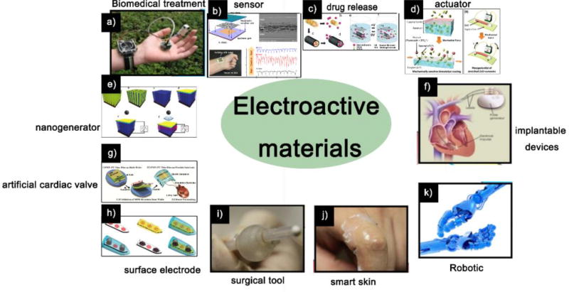
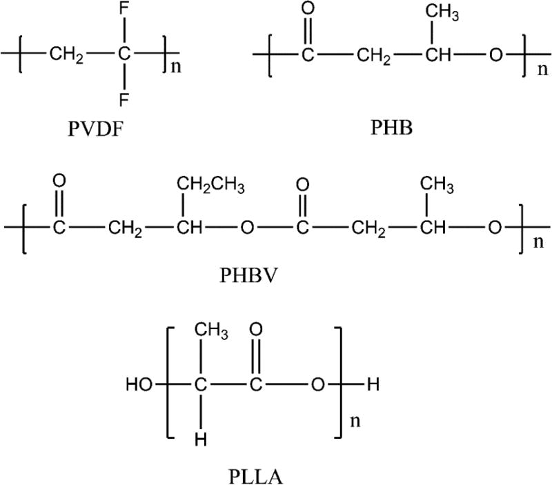
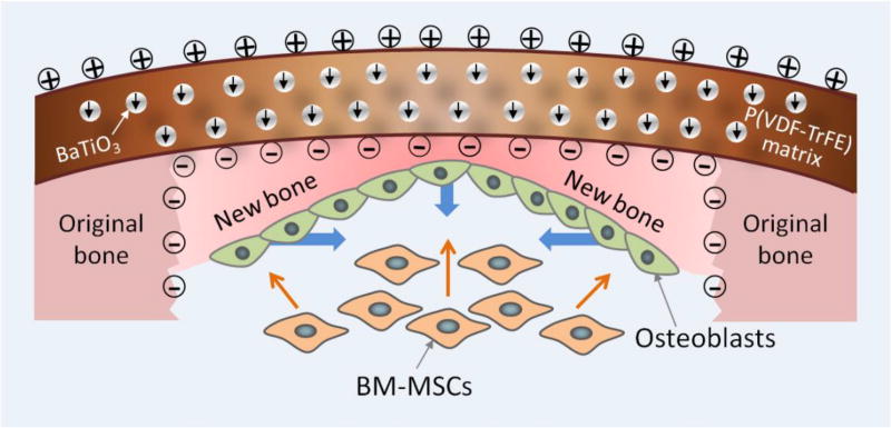
Human body motion can generate a biological electric field and a current, creating a voltage gradient of -10 to -90 mV across cell membranes. In turn, this gradient triggers cells to transmit signals that alter cell proliferation and differentiation. Several cell types, counting osteoblasts, neurons and cardiomyocytes, are relatively sensitive to electrical signal stimulation. Employment of electrical signals in modulating cell proliferation and differentiation inspires us to use the electroactive polymers to achieve electrical stimulation for repairing impaired tissues. Electroactive polymers have found numerous applications in biomedicine due to their capability in effectively delivering electrical signals to the seeded cells, such as biosensing, tissue regeneration, drug delivery, and biomedical implants. Here we will summarize the electrical characteristics of electroactive polymers, which enables them to electrically influence cellular function and behavior, including conducting polymers, piezoelectric polymers, and polyelectrolyte gels. We will also discuss the biological response to these electroactive polymers under electrical stimulation. In particular, we focus this review on their applications in regenerating different tissues, including bone, nerve, heart muscle, cartilage and skin. Additionally, we discuss the challenges in tissue regeneration applications of electroactive polymers. We conclude that electroactive polymers have a great potential as regenerative biomaterials, due to their ability to stimulate desirable outcomes in various electrically responsive cells.

Keywords: Conducting Polymers; Electroactive Polymers; Piezoelectric Polymers; Polyelectrolyte Gels; Tissue Regeneration.


Figures

Figure 1
Figure 1
The applications of electroactive materials. a) Piezoelectric materials used for biomedical treatment application; b) Piezoelectric sensor; c) Drug release from the PEDOT nanotubes was achieved through the contraction or expansion of the PEDOT nanotubes under external electrical stimulation; d) ZnO nanowire actuator resulted in mechanical stretching, which activate the enzymes; e) Potential of PVDF piezoelectric nanogenerator generated by sonic wave; f) Using piezoelectric materials in implantable devices; g) Use of piezoelectric materials in artificial cardiac valves; h) Conducting polymers on neural microelectrodes; i) Piezoelectric materials as surgical tools; j) Piezoelectric materials as smart skins; k) Piezoelectric materials as Robotics. Sources: (a, f, i, j, k) [19], Copyright 2013, All reproduced with permission from Elsevier Ltd; (b) [20], Copyright 2016, (e) [23], Copyright 2011, Both reproduced with permission from the American Chemical Society; (c) [21], Copyright 2006, (d) [22], Copyright 2012, (g) [24], Copyright 2014, (h) [25], Copyright 2012, All reproduced with permission from John Wiley and Sons.
Figure 2
Figure 2
Tissues in the human body are mechanically and/or electrically sensitive to specific environment, indicating that the electroactive materials have potential applications in tissue regeneration.
Figure 3
Figure 3
Structures of some representative biomedical CPs.
Figure 4
Figure 4
Representative structures of some biomedical piezoelectric polymers.
Figure 5
Figure 5
Possible mechanism of cell interactions with reduced and oxidized PEDOT. When the cells grew on the reduced films, fibronectin was extended to present RGD sites for controlling cell adhesion. When the cells grew on the oxidized films, fibronectin became more compacted to hide RGD sites, preventing cell adhesion. [117], Copyright 2012, reproduced with permission from the American Chemical Society.
Figure 6
Figure 6
Cell fate on different nanostructured conducting polymer PPy substrates. (A) Immunofluorescence stained bone forming cells after the cells were cultured on TCA-doped PPy nanotubes in their original state (a), potential-off states (b, d and f) and potential-on states (c, e and g); (B) Schematic and SEM image of cells on an oxidized and reduced PPy nanoarrays. The cells on oxidized PPy nanoarrays presented more and longer filopodia. They began to detach from the nanoarrays when the nanoarrays were reduced. Sources: (A) [152], Copyright 2014, reproduced with permission from the John Wiley and Sons; (B) [154], Copyright 2016, reproduced with permission from the John Wiley and Sons.
Figure 7
Figure 7
SEM images of SCs cultured on PPy/chitosan films. Twenty hours later, SCs were spindle shaped (A) and in the division stage on PPy/chitosan membranes (B). SCs are shown from the conductive films without ES (M−ES) group (C) and conductive films with ES (M+ES) group: (D) 100 mV/mm, (E) 600 mV/mm, and (F) 1000 mV/mm. [47], Copyright 2009, reproduced with permission from the John Wiley and Sons.
Figure 8
Figure 8
Cardiac differentiation on day 14 from embryonic stem cells. (a–b) Typical images of unstimulated (a) and stimulated EBs (b). Red: areas where cardiac/ventricular differentiation occurred. Blue: nuclear DNA. It should be noted that red areas are corresponding to spontaneously contractile regions. [58], Copyright 2011, reproduced with permission from the Elsevier.
Figure 9
Figure 9
The design and synthesis of conducting polymers to mimic cell membranes. (a) General idea of the biomimetic design so that interactions between cells and ECM can be mimicked. (b–d) Morphologies of differentiated PC12 cells that were cultured in NGF-supplemented medium for five days on different substrates, including (b) a PEDOT film, (c) a biomimetic PEDOT film, and (d) the biomimetic PEDOT film under electrical stimulation. The insets are magnified images. Scale bars=200 µm. [214], Copyright 2014, reproduced with permission from the Nature Publishing Group.
Figure 10
Figure 10
Spinal cord neurons immunostained with a mouse anti-MAP2 antibody on stimulated piezoelectric PVDF after 5 days in vitro. Scale bar =15 µm. [55], Copyright 2012, reproduced with permission from the Springer.
Figure 11
Figure 11
Characterization of mES-CM and mES-EC cells grown on PVDF scaffolds for 6 days. (A) mES-CM stained by a live/dead assay, Scale bar=50 µm. (B) Strong cTnT-eGFP expression, Scale bar=20 µm. (C) Protein expression of in mES-CM grown in 2D substrates or on PVDF scaffolds. (D) mES-EC stained by a live/dead assay, Scale bar=50 µm. (E) Uptake of LDL (red) by mES-EC grown on PVDF scaffolds, Scale bar=50 µm. (F) Protein expression in mES-EC. Arrows represent principle fiber axis. [240], Copyright 2016, reproduced with permission from the John Wiley and Sons.
Figure 12
Figure 12
Illustration of biomimetic electric microenvironment created by BTO NP/P(VDF-TrFE) composite membranes encouraging bone defect repair. Electrical dipoles of BTO NPs are reoriented in the direction of poling electric field after corona poling treatment, and consequently induced charges on the membrane. When the composite membranes are implanted like native periosteum covering the bone defect, endogenous BMSCs can be recruited by galvanotaxis and induced to differentiate into osteoblasts. Consequently, the electric microenvironment in the membrane resulted in swift bone regeneration and mature bone formation. The short black arrows denote the direction of electrical dipole in BTO NPs. The blue thick arrows denote the direction of new bone growth. The orange thin arrows denote the recruitment and osteogenic differentiation of BMSCs. [253], Copyright 2016, reproduced with permission from the American Chemical Society.

References

    1. Jiang T, Carbone EJ, Lo KWH, Laurencin CT. Electrospinning of polymer nanofibers for tissue regeneration. Prog Polym Sci. 2015;46:1–24.
    1. Balint R, Cassidy NJ, Cartmell SH. Conductive polymers: towards a smart biomaterial for tissue engineering. Acta Biomater. 2014;10:2341–53. - PubMed
    1. Ohki T, Yamato M, Ota M, Takagi R, Kondo M, Kanai N, Okano T, Yamamoto M. Application of regenerative medical technology using tissue-engineered cell sheets for endoscopic submucosal dissection of esophageal neoplasms. Digest Endosc. 2015;27:182–8. - PubMed
    1. Mooney E, Mackle JN, Blond DJ, O'Cearbhaill E, Shaw G, Blau WJ, Barry FP, Barron V, Murphy JM. The electrical stimulation of carbon nanotubes to provide a cardiomimetic cue to MSCs. Biomaterials. 2012;33:6132–9. - PubMed
    1. Ghasemi-Mobarakeh L, Prabhakaran MP, Morshed M, Nasr-Esfahani MH, Baharvand H, Kiani S, Al-Deyab SS, Ramakrishna S. Application of conductive polymers, scaffolds and electrical stimulation for nerve tissue engineering. J Tissue Eng Regener Med. 2011;5:e17–35. - PubMed