. 2018 Jun 26:12:1905-1915.
doi: 10.2147/DDDT.S161113. eCollection 2018.

The mechanism of botulinum A on Raynaud syndrome

Affiliations

The mechanism of botulinum A on Raynaud syndrome

Yanwen Zhou et al. Drug Des Devel Ther. .

Abstract

Background: Botulinum neurotoxin type A (BoNT/A) is emerging as a treatment modality for Raynaud's phenomenon (RP). However, the mechanism of the role of BoNT/A in antagonizing the constriction of arteriola in RP remains unclear.

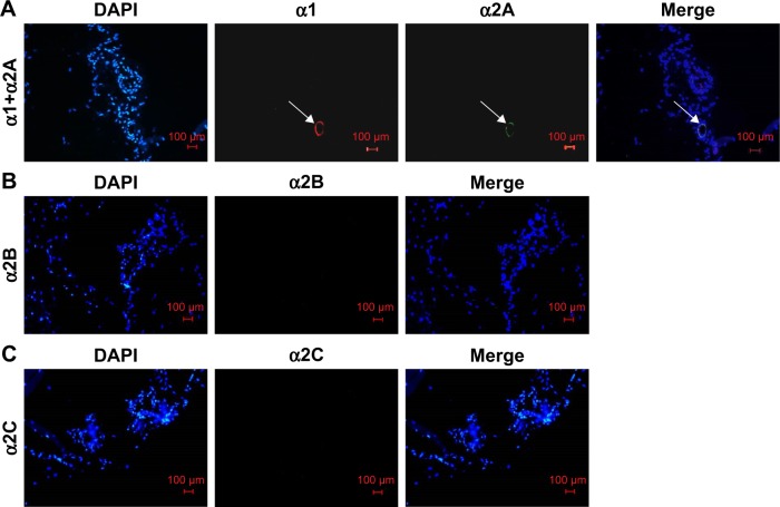
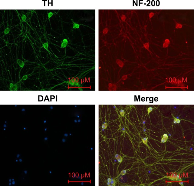
Materials and methods: We tested the constriction of arteriole diameter and the distribution of adrenergic receptors on the rat cremaster modle. Moreover, we measured the secretion of norepinephrine (NE), protein level changes and related receptors on cultured rat superior cervical ganglia neurons(SCGNs), a model of sympathetic neuron.

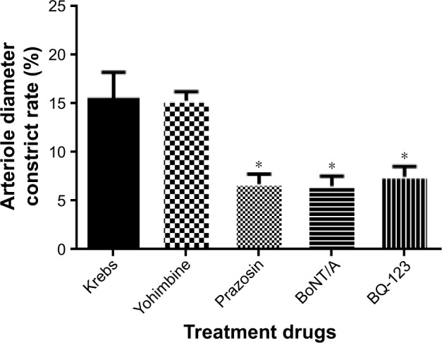
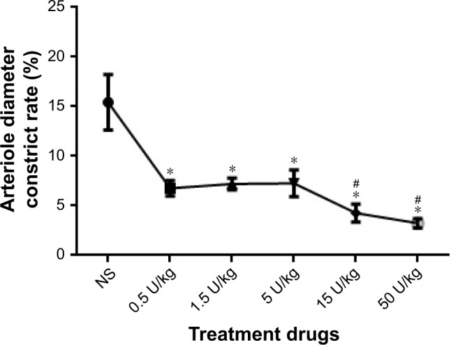
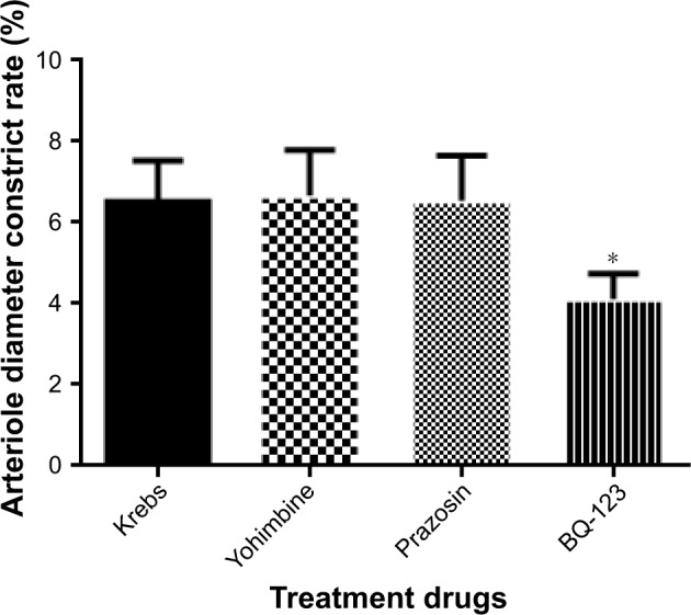
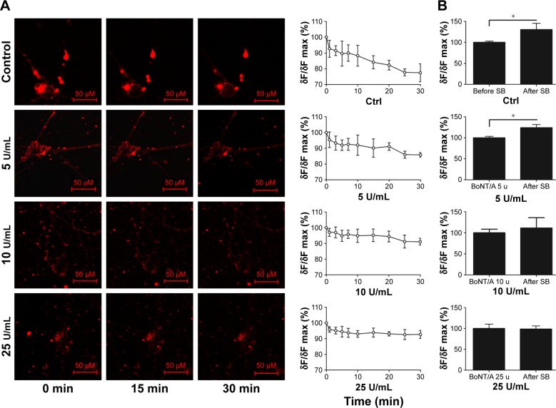
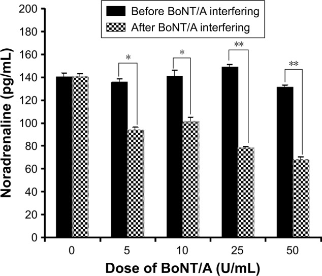
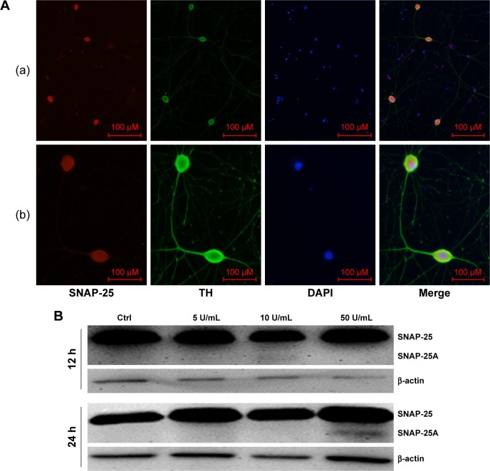
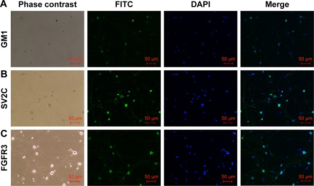
Results: Based on our results, the inhibition of arteriole vasoconstriction was increased with increasing doses of BoNT/A. BoNT/A, prazosin, and BQ123 treatment can result in significant inhibition of arteriole vasoconstriction with the same electrical stimulation. The inhibition effect of prazosin was equivalent to BoNT/A, while BQ123 has a synergistic effect with BoNT/A. After treating SCGNs using BoNT/A for 30 min, the decrease in fluorescence intensity of FM1-43 slowed down which was correlated with the doses of BoNT/A. Furthermore, release of NE in the supernatant was significantly decreased as measured by enzyme-linked immunosorbent assay, 24 h after a high dose of BoNT/A (25 µ/mL). Cleaved-SNAP-25 was detected by Western blotting 24 h following BoNT/A (50 µ/mL) treatment. Moreover, receptor SV2C, GM1, and FGFR3 were detected on sympathetic neurons, similarly to cholinergic neurons.

Conclusion: Our study showed that BoNT/A could significantly inhibit electrical stimulation-induced arteriole vasoconstriction through the sympathetic pathway. The mechanism was similar to the cholinergic one, in which the vesicle release of sympathetic neurons could be inhibited by cleavage of SNAP-25. The end result was blocked vesicle fusion with the presynaptic membrane after BoNT/A treatment, inhibiting the release of the NE.

Keywords: FGFR3; GM1; Raynaud’s phenomenon; SNAP-25; SV2C; arteriole diameter constrict rate; botulinum neurotoxin type A; sympathetic neuron; vesicle cycle; α-adrenoceptor.


Conflict of interest statement

Disclosure The authors report no conflicts of interest in this work.

Figures

Figure 1
Figure 1
The effects of prazosin (0.1 µM), Yohimbine (1 µM), BQ-123 (1 µM), and BoNT/A (5 U/kg) on arteriole diameter constriction rate. Notes: *p<0.05 vs Krebs solution. Data expressed as mean±SD from five independent experiments. Validated rat cremaster model treated with buffered modified Krebs solution regarded as control. Abbreviation: BoNT/A, botulinum neurotoxin type A.
Figure 2
Figure 2
BoNT/A led to a dose-dependent and time-dependent vasodilatory response. Notes: *p<0.05, 0.5 U/kg, 1.5 U/kg, and 5 U/kg vs control; #p<0.001, 15 U/kg and 50 U/kg vs control. Abbreviations: BoNT/A, botulinum neurotoxin type A; NS, normal saline.
Figure 3
Figure 3
Pretreatment with BoNT/A (5 U/kg) and the functional consequences of prazosin (0.1 µM), Yohimbine (1 µM), and BQ-123 (1 µM) treatments on arteriole diameter constriction rate. Notes: *p<0.05 vs Krebs solution. Data expressed as mean±SD from five independent experiments. Validated rat cremaster model treated with buffered modified Krebs solution and BoNT/A regarded as control. Abbreviation: BoNT/A, botulinum neurotoxin type A.
Figure 4
Figure 4
Distribution of α1 and α2 receptors on the rat cremaster arteriole. Notes: (A) α1 and α2A receptors were coexpressed on rat cremaster arteriole (white arrow). (B) α2B receptors were not found on rat cremaster arteriole. (C) α2C receptors were not found on rat cremaster arteriole. The white arrows indicate α1 and α2A receptors on the rat cremaster arteriole (magnification ×200, bar=100 µm). Abbreviation: DAPI, 4′,6-diamidino-2-phenylindole.
Figure 5
Figure 5
Identification of primary sympathetic neurons by immunofluorescence. Notes: The cells derived from SCGs of newborn (1–3 days) rats were TH (green) and NF-200 (red) immunopositive. Overlayed images showed that NF-200 (red), TH, and DAPI (blue) completely overlapped (magnification ×200, bar=100 µm). Abbreviations: DAPI, 4′,6-diamidino-2-phenylindole; NF-200, neurofilament-200; TH, tyrosine hydroxylase.
Figure 6
Figure 6
The inhibition of vesicle cycle by BoNT/A. Notes: (A)The vesicle cycle of sympathetic neuron after injection of BoNT/A with different doses in 30-min. FM1-43 together with SB and 0 U/mL, 5 U/mL, 10 U/mL, 25 U/mL BoNT/A were added in different groups. Leica TCS SP5 LSCM was used to observe the changes of fluorescence intensity in 30-min. Live-cell staining images of sympathetic cells labeled with FM1-43(red) in 0,15,30 min are shown. The chart showed the changes of fluorescence intensity at different time points within 30 min(magnification: 400×, bar: 100 µm). (B) The effect of different doses of BoNT/A on the vesicle cycle after 1 hours of injection. The charts showed changes of fluorescence intensity when stimulated with SB after 1 hour of BoNT/A (0 U/mL, 5 U/mL, 10 U/mL, 25 U/mL) intervention in each group. *p<0.05. Abbreviations: BoNT/A, botulinum neurotoxin type A; Ctrl, control; SB, stimulation buffer.
Figure 7
Figure 7
NA release decreased after BoNT/A treatment. Notes: *p<0.05, the concentration of NE decreased remarkably in 5 U/mL and 10 U/mL BoNT/A-treated group. **p<0.01, the concentration of NA decreased remarkably in 25 U/mL and 50 U/mL BoNT/A-treated group. Abbreviations: BoNT/A, botulinum neurotoxin type A; NE, noradrenaline.
Figure 8
Figure 8
The target protein of BoNT/A in sympathetic neurons is SNAP-25. Notes: (A) The distribution of SNAP-25 in primary cultured sympathetic neurons. The entire sympathetic neuron was immunopositive with SNAP-25 (red) except its nucleus ([a] magnification ×100, bar=100 µm; [b]magnification ×400, bar=100 µm). (B) After a high dose treatment of BoNT/A (50 U/mL) for 24 hours, cleaved SNAP-25 protein was detected. Abbreviations: BoNT/A, botulinum neurotoxin type A; DAPI, 4′,6-diamidino-2-phenylindole; SNAP-25, synaptosomal-associated protein 25; TH, tyrosine hydroxylase.
Figure 9
Figure 9
GM1, SV2C, and FGFR3 are located on sympathetic neurons. Note: The cells were immunopositive for GM1 (A), SV2C (B) and FGFR3 (C) (green fluorescence). Blue fluorescence indicates DAPI. The images were achieved from three separate experiments (magnification ×100, bar=50 µm). Abbreviations: DAPI, 4′,6-diamidino-2-phenylindole; GM1, gangliosidosis-1; FGFR3, fibroblast growth factor receptor 3; FITC, fluorescein isothiocyanate; SV2C, synaptic vesicle glycoprotein 2C.

References

    1. Herrick AL. The pathogenesis, diagnosis and treatment of Raynaud phenomenon. Nat Rev Rheumatol. 2014;8:469–479. - PubMed
    1. Pope JE. Raynaud’s phenomenon (primary) BMJ Clin Evid. 2013;10:1119. - PMC - PubMed
    1. Pope JE. The diagnosis and treatment of Raynaud’s phenomenon: a practical approach. Drugs. 2007;67(4):517–525. - PubMed
    1. Herrick AL. Pathogenesis of Raynaud’s phenomenon. Rheumatology. 2005;44:587–596. - PubMed
    1. Levien TL. Advances in the treatment of Raynaud’s phenomenon. Vasc Health Risk Manag. 2010;6:167–177. - PMC - PubMed

MeSH terms