. 2018 Jul;42(3):389-399.
doi: 10.1016/j.jgr.2018.02.007. Epub 2018 Feb 21.

Antimelanogenesis and skin-protective activities of Panax ginseng calyx ethanol extract

Affiliations

Antimelanogenesis and skin-protective activities of Panax ginseng calyx ethanol extract

Jeong-Ook Lee et al. J Ginseng Res. 2018 Jul.

Abstract

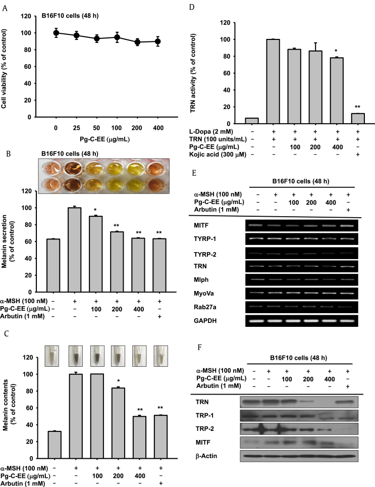
Background: The antioxidant effects of Panax ginseng have been reported in several articles; however, little is known about the antimelanogenesis effect, skin-protective effect, and cellular mechanism of Panax ginseng, especially of P. ginseng calyx. To understand how an ethanol extract of P. ginseng berry calyx (Pg-C-EE) exerts skin-protective effects, we studied its activities in activated melanocytes and reactive oxygen species (ROS)-induced keratinocytes.

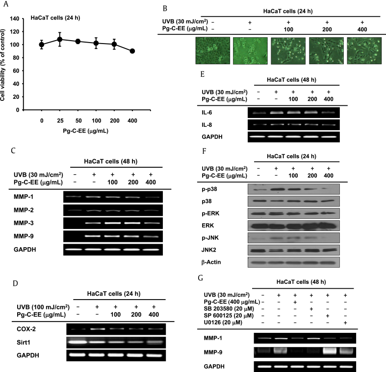
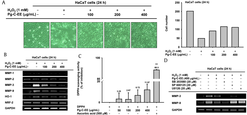
Methods: To confirm the antimelanogenesis effect of Pg-C-EE, we analyzed melanin synthesis and secretion and messenger RNA and protein expression levels of related genes. Ultraviolet B (UVB) and hydrogen peroxide (H2O2) were used to induce cell damage by ROS generation. To examine whether this damage is inhibited by Pg-C-EE, we performed cell viability assays and gene expression and transcriptional activation analyses.

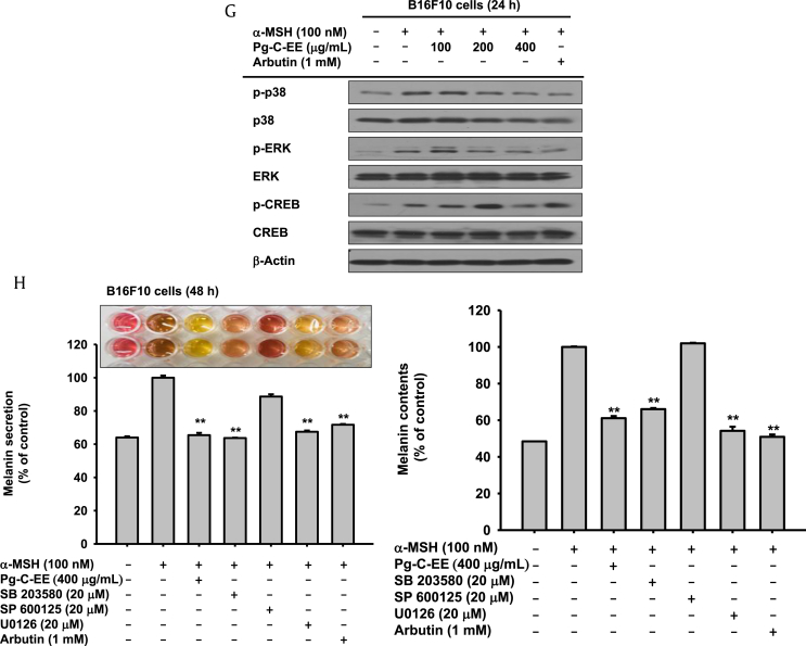
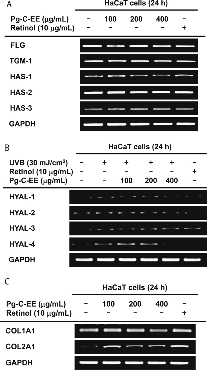
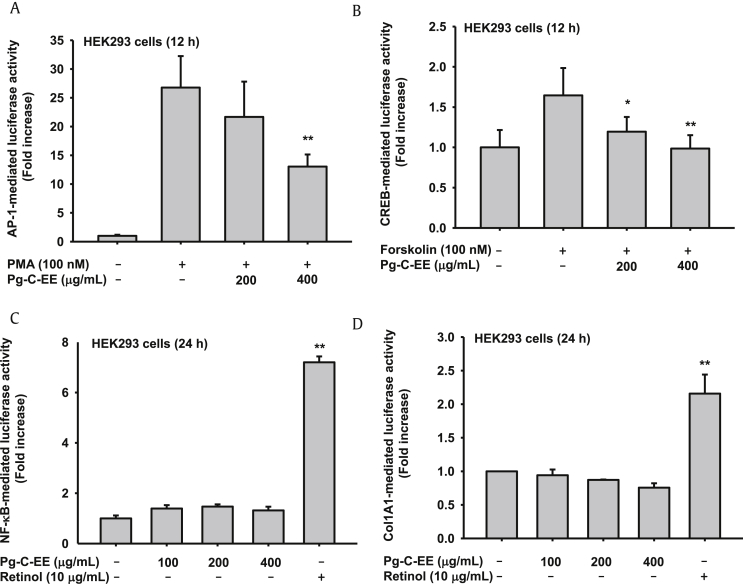
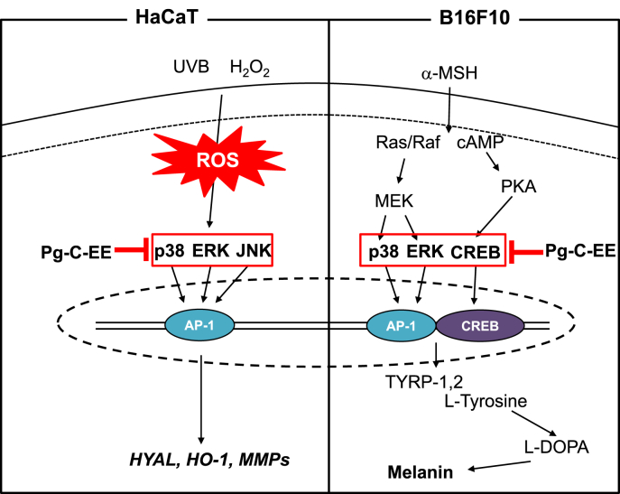
Results: Pg-C-EE inhibited melanin synthesis and secretion by blocking activator protein 1 regulatory enzymes such as p38, extracellular signal-regulated kinases (ERKs), and cyclic adenosine monophosphate response element-binding protein. Pg-C-EE also suppressed ROS generation induced by H2O2 and UVB. Treatment with Pg-C-EE decreased the expression of matrix metalloproteinases, mitogen-activated protein kinases, and hyaluronidases and increased the cell survival rate.

Conclusion: These results suggest that Pg-C-EE may have antimelanogenesis properties and skin-protective properties through regulation of activator protein 1 and cyclic adenosine monophosphate response element-binding protein signaling. Pg-C-EE may be used as a skin-improving agent, with moisture retention and whitening effects.

Keywords: Antimelanogenesis; Calyx of berry; Matrix metalloproteinases; Panax ginseng; Skin protective.


Figures

Fig. 1
Fig. 1
Antimelanogenesis effect of Pg-C-EE on B16F10 cells. (A) B16F10 cell viability was measured by MTT assay in the Pg-C-EE–treated group. (B and C) B16F10 cells were treated with α-MSH (100 nM) and various concentrations of Pg-C-EE (100, 200, and 400 μg/mL), or arbutin (1 mM) for 48 h. Melanin secretion was measured by absorbance of cell supernatant (O.D) at 475, nm and melanin content was measured by absorbance of cell pellets (O.D) at 405 nm. (D) The effects of Pg-C-EE (100-400 μg/mL) or kojic acid (300 μM) on tyrosinase enzyme activity were confirmed by treatment with L-DOPA and detection of tyrosinase at 475 nm. (E) mRNA expression of genes related to melanin synthesis (MITF, TYRP-1, TYRP-2, and tyrosinase) and melanin secretion (MLPH, MyoVa, and Rab27a) were determined in B16F10 cells treated with Pg-C-EE (100 μg/mL, 200 μg/mL, and 400 μg/mL), arbutin (1 mM), and α-MSH (100 nM) using RT-PCR. (F) The levels of melanogenesis-related protein (tyrosinase, TRP-1, TRP-2, and MITF) were confirmed by immunoblot analysis of B16F10 cells treated with Pg-C-EE (100 μg/mL, 200 μg/mL, and 400 μg/mL), arbutin (1 mM), and α-MSH (100 nM) for 48 h. (G) Upstream proteins associated with melanogenesis were identified by treatment of B16F10 cells with Pg-C-EE (100 μg/mL, 200 μg/mL, and 400 μg/mL), arbutin (1 mM), and α-MSH (100 nM) for 24 h. (H) Levels of melanin secretion and content were determined from supernatants and pellets of B16F10 cells treated with Pg-C-EE (400 μg/mL), SB203580 (p38 inhibitor, 20 μM), SP600125 (JNK inhibitor, 20 μM), U0126 (ERK inhibitor, 20 μM), or arbutin (1 mM) with α-MSH (100 nM) for 48 h. *p < 0.05 and **p < 0.01 compared with control. α-MSH, α-melanocyte–stimulating hormone; cAMP, cyclic adenosine monophosphate; CREB, cAMP response element binding; ERK, extracellular signal-regulated kinase; JNK, c-Jun N-terminal kinase; GAPDH, glyceraldehyde 3-phosphate dehydrogenase; L-DOPA, L-dopamine; MITF, microphthalmia-associated transcription factor; mRNA, messenger RNA; MLPH, melanophilin; MTT, 3-(4-5-dimethylthiazol-2-yl)-2,5-diphenyltetrazolium bromide; MyoVa, myosin Va; Pg-C-EE, Panax ginseng calyx ethanol extract; RT-PCR, reverse transcription polymerase chain reaction; TRN, tyrosinase; TYRP, tyrosinase-related protein.
Fig. 1
Fig. 1
Antimelanogenesis effect of Pg-C-EE on B16F10 cells. (A) B16F10 cell viability was measured by MTT assay in the Pg-C-EE–treated group. (B and C) B16F10 cells were treated with α-MSH (100 nM) and various concentrations of Pg-C-EE (100, 200, and 400 μg/mL), or arbutin (1 mM) for 48 h. Melanin secretion was measured by absorbance of cell supernatant (O.D) at 475, nm and melanin content was measured by absorbance of cell pellets (O.D) at 405 nm. (D) The effects of Pg-C-EE (100-400 μg/mL) or kojic acid (300 μM) on tyrosinase enzyme activity were confirmed by treatment with L-DOPA and detection of tyrosinase at 475 nm. (E) mRNA expression of genes related to melanin synthesis (MITF, TYRP-1, TYRP-2, and tyrosinase) and melanin secretion (MLPH, MyoVa, and Rab27a) were determined in B16F10 cells treated with Pg-C-EE (100 μg/mL, 200 μg/mL, and 400 μg/mL), arbutin (1 mM), and α-MSH (100 nM) using RT-PCR. (F) The levels of melanogenesis-related protein (tyrosinase, TRP-1, TRP-2, and MITF) were confirmed by immunoblot analysis of B16F10 cells treated with Pg-C-EE (100 μg/mL, 200 μg/mL, and 400 μg/mL), arbutin (1 mM), and α-MSH (100 nM) for 48 h. (G) Upstream proteins associated with melanogenesis were identified by treatment of B16F10 cells with Pg-C-EE (100 μg/mL, 200 μg/mL, and 400 μg/mL), arbutin (1 mM), and α-MSH (100 nM) for 24 h. (H) Levels of melanin secretion and content were determined from supernatants and pellets of B16F10 cells treated with Pg-C-EE (400 μg/mL), SB203580 (p38 inhibitor, 20 μM), SP600125 (JNK inhibitor, 20 μM), U0126 (ERK inhibitor, 20 μM), or arbutin (1 mM) with α-MSH (100 nM) for 48 h. *p < 0.05 and **p < 0.01 compared with control. α-MSH, α-melanocyte–stimulating hormone; cAMP, cyclic adenosine monophosphate; CREB, cAMP response element binding; ERK, extracellular signal-regulated kinase; JNK, c-Jun N-terminal kinase; GAPDH, glyceraldehyde 3-phosphate dehydrogenase; L-DOPA, L-dopamine; MITF, microphthalmia-associated transcription factor; mRNA, messenger RNA; MLPH, melanophilin; MTT, 3-(4-5-dimethylthiazol-2-yl)-2,5-diphenyltetrazolium bromide; MyoVa, myosin Va; Pg-C-EE, Panax ginseng calyx ethanol extract; RT-PCR, reverse transcription polymerase chain reaction; TRN, tyrosinase; TYRP, tyrosinase-related protein.
Fig. 2
Fig. 2
Antiphotoaging effect of Pg-C-EE against UVB irradiation in HaCaT cells. (A) Viability of HaCaT cells was measured by MTT assay in Pg-C-EE–treated group. (B) HaCaT cells were treated with Pg-C-EE (100 μg/mL, 200 μg/mL, and 400 μg/mL) and UVB irradiation (30 mJ/cm2) for 24 h. Cytoprotective effects of Pg-C-EE against UVB were observed. (C) mRNA expression of MMP-1, MMP-2, MMP-3, and MMP-9 in HaCaT cells treated with Pg-C-EE (100 μg/mL, 200 μg/mL, and 400 μg/mL) and UVB (30 mJ/cm2) was determined by RT-PCR. (D) mRNA expression of COX-2 and Sirt1 in HaCaT cells treated with Pg-C-EE (100 μg/mL, 200 μg/mL, and 400 μg/mL) and UVB (30 mJ/cm2) was determined by RT-PCR. (E) mRNA expression of IL-6 and IL-8 in HaCaT cells treated with Pg-C-EE (100 μg/mL, 200 μg/mL, and 400 μg/mL) and UVB (30 mJ/cm2) was determined by RT-PCR. (F) The levels of phospho and total forms of MAPK (p38, ERK, and JNK) in whole cell lysates were determined by immunoblot analysis after treatment of HaCaT cells with Pg-C-EE (100 μg/mL, 200 μg/mL, and 400 μg/mL) and UVB (30 mJ/cm2) for 48 h. (G) The effects of MAPK inhibitors SB203580 (20 μM), SP600125 (20 μM), and U0126 (20 μM) on expression of MMP-9 in UVB-irradiated HaCaT cells were identified by RT-PCR. COX-2, cyclooxygenase-2; ERK, extracellular signal-regulated kinase; IL-6, interleukin-6; JNK, c-Jun N-terminal kinase; MAPK, mitogen-activated protein kinase; MMP, matrix metalloproteinase; mRNA, messenger RNA; MTT, 3-(4-5-dimethylthiazol-2-yl)-2,5-diphenyltetrazolium bromide; Pg-C-EE, Panax ginseng calyx ethanol extract; RT-PCR, reverse transcription polymerase chain reaction; Sirt1, NAD-dependent protein deacetylase sirtuin-1; UVB, ultraviolet B.
Fig. 3
Fig. 3
Antioxidant effect of Pg-C-EE against H2O2-induced damage in HaCaT cells. (A) Images of HaCaT cells treated with Pg-C-EE (100 μg/mL, 200 μg/mL, and 400 μg/mL) and H2O2 (1 mM) for 24 h were obtained with a camera attached to the microscope. The number of cells in each image was counted (right panel). (B) Expression of MMP-1, MMP-2, MMP-3, MMP-9, HO-1, and NRF2 was confirmed by RT-PCR analysis of HaCaT cells treated with Pg-C-EE (100 μg/mL, 200 μg/mL, and 400 μg/mL) and H2O2 (1 mM) for 24 h. (C) The antioxidative activity of Pg-C-EE (50 μg/mL, 100 μg/mL, 200 μg/mL, and 400 μg/mL) and ascorbic acid (500 μM) was measured by DPPH assay. (D) The effects of MAPK inhibitors SB203580 (20 μM), SP600125 (20 μM), and U0126 (20 μM) on expression of MMP-3 in H2O2 (1 mM)-treated HaCaT cells were identified by RT-PCR. MAPK inhibitors were used to confirm the cytoprotective effect by MTT assay. DPPH, 2,2-diphenyl-1-picrylhydrazyl; H2O2, hydrogen peroxide; HO-1, heme oxygenase-1; MAPK, mitogen-activated protein kinase; MMP, matrix metalloproteinase; MTT, 3-(4-5-dimethylthiazol-2-yl)-2,5-diphenyltetrazolium bromide; NRF-2, nuclear factor (erythroid-derived 2)-like 2; Pg-C-EE, Panax ginseng calyx ethanol extract; RT-PCR, reverse transcription polymerase chain reaction.
Fig. 4
Fig. 4
Effect of Pg-C-EE on moisture retention and collagen synthesis in HaCaT cells. (A) Expression of NMF synthesis–related genes (FLG, TGM-1, and HAS-1, -2, and -3) was measured by RT-PCR in HaCaT cells treated with Pg-C-EE (100 μg/mL, 200 μg/mL, and 400 μg/mL) and retinol (10 μg/mL) for 24 h. (B) mRNA expression of HYAL-1, -2, -3, and 4 was determined by RT-PCR in HaCaT cells after irradiation with UVB (30 mJ/cm2) for 24 h and treatment with Pg-C-EE (100 μg/mL, 200 μg/mL, and 400 μg/mL) and retinol (10 μg/mL). (C) Col1A1 and Col2A1 mRNA expression was analyzed using RT-PCR in HaCaT cells treated with Pg-C-EE (100 μg/mL, 200 μg/mL, and 400 μg/mL) and retinol (10 μg/mL) for 24 h. Col1A1, collagen, type I, alpha 1; Col2A1, collagen, type II, alpha 1; FLG, filaggrin; HAS, hyaluronic acid synthase; HYAL, hyaluronidase; NMF, natural moisturizing factor; Pg-C-EE, Panax ginseng calyx ethanol extract; RT-PCR, reverse transcription polymerase chain reaction; TGM-1, transglutaminase-1; UVB, ultraviolet B.
Fig. 5
Fig. 5
The effect of Pg-C-EE on transcription factor–mediated luciferase activity in Human embryonic kidney (HEK) HEK293 cells. (A) HEK293 cells were transfected with AP-1-Luc (1 μg/mL) and β-gal plasmid in the presence or absence of PMA (100 nM) and Pg-C-EE (200 μg/mL and 400 μg/mL) for 12 h. (B) CREB promoter binding activity was determined by a reporter gene assay with CREB-Luc construct (1 μg/mL) and β-gal plasmid (as a transfection control) in the presence or absence of forskolin (100 nM) and Pg-C-EE (200 μg/mL and 400 μg/mL) for 12 h. (C) HEK cells were transfected with NF-κB-Luc and β-gal plasmid construct with Pg-C-EE (0 μg/mL, 100 μg/mL, 200 μg/mL, and 400 μg/mL) and retinol (10 μg/mL) for 24 h. (D) HEK cells were transfected with Col1A1-Luc and β-gal plasmid construct with Pg-C-EE (0 μg/mL, 100 μg/mL, 200 μg/mL, and 400 μg/mL) and retinol (10 μg/mL) for 24 h. *p < 0.05 compared with control. **p < 0.01 compared with control. AP-1, activator protein 1; β-gal, β-galactosidase; cAMP, cyclic adenosine monophosphate; Col1A1, collagen, type I, alpha 1; CREB, cAMP response element binding; Pg-C-EE, Panax ginseng calyx ethanol extract; PMA, phorbol 12-myristate 13-acetate; NF-κB, nuclear factor-κB.
Fig. 6
Fig. 6
Pathway by which Pg-C-EE inhibits melanogenesis and ROS generation. α-MSH, α-melanocyte–stimulating hormone; AP-1, activator protein 1; cAMP, cyclic adenosine monophosphate; CREB, cAMP response element binding; ERK, extracellular signal-regulated kinase; HO-1, heme oxygenase-1; HYAL, hyaluronidase; JNK, c-Jun N-terminal kinase; L-DOPA, L-dopamine; MMP, matrix metalloproteinase; Pg-C-EE, Panax ginseng calyx ethanol extract; PKA, Protein kinase A; ROS, reactive oxygen species; TYRP, tyrosinase-related protein; UVB, ultraviolet B.

References

    1. Murphy K., Weaver C. Janeway's immunobiology. Garland Science. 2016
    1. Wilkinson P., Millington R. Cambridge university press; Cambridge (GB)[etc.]: 1983. Skin (Digitally printed version ed.)
    1. Kumar C.M., Sathisha U., Dharmesh S., Rao A.A., Singh S.A. Interaction of sesamol (3, 4-methylenedioxyphenol) with tyrosinase and its effect on melanin synthesis. Biochimie. 2011;93:562–569. - PubMed
    1. Laskin J.D., Piccinini L., Engelhardt D.L., Weinstein I.B. Control of melanin synthesis and secretion by B16/C3 melanoma cells. J Cell Physiol. 1982;113:481–486. - PubMed
    1. Kim S.S., Kim M.-J., Choi Y.H., Kim B.K., Kim K.S., Park K.J., Park S.M., Lee N.H., Hyun C.-G. Down-regulation of tyrosinase, TRP-1, TRP-2 and MITF expressions by citrus press-cakes in murine B16 F10 melanoma. Asian Pac J Trop Biomed. 2013;3:617–622. - PMC - PubMed

LinkOut - more resources