. 2018 Jul;14(2):181-185.
doi: 10.1007/s11420-017-9585-1. Epub 2017 Nov 27.

Tendon Excision Following Distal Semitendinosus Injury in the Elite Athlete: A Surgical Technique

Affiliations

Tendon Excision Following Distal Semitendinosus Injury in the Elite Athlete: A Surgical Technique

Brian J Rebolledo et al. HSS J. 2018 Jul.

Abstract

Background: Hamstring injuries can present in numerous forms, some of which can lead to persistent pain, loss of function, and delay in return to sport. Although most are treated conservatively, proximal and distal tendon avulsion injuries have become more commonly treated with surgery. Distal semitendinosus avulsion injuries have been largely reported in the elite athlete population. While conservative management has been utilized, failure in this group can significantly impact a future career.

Purpose: The purpose of the manuscript is to describe our approach of surgical tendon excision for distal semitendinosus injury in an elite athlete.

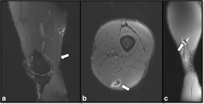
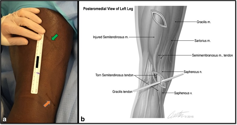
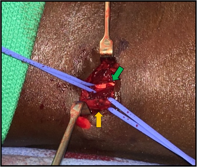
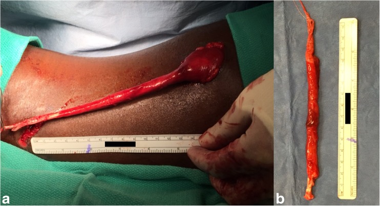
Methods: We highlight a two-incision technique to isolate the avulsed tendon, followed by exteriorization and tendon excision. In addition, we provide insight on clinical and imaging findings to help guide management.

Results: This technique provides a reliable and effective surgical option for managing these rare injuries of the distal semitendinosus, along with outlining rehabilitation goals in the postoperative period.

Conclusion: In this setting, we present a detailed surgical technique to excise the injured distal semitendinosus tendon to promote recovery and potentially allow for earlier return to play.

Keywords: Distal semitendinosus; Tendon excision.


Conflict of interest statement

Compliance with Ethical StandardsBrian J. Rebolledo, MD, and Timothy R. McAdams, MD, declare that they have no conflict of interest. Daniel E. Cooper, MD, reports receiving fees as a consultant from Arthrex and Stryker and royalties from Stryker.Exemption was granted from the Institutional Review Board at Stanford University, where the surgery was performed.Disclosure forms provided by the authors are available with the online version of this article.

Figures

Fig. 1
Fig. 1
Preoperative magnetic resonance imaging (MRI) depicting the distal semitendinosus injury. Sagittal T2, fat suppressed MRI image showing proximal retraction of the distal semitendinosus tendon (arrow) (a). Axial T2, fat suppressed MRI image showing injury to the semitendinosus with surrounding scar formation (arrow) (b). Coronal T2, fat suppressed MRI depicting the injury at the musculotendinous junction of the semitendinosus (arrow) (c).
Fig. 2
Fig. 2
A two-incision surgical technique is utilized for isolating the injured semitendinosus. Using preoperative measurements with magnetic resonance imaging, surgical incisions are marked at the level of the retracted distal tendon (orange arrow) and proximal musculotendinous junction (green arrow) of the semitendinosus over the posteromedial thigh (a). An artistic rendering of the surgical approach, with relevant anatomy encountered through both incisions (b).
Fig. 3
Fig. 3
Distal approach for identifying the injured distal semitendinosus tendon. This highlights the gracilis tendon (green arrow) and saphenous nerve and vein (orange arrow), which should be isolated and protected during dissection.
Fig. 4
Fig. 4
Intraoperative images during tendon excision. The injured semitendinosus is exteriorized from the proximal incision to perform tenotomy at the musculotendinous junction (a). The excised tendon with minimizing proximal muscle disruption (b).

References

    1. Ali K, Leland JM. Hamstring strains and tears in the athlete. Clin Sports Med. 2012;31:263–272. doi: 10.1016/j.csm.2011.11.001. - DOI - PubMed
    1. Cohen SB, Towers JD, Zoga A, Irrgang JJ, Makda J, Deluca PF, Bradley JP. Hamstring injuries in professional football players: magnetic resonance imaging correlation with return to play. Sports Health. 2011;3:423–30. doi: 10.1177/1941738111403107. - DOI - PMC - PubMed
    1. Comin J, Malliaras P, Baquie P, Barbour T, Connell D. Return to competitive play after hamstring injuries involving disruption of the central tendon. Am J Sports Med. 2013;41:111–5. doi: 10.1177/0363546512463679. - DOI - PubMed
    1. Cooper DE, Conway JE. Distal semitendinosus ruptures in elite-level athletes: low success rates of nonoperative treatment. Am J Sports Med. 2010;38:1174–1178. doi: 10.1177/0363546509361016. - DOI - PubMed
    1. Elliott MCCW, Zarins B, Powell JW, Kenyon CD. Hamstring muscle strains in professional football players: a 10-year review. Am J Sports Med. 2011;39:843–850. doi: 10.1177/0363546510394647. - DOI - PubMed