

Anatomical Variations of the Middle Turbinate Concha Bullosa and its Relationship with Chronic Sinusitis: A Prospective Radiologic Study
- PMID: 29983772
- PMCID: PMC6033609
- DOI: 10.1055/s-0038-1625978
Anatomical Variations of the Middle Turbinate Concha Bullosa and its Relationship with Chronic Sinusitis: A Prospective Radiologic Study
Abstract
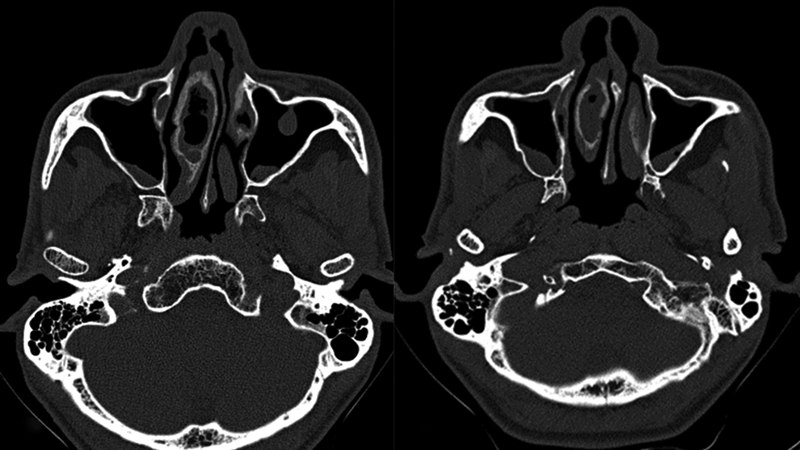
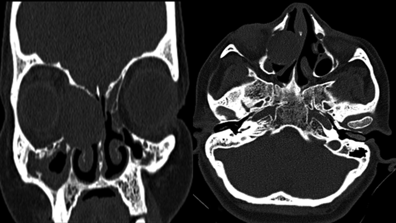
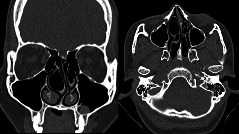
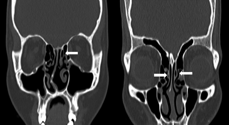
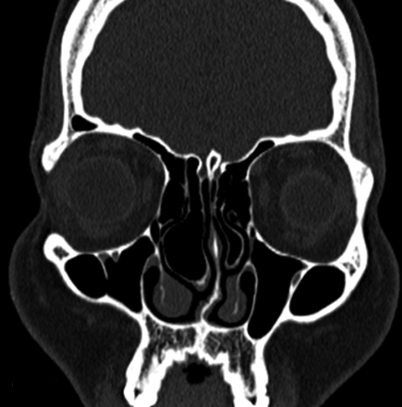
Introduction A pneumatized turbinate, also called concha bullosa, is a normal anatomical variant of the paranasal sinus region. Depending on the site of pneumatization, the concha is classified into extensive, bulbous or lamellar type. The middle turbinate concha bullosa has been implicated as a possible etiological factor in chronic sinusitis. Objectives The aim of this study was to investigate the anatomical variations of the concha bullosa, based on paranasal sinus imaging, and its possible association with sinusitis. Methods This prospective descriptive study was performed at the Department of ENT and Head Neck Surgery over a period of one year, from 2016 to 2017. We studied the computed tomography scans of the nose and paranasal sinuses- in axial, coronal and sagittal planes-of patients who had symptoms of nasal obstruction, or headache and features of chronic sinusitis. Results Out of the 202 scans studied, the prevalence of concha bullosa was 31.7%. The concha was bilateral in 35 (54.7%) patients and unilateral in 29 (45.3%) patients. Out of 99 conchae, 54 were on the right side and 45 were on left side. Ipsilateral sinusitis was found in 40.4% of the sides in the scans of subjects with concha. There was no statistically significant association between any type of middle turbinate concha with sinusitis, but sinusitis was more predominant with the extensive type of concha ( p > 0.05). Conclusion Multiple air cells, mucocele, pyocele and inflammatory mucosal thickenings in the concha are relatively rare. Detailed knowledge of anatomic variations of the concha bullosa is imperative for the radiologists and the operating surgeons.
Keywords: mucocele; sinusitis; turbinates.
Figures
References
-
- Joe J K, Ho S Y, Yanagisawa E.Documentation of variations in sinonasal anatomy by intraoperative nasal endoscopy Laryngoscope 2000110(2 Pt 1):229–235. - PubMed
-
- Al-Sebeih K H, Bu-Abbas M H. Concha bullosa mucocele and mucopyocele: a series of 4 cases. Ear Nose Throat J. 2014;93(01):28–31. - PubMed
-
- Unlü H H, Akyar S, Caylan R, Nalça Y. Concha bullosa. J Otolaryngol. 1994;23(01):23–27. - PubMed
-
- Rak K M, Newell J D, II, Yakes W F, Damiano M A, Luethke J M. Paranasal sinuses on MR images of the brain: significance of mucosal thickening. AJR Am J Roentgenol. 1991;156(02):381–384. - PubMed
LinkOut - more resources
Full Text Sources
Other Literature Sources
Research Materials
Miscellaneous