

Coregistrating magnetic source and magnetic resonance imaging for epilepsy surgery in focal cortical dysplasia
- PMID: 29984157
- PMCID: PMC6029564
- DOI: 10.1016/j.nicl.2018.04.034
Coregistrating magnetic source and magnetic resonance imaging for epilepsy surgery in focal cortical dysplasia
Abstract
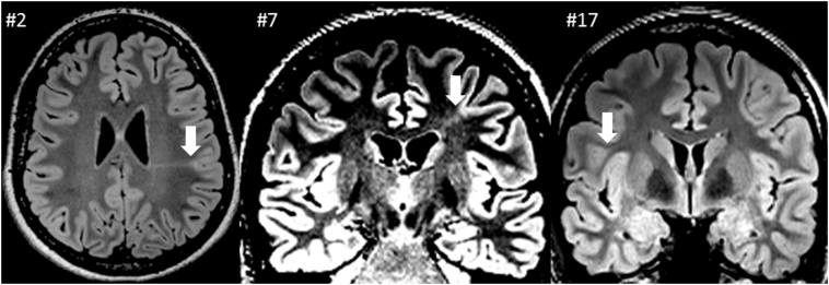
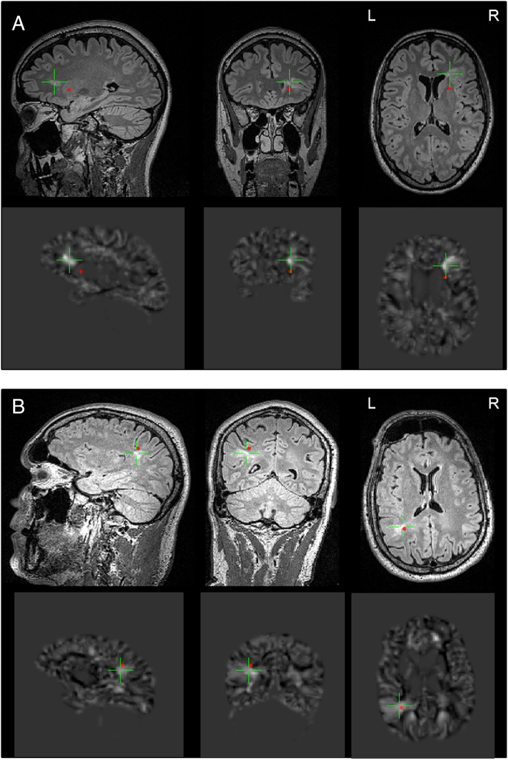
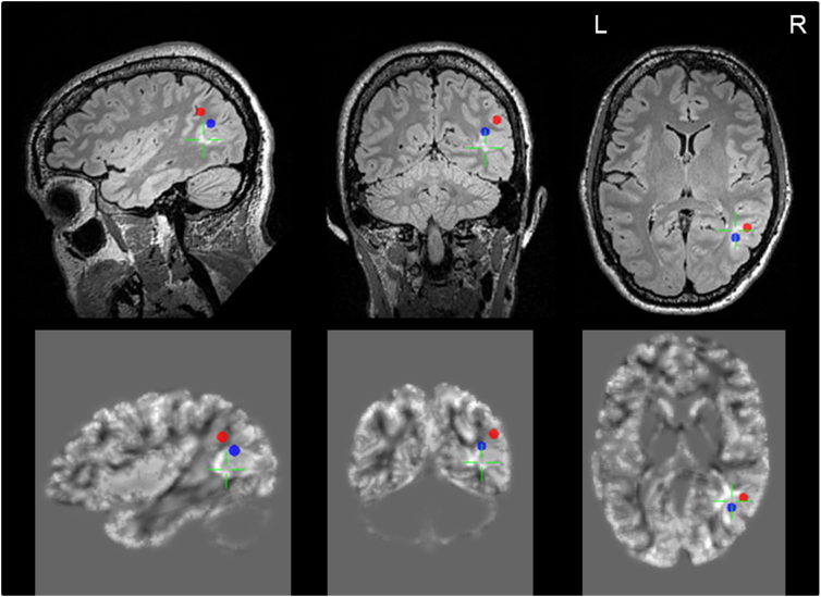
Background: Epilepsy surgery for focal cortical dysplasia type II (FCD II) offers good chances for seizure freedom, but remains a challenge with respect to lesion detection, defining the epileptogenic zone and the optimal resection strategy. Integrating results from magnetic source imaging from magnetoencephalography (MEG) with magnetic resonance imaging (MRI) including MRI postprocessing may be useful for optimizing these goals.
Methods: We here present data from 21 adult FCD II patients, investigated during a 10 year period and evaluated including magnetic source imaging. 16 patients had epilepsy surgery, i.e. histopathologically verified FCD II, and a long follow up. We present our analysis of epileptogenic zones including MEG in relation to structural data according to MRI data and relate these results to surgical outcomes.
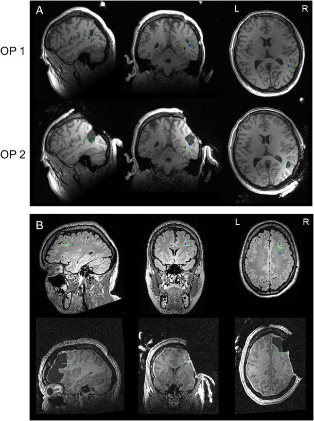
Results: FCD II in our cohort was characterized by high MEG yield and localization accuracy and MEG showed impact on surgical success-rates. MEG source localizations were detected in 95.2% of patients and were as close as 12.3 ± 8,1 mm to the MRI-lesion. After a mean follow up of >3 years, we saw >80% Engel I outcomes, with more favourable outcomes when the MEG source was completely resected (Fishers exact test 0,033).
Conclusion: We argue for a high value of conducting a combined MEG-MRI approach in the presurgical workup and the resection strategy in patients with FCD II related epilepsy.
Keywords: Focal cortical dysplasia; Magnetic source imaging, epilepsy surgery.
Figures
References
-
- Abdallah C., Maillard L.G., Rikir E., Jonas J., Thiriaux A., Gavaret M., Bartolomei F., Colnat-Coulbois S., Vignal J.P., Koessler L. Localizing value of electrical source imaging: frontal lobe, malformations of cortical development and negative MRI related epilepsies are the best candidates. Neuroimage Clin. 2017;16:319–329. - PMC - PubMed
-
- Antel S.B., Collins D.L., Bernasconi N., Andermann F., Shinghal R., Kearney R.E., Arnold D.L., Bernasconi A. Automated detection of focal cortical dysplasia lesions using computational models of their MRI characteristics and texture analysis. NeuroImage. 2003;19:1748–1759. - PubMed
-
- Bartolomei F., Trébuchon A., Bonini F., Lambert I., Gavaret M., Woodman M., Giusiano B., Wendling F., Bénar C. What is the concordance between the seizure onset zone and the irritative zone? A SEEG quantified study. Clin. Neurophysiol. 2016;127:1157–1162. - PubMed
-
- Bast T., Oezkan O., Rona S., Stippich C., Seitz A., Rupp A., Fauser S., Zentner J., Rating D., Scherg M. EEG and MEG source analysis of single and averaged interictal spikes reveals intrinsic epileptogenicity in focal cortical dysplasia. Epilepsia. 2004;45:621–631. - PubMed
Publication types
MeSH terms
Supplementary concepts
LinkOut - more resources
Full Text Sources
Other Literature Sources
Medical
