Review
. 2018 Jun 27:5:61-73.
doi: 10.2147/JHC.S156701. eCollection 2018.

Role of Wnt/β-catenin signaling in hepatocellular carcinoma, pathogenesis, and clinical significance

Affiliations
Review

Role of Wnt/β-catenin signaling in hepatocellular carcinoma, pathogenesis, and clinical significance

Ahmed M Khalaf et al. J Hepatocell Carcinoma. .

Abstract

Hepatocellular carcinoma (HCC) is one of the most common primary hepatic malignancies and one of the fastest-growing causes of cancer-related mortality in the United States. The molecular basis of HCC carcinogenesis has not been clearly identified. Among the molecular signaling pathways implicated in the pathogenesis of HCC, the Wnt/β-catenin signaling pathway is one of the most frequently activated. A great effort is under way to clearly understand the role of this pathway in the pathogenesis of HCC and its role in the transition from chronic liver diseases, including viral hepatitis, to hepatocellular adenomas (HCAs) and HCCs and its targetability in novel therapies. In this article, we review the role of the β-catenin pathway in hepatocarcinogenesis and progression from chronic inflammation to HCC, the novel potential treatments targeting the pathway and its prognostic role in HCC patients, as well as the imaging features of HCC and their association with aberrant activation of the pathway.

Keywords: Wnt/β-catenin; gadoxetic acid-enhanced magnetic resonance imaging; hepatocellular carcinoma; molecular therapy.


Conflict of interest statement

Disclosure The authors report no conflicts of interest in this work.

Figures

Figure 1
Figure 1
Carcinogenesis in HCC with β-catenin mutation with different β-catenin mutants identified, leading to different levels of β-catenin activation depending on the state of tumor progression. Reproduced with permission from Rebouissou S, Franconi A, Calderaro J, et al. Genotype-phenotype correlation of CTNNB1 mutations reveals different β-catenin activity associated with liver tumor progression. Hepatology. 2016;64(6):2047–2061. Copyright John Wiley and Sons. Abbreviation: HCC, hepatocellular carcinoma.
Figure 2
Figure 2
Hematoxylin-eosin staining showing moderately-differentiated HCC (A). At immunohistochemical staining (B), the tumor shows expression of β-catenin protein in the nucleus and/or cytoplasm. Magnification ×200. Abbreviation: HCC, hepatocellular carcinoma.
Figure 3
Figure 3
Overview of Wnt/β-catenin signaling. Notes: In the off-state, or absence of Wnt, cytoplasmic β-catenin forms a complex with APC, axin, CKI, and GSK-3 and then is targeted for proteosomal degradation while Wnt target genes are repressed by TCF/TLE and HDAC. In the on-state, or presence of Wnt, a receptor complex forms between Frizzled and lipoprotein receptor-related protein families, which leads to accumulation of β-catenin in the cytoplasm and nucleus, where it serves as a coactivator for T cell-factor proteins to activate Wnt-responsive genes. Abbreviations: CKI, cyclin-dependent kinase inhibitor; GSK-3, glycogen synthase kinase 3; TCF/TLE, T cell-factor proteins/transducin like enhancer of split; HDAC, histone deacetylases; LRP, low-density-lipoprotein receptor-related protein; APC, adenomatous polyposis coli.
Figure 4
Figure 4
HCCs with β-catenin mutation but not CK19 expression had the best 5-year survival rate, while HCCs with CK19 expression but not β-catenin mutation had the worst 5-year survival rate (P = 0.0002). Notes: (+) = presence of CK19 expression or β-catenin mutation. (−) = absence of CK19 expression or β-catenin mutation. Reprinted by permission from Journal of Gastrointestinal Surgery, Springer Nature, Role of p53 and β-catenin mutations in conjunction with CK19 expression on early tumor recurrence and prognosis of hepatocellular carcinoma, Yuan RH, Jeng YM, Hu RH, et al, copyright 2010. Abbreviations: HCC, hepatocellular carcinoma; CK19, cytokeratin 19.
Figure 5
Figure 5
Relationship between β-catenin, MEN1, and NCOR1 mutations and the Wnt signaling pathway summarized.
Figure 6
Figure 6
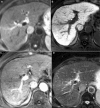
Axial images in a 72-year-old man with HCC. Notes: HCC with β-catenin mutation, hepatitis C, and cirrhosis showing increased tumor intensity on diffusion-weighted imaging (A) and retention of contrast on 20-minute hepatobiliary phase (B). The lesion is seen extending from the caudate lobe and compressing the inferior vena cava with adequate contrast uptake on arterial phase (C), the lesion is also seen to be slightly hyperintense on T2-weighted images (D). Abbreviation: HCC, hepatocellular carcinoma.
Figure 7
Figure 7
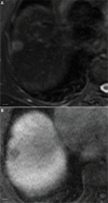
Axial image in HCV-related hepatitis showing HCC without β-catenin mutation. Notes: The images show hyperintensity on T2-weighted images (A) and definite hypointensity on 20-minute hepatobiliary phase compared with background liver (B). Scale bar=1 cm. Abbreviations: HCV, hepatitis C virus; HCC, hepatocellular carcinoma.
Figure 8
Figure 8
Relationship between expression of different genes and the tumor enhancement patterns in hepatobiliary phase images of EOB-MRI. Notes: (A) hyperintense nodule in hepatobiliary-phase (arrow) showed strong expression of (C) BSEP, (D) OATP1B1/1B3, and (E) OATP1B3. (F) markedly hypointense nodule (arrow) showed very low expression of (H) BSEP, (I) OATP1B1/1B3, and (J) OATP1B3. (K) slightly hypointense nodule (arrows) showed strong expression of (N) OATP1B1/1B3 but weak expression of (M) BSEP and (O) OATP1B3. (B, G, and L) are H&E staining of correspondent HCC nodules. Scale bars=0.5 mm. Reprinted from Journal of Hepatology, 61(5), Ueno A, Masugi Y, Yamazaki K, et al, OATP1B3 expression is strongly associated with Wnt/β-catenin signalling and represents the transporter of gadoxetic acid in hepatocellular carcinoma, 80–1087, Copyright (2014), with permission from Elsevier. Abbreviation: EOB-MRI, gadoxetic acid-enhanced magnetic resonance imaging; H&E, hematoxylin and eosin staining; N, non-tumoural liver; T, tumor; HCC, hepatocellular carcinoma.

References

    1. Li P, Cao Y, Li Y, Zhou L, Liu X, Geng M. Expression of Wnt-5a and B-catenin in primary hepatocellular carcinoma. Int J Clin Exp Pathol. 2014;7(6):3190–3195. - PMC - PubMed
    1. Shanbhogue AK, Prasad SR, Takahashi N, Vikram R, Sahani DV. Recent advances in cytogenetics and molecular biology of adult hepatocel-lular tumors: implications for imaging and management. Radiology. 2011;258(3):673–693. - PubMed
    1. Waisberg J, Saba GT. Wnt−/−β-catenin pathway signaling in human hepatocellular carcinoma. World J Hepatol. 2015;7(26):2631–2635. - PMC - PubMed
    1. Dahmani R, Just P-A, Perret C. The Wnt/β-catenin pathway as a therapeutic target in human hepatocellular carcinoma. Clin Res Hepatol Gastroenterol. 2011;35(11):709–713. - PubMed
    1. Oishi N, Wang XW. Novel therapeutic strategies for targeting liver cancer stem cells. Int J Biol Sci. 2011;7(5):517–535. - PMC - PubMed

LinkOut - more resources