Review
. 2018 Nov 14;118(21):10659-10709.
doi: 10.1021/acs.chemrev.8b00163. Epub 2018 Jul 9.

Blue-Light Receptors for Optogenetics

Affiliations
Review

Blue-Light Receptors for Optogenetics

Aba Losi et al. Chem Rev. .

Abstract

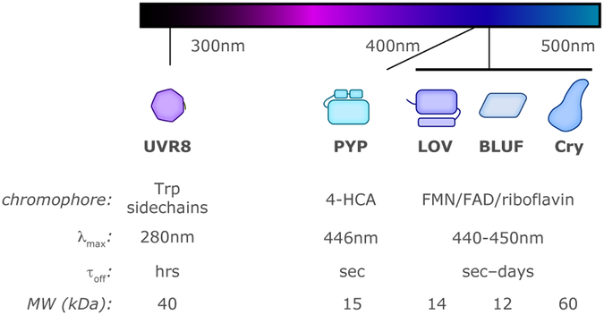
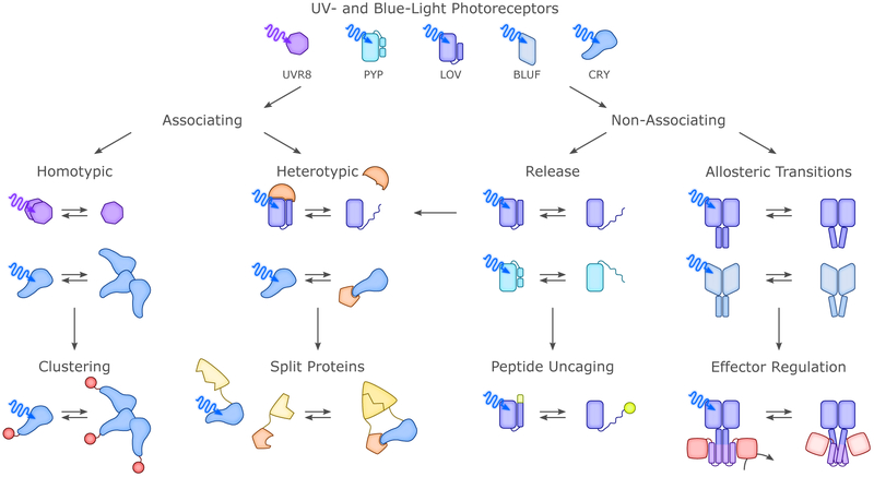
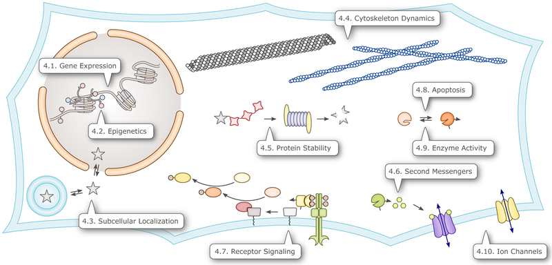
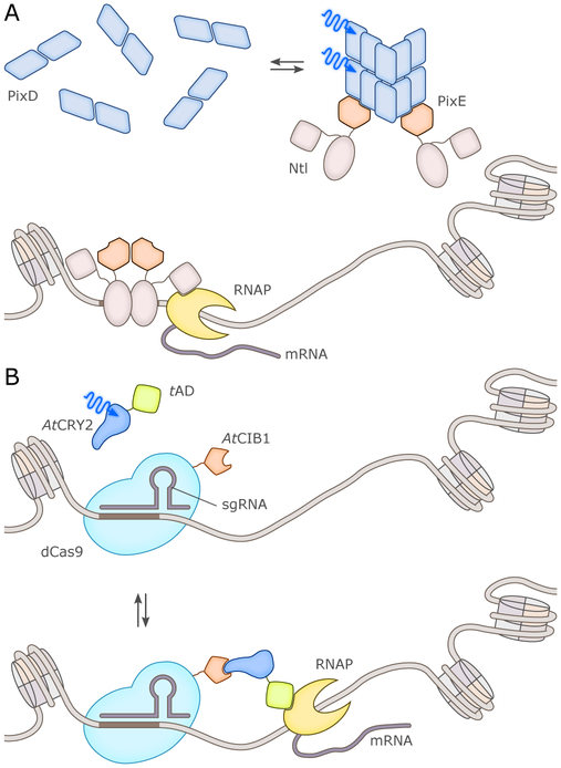
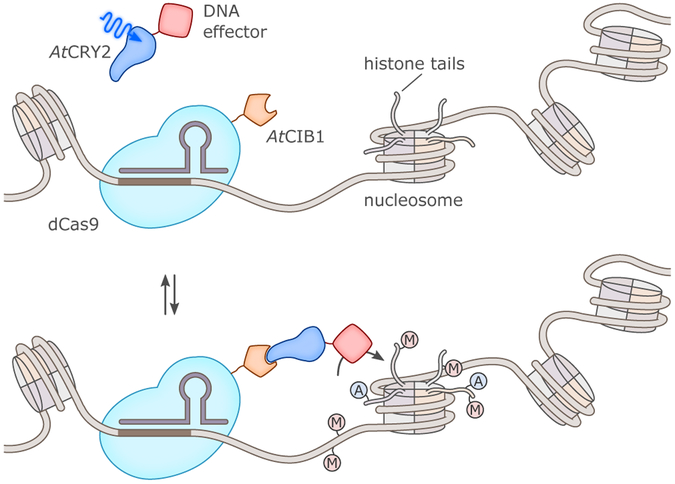
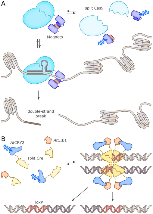
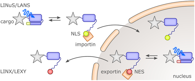
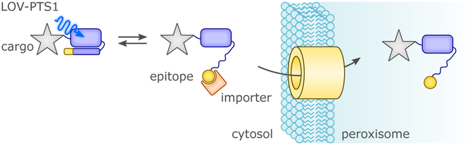
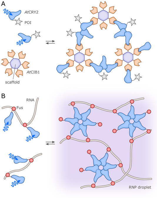
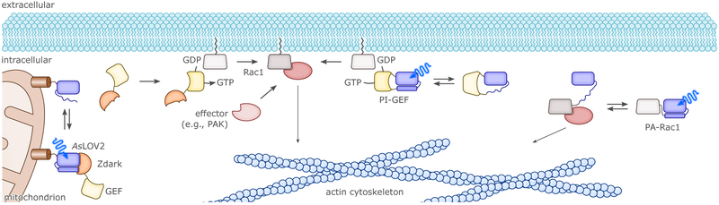
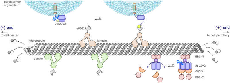
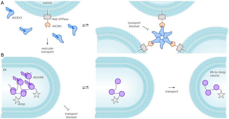
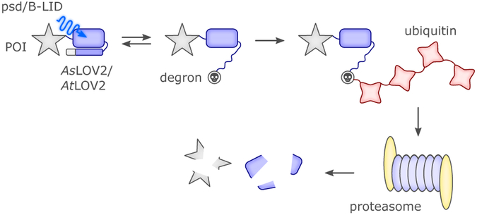
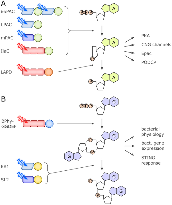
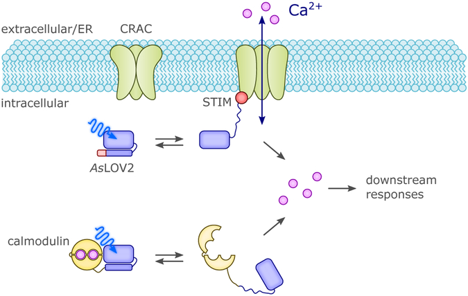
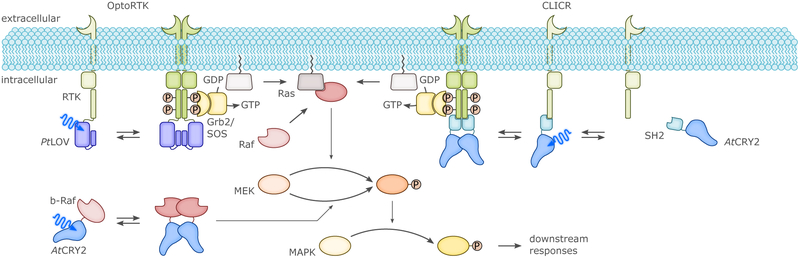
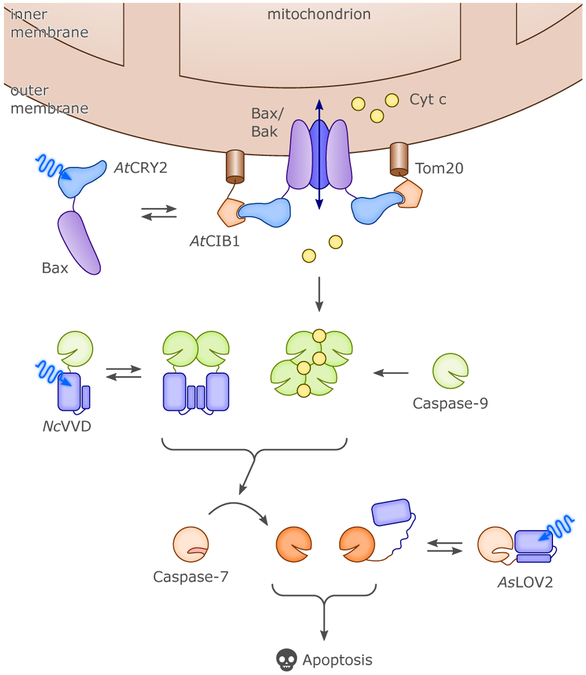
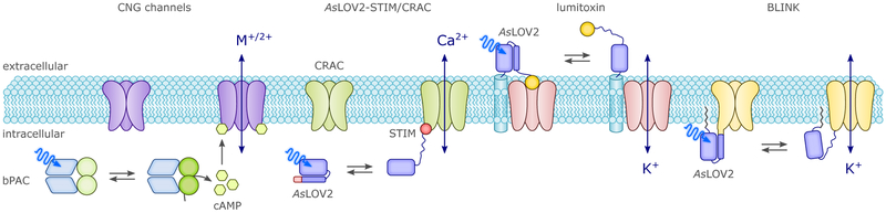
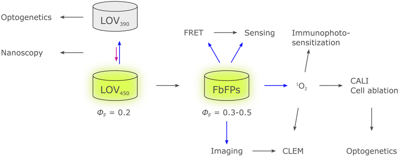
Sensory photoreceptors underpin light-dependent adaptations of organismal physiology, development, and behavior in nature. Adapted for optogenetics, sensory photoreceptors become genetically encoded actuators and reporters to enable the noninvasive, spatiotemporally accurate and reversible control by light of cellular processes. Rooted in a mechanistic understanding of natural photoreceptors, artificial photoreceptors with customized light-gated function have been engineered that greatly expand the scope of optogenetics beyond the original application of light-controlled ion flow. As we survey presently, UV/blue-light-sensitive photoreceptors have particularly allowed optogenetics to transcend its initial neuroscience applications by unlocking numerous additional cellular processes and parameters for optogenetic intervention, including gene expression, DNA recombination, subcellular localization, cytoskeleton dynamics, intracellular protein stability, signal transduction cascades, apoptosis, and enzyme activity. The engineering of novel photoreceptors benefits from powerful and reusable design strategies, most importantly light-dependent protein association and (un)folding reactions. Additionally, modified versions of these same sensory photoreceptors serve as fluorescent proteins and generators of singlet oxygen, thereby further enriching the optogenetic toolkit. The available and upcoming UV/blue-light-sensitive actuators and reporters enable the detailed and quantitative interrogation of cellular signal networks and processes in increasingly more precise and illuminating manners.


Figures

Figure 1
Figure 1
Overview of the five types of soluble blue-/UV-light-sensitive photosensory proteins and protein domains utilized in optogenetic applications. Typical characteristics for members of each family are listed, including chromophores, wavelength of maximum sensitivity (γmax), typical time constants for thermal reversion of photoactivated state (τoff) and molecular weight (MW) of minimal sensory fragment minus effectors.
Figure 2
Figure 2
Fundamental aspects of photoactivation of LOV domains. (A) Structure of AsLOV2,, showing the location of the LOV α/β “core” domain surrounding the FMN chromophore and effector A’α and Jα helices on adjacent faces of the β-sheet. (B) Simplified LOV photocycle, demonstrating the effect of blue light to interconvert between noncovalent protein complex with oxidized flavin and covalently-attached, reduced flavin. Residue numberings apply to AsLOV2. (C) Example of a more complex LOV-effector arrangement within EL222, a LOV-helix-turn-helix (HTH) protein, where an effector helix (more distantly located within the primary structure) from within the HTH domain structurally and functionally replaces the Jα helix of AsLOV2.
Figure 3
Figure 3
Fundamental aspects of photoactivation of BLUF domains. (A) Structure of the BLUF domain of Klebsiella pneumoniae BlrP1,, exemplifying location of the BLUF α/β “core” domain surrounding the FAD chromophore and effector C-terminal helices. (B) Simplified BLUF photocycle, showing how photochemically-driven effects including altered hydrogen-bonding of a nearby glutamine lead to altered protein/FAD interactions. (C) Example of a more complex BLUF-effector arrangement within full-length BlrP1, a BLUF-EAL enzyme involved in c-di-GMP breakdown.
Figure 4
Figure 4
Fundamental aspects of photoactivation of cryptochromes. (A) Structure of the photolyase homology region (PHR) of A. thaliana CRY1, showing the location of the bound FAD chromophore within the highly-helical C-terminal domain. The locations of the N- and C-termini are also indicated, as these have been implicated in CIB1 binding and homooligomerization in the homologous A. thaliana CRY2 (AtCRY2) protein widely used for optogenetic applications. CCT = CRY C-Terminal region. (B) Simplified cryptochrome photocycle, showing the oxidized FAD chromophore present in the inactive dark-adapted state and photochemically-generated anionic and neutral semiquinone states.
Figure 5
Figure 5
Allostery and engineering of UV-light- and BL-sensitive photoreceptors. Despite the rich diversity of these photoreceptors, their signal transduction mechanisms largely fall into but a few classes. In case of the associating photoreceptors, the transition between dark-adapted and light-adapted states entails a change in oligomeric state, either in homotypic or heterotypic manner. Light-modulated oligomerization has proven a particularly versatile approach for engineering novel optogenetic actuators as detailed in section 4. Among the non-associating photoreceptors, we identify light-modulated order-disorder transitions, as exemplified by the Jα helix unfolding in AsLOV2, and changes in tertiary and quaternary structure as prevalent mechanisms. Both types of mechanisms have lent themselves to the engineering of novel photoreceptors (cf. sec. 4.).
Figure 6
Figure 6
Overview of cellular processes that have been optogenetically controlled via photoreceptors sensitive to UV and blue light. The callouts direct to the sections that discuss the corresponding applications.
Figure 7
Figure 7
Several optogenetic systems achieved BL control over transcription in prokaryotes. In LOV-TAP, the E. coll Trp repressor and the AsLOV2 were fused such that mutually exclusive folding of a shared α helix resulted; BL exposure allowed the repressor to correctly fold and bind to DNA. The homodimeric YF1 and the monomeric EL346 are LOV histidine kinases that phosphorylate cognate response regulators in BL-dependent manner; when phosphorylated, the response regulators bind DNA and activate transcription. In EL222, BL absorption by a LOV photosensor prompts dimerization and DNA binding of an associated helix-turn-helix effector, leading to transcriptional activation. The LEVI approach is based on the NcVivid LOV sensor; BL-induced homodimerization rescues the repressional activity of the truncated LexA repressor. Based on the Magnets system for BL-induced heterodimerization, BL-activated split variants of the phage T7 polymerase were engineered.,
Figure 8
Figure 8
BL-dependent of control of transgene transcription in eukaryotes was realized with single-chain constructs (panels A and C) and with split transcription factors (TF) (panel B). (A) The bacterial LOV receptor EL222 was linked to a eukaryotic trans-activating domain (tAD) to achieve light-dependent control of transgenes in eukaryotic cells. The GAVPO approach makes use of the homodimerization reaction NcVivid undergoes upon BL exposure. By linking a DNA-binding domain (DBD) and a tAD to NcVivid, a chimeric TF was obtained that in darkness is monomeric and unable to bind to the DNA operator sequence. BL induced dimerization, DNA binding and transcriptional activation. (B) In several studies, split-TF systems were generated, as exemplarily shown for two specific scenarios. (top) Several approaches relied on linking AtCRY2 to a DBD such that upon BL application a tAD, linked to AtCIB1, could be recruited to induce gene expression., (bottom) Conceptually similar approaches were realized for AtUVR8 which forms a homodimer in the dark but dissociates upon UV-light exposure., In its monomerized form, AtUVR8 can then bind to AtCOP1. By linking the two proteins to a DBD and tAD, respectively, UV-light-dependent control of transcription was achieved. (C) AtCRY2 was fused with both a DBD and a tAD to yield a single-chain TF. BL induced nuclear clearing of this TF, accompanied by downregulation of transcription.
Figure 9
Figure 9
BL-dependent expression from endogenous eukaryotic promoters. (A) In the PICCORO approach, an endogenous transcription factor, e.g., Ntl, was fused to the PixE protein which in the dark associates with the homodecameric BLUF photoreceptor PixD. Upon BL absorption, PixD disassembled into homodimers and dissociated from the PixE-Ntl fusion protein, thus allowing Ntl to bind its endogenous operator site and activate transcription. (B) Programmable DNA-binding proteins, e.g., the TALEs or the cleavage-deficient dCas9, allow to specifically designate endogenous promoters. Transcriptional activation of these promoters was achieved by BL-dependent recruitment of a trans-activating domain (tAD), for example via the AtCRY2:AtCIB1 pair.,
Figure 10
Figure 10
Optogenetic control of epigenetics. TALE proteins and cleavage-deficient dCas9 serve as inert DNA-binding modules that can be programmed to specifically locate to unique target sites in eukaryotic genomes. Linkage of dCas9 with AtCIB1 allows BL-dependent recruitment of DNA effector enzymes that are covalently coupled to AtCRY2. Suitable effectors include DNA and histone methylases, histone (de)acetylases as well as chromatin remodeling enzymes. For example, acetylation and methylation (indicated by ‘A’ and ‘M’, respectively) serve as epigenetic marks and modulate transcriptional activity. For clarity, histone N-terminal tails are only drawn for one nucleosome unit.
Figure 11
Figure 11
Optogenetic control of DNA recombination. (A) The programmable DNA endonuclease Cas9 was split into two parts which could be reassembled in BL-dependent manner via the Magnets LOV receptors. The reconstituted Cas9 enzyme mediated double-strand breaks at defined genomic sites, thus triggering non-homologous end joining and homology-directed repair. (B) Light-regulated recombination was also achieved by splitting the Cre recombinase into two fragments which were linked with AtCRY2 and AtCIB1, respectively.,, BL induced fragment assembly and restoration of activity, thereby enabling recombination at loxP sites.
Figure 12
Figure 12
BL control of nuclear import and export processes was achieved in the LINuS/LANS and LINX/LEXY approaches by embedding corresponding trafficking signal peptides in the Jα helix of the AsLOV2 photosensor. BL-induced unfolding prompted exposure of the signal peptides and caused nuclear import and export, respectively, of cargo proteins.
Figure 13
Figure 13
The LOV-PTS1 strategy is based on the AsLOV2 photosensor to which a peroxisomal trafficking epitope was appended. BL-induced Jα unfolding relieved caging of the epitope and promoted peroxisomal import of cargo proteins.
Figure 14
Figure 14
Optically induced compartments. (A) In the LARIAT method, AtCIB1 is conjugated to a multimeric scaffold protein such that upon BL-induced association with AtCRY2 clusters formed. Proteins of interest (POI) can be sequestered into said clusters either via direct coupling to AtCRY2 or via adapter proteins. For clarity, not all AtCRY2 molecules are shown with attached POI. The related LINC approach does away with AtCIB1 and instead exploits the ability of AtCRY2 to form homooligomers upon BL absorption. (B) The BL-induced clustering of AtCRY2 also underpins a strategy for optogenetically controlling ribonucleoprotein (RNP) droplets. To this end, AtCRY2 was fused to the unstructured RNA-binding protein FUS to allow light-induced liquid-liquid phase transition and formation of RNP droplets.
Figure 15
Figure 15
Optogenetic control of cytoskeleton dynamics. The activity of a soluble form of Rac1 was put under BL control via linkage to the AsLOV2 photosensor such that steric occlusion of the active site resulted. In the resultant PA-Rac1, BL-induced Jα unwinding triggered dissociation of the AsLOV2 and Rac1 moieties, thus restoring access to the active site and eliciting downstream effects on the cytoskeleton. Other approaches targeted the guanine nucleotide exchange factors (GEF) that act on Rac1, drive the exchange of bound GDP for GTP and thereby activate Rac1. Several strategies, including the BL-dependent interaction between AsLOV2 and Zdark, were harnessed to regulate access to the plasma membrane and exchange activity of the GEFs. In the PI-GEF strategy, several GEFs were also subjected to BL control via insertion of AsLOV2 in surface-exposed protein loops. Note that the examples shown here are paradigmatic for numerous related approaches by which the activity of the Rho-family GTPases Rac1, Cdc42 and RhoA was put under BL control, cf. sec. 4.4. for details.
Figure 16
Figure 16
Optogenetic control of microtubule stability and transport. Using the TULIP system for BL-induced heterodimerization, kinesin motors could be recruited to desired organelles, e.g., peroxisomes, which were then transported to the (+) end of microtubules. The principal concept extends to dyneins which move to the (−) end and to myosins which move along actin filaments (not shown). The polymerization dynamics of microtubules was modulated by using a split version of the end-binding protein EB1. In darkness, the two halves of split-EB1 were held together via the AsLOV2:Zdark interaction but BL prompted AsLOV2 Jα unwinding and dissociation of the EB1 fragments.
Figure 17
Figure 17
Optogenetic control of vesicular transport. (A) AtCIB1 was linked to different Rab GTPases that orchestrate vesicular transport. BL induced AtCRY2 to form clusters and to bind AtCIB1, thereby gumming up the vesicular transport machinery. (B) The secretory export of cargo proteins could be modulated in UV-light-dependent manner by linking them to one or several copies of the homodimeric AtUVR8. Formation of higher-order assemblies resulted in retention in the endoplasmic reticulum. UV light prompted AtUVR8 dissociation and resolution of these assemblies, and transport of the cargo ensued.
Figure 18
Figure 18
The intracellular half life of POIs was optogenetically regulated with the psd and B-LID strategies. BL stimulated unfolding of the Jα helix of AtLOV2 or AsLOV2, thereby increasing the exposure of an embedded degron epitope. The cellular ubiquitin/proteasome machinery then degraded the POI and the attached LOV2 module.
Figure 19
Figure 19
Optogenetic actuators for controlling cyclic-nucleotide second messengers. (A) A palette of photoactivated adenylate cyclases (PACs) responsive to BL,,, or red light catalyze the formation of cAMP or cGMP. In eukaryotic cells, cAMP binds to and thus activates CNG channels, PKA, Epac and popeye-domain-containing proteins (PODCP). The red-light-activated PDE LAPD mediates the hydrolytic breakdown of cAMP and cGMP. (B) C-di-GMP is a versatile second messenger involved in numerous physiological adaptations of bacteria. Red-light-activated GGDEF enzymes produce c-di-GMP and achieve optogenetic control over physiology and gene expression in bacteria., In eukaryotes, c-di-GMP triggers the STING response as part of the vertebrate innate immune system. BL-activated EAL enzymes,, catalyze the hydrolysis of c-di-GMP. For clarity, all photoreceptors in panels (A) and (B) are drawn as monomers although they are active as homodimers. BLUF, LOV and bacteriophytochrome photosensors are denoted as parallelograms, rectangles and tripartite shapes, respectively; colored circles denote cyclases and phosphodiesterases specific for cyclic nucleotides.
Figure 20
Figure 20
Intracellular calcium concentrations could be perturbed with BL-sensitive photoreceptors. On the one hand, the opening of Ca2+-specific CRAC channels in the plasma membrane or the endoplasmic/sarcoplasmic reticulum was gated via interactions with the STIM peptide.,, When interleaved with Jα of AsLOV2, the exposure of STIM could be controlled by BL exposure. Alternatively, the STIM epitope was fused to AtCRY2 such that BL-induced clustering resulted in translocation to the membrane and CRAC gating (not shown). A different strategy was pursued in the construction of a fusion protein between the AsLOV2 photosensor and the calcium-binding calmodulin. BL prompted Jα unfolding, destabilization of the calmodulin module and release of bound Ca2+ ions.
Figure 21
Figure 21
Receptor tyrosine kinase (RTK) signaling was subjected to BL-dependent optogenetic control as exemplarily illustrated for the MAPK/ERK pathway. In several approaches,,, OptoRTKs were constructed by appending an associating photoreceptor, e.g., the LOV domain of P. tricornutum aureochrome, to the intracellular C terminus of an RTK. BL then induced homodimerization of the chimeric receptor and activation of the downstream signaling cascade. In the CLICR strategy, endogenous RTKs could be activated upon BL exposure via an adapter protein consisting of AtCRY2 and an SH2 domain that specifically binds to the C termini of RTKs. The MAPK/ERK pathway was also targeted at lower tiers., On the one hand, the Raf kinase can be activated by recruiting it in BL-dependent manner to the plasma membrane (not shown). On the other hand, the B-Raf isoform can be activated away from the membrane in the cytosol by homodimerization or association with the isoform c-Raf. To optogenetically control these processes, the BL-dependent oligomerization of AtCRY2 or its interaction with AtCIB1 was harnessed.
Figure 22
Figure 22
Optogenetic control of phosphatidylinositol signaling. In the CLICR strategy, endogenous receptor tyrosine kinases (RTKs) were put under BL control via AtCRY2-mediated clustering, and the PI3K/Akt signal pathway could be optogenetically manipulated. Once activated by the RTK, the PI3K kinase phosphorylates phosphatidylinositol (PI) to produce the phosphoinositides PIP2 and PIP3. In turn, the Akt kinase binds to PIP3, is thereby activated and elicits downstream responses. Optogenetic intervention in the pathway was also accomplished at the level of PI3K via AtCRY2:AtCIB1-mediated membrane recruitment and concomitant activation., Likewise, the Akt kinase could be directly controlled by translocating it to the membrane upon BL exposure, again using the AtCRY2:AtCIB1 system.
Figure 23
Figure 23
Apoptosis, the programmed cell death, was optogenetically controlled at several tiers with BL-responsive photoreceptors. Covalent fusion of Bax with AtCRY2 allowed its BL-regulated recruitment to AtCIB1 which was connected to the Tom20 protein residing in the outer membrane of the mitochondrion. Oligomerization and assembly with Bak contributed to pore formation and outflow of cytochrome c from the mitochrondrial intermembrane compartment; in the cytosol, cytochrome c promoted oligomerization and activation of the initiator caspase-9. In an alternative approach, the activity of caspase-9 was directly controlled via coupling to the NcVivid photosensor which undergoes BL-induced homodimerization. Activated caspase-9 proteolytically activated downstream executioner caspases, e.g., caspase-7. The latter could be subjected to direct BL control by linkage to the AsLOV2 photosensor such that steric hindrance of the active site resulted. BL promoted AsLOV2 Jα unfolding and restored catalytic activity of caspase-7. Executioner caspases then acted on numerous downstream targets to elicit apoptosis.
Figure 24
Figure 24
Optogenetic regulation of membrane potential and ion flux. BL-sensitive photoreceptors mediate optogenetic perturbation of membrane potential and thus supplement the light-gated channelrhodopsins. Several PACs catalyze the formation of cAMP and cGMP upon BL exposure,,,, cf. Fig. 19, and can be combined with CNG channels to optogenetically control ion flux across the plasma membrane. Depending upon CNG channel, different mono- and divalent cations (M+/2+) are specifically conducted. The gating of CRAC channels was optogenetically controlled by embedding a stimulatory peptide derived from the STIM protein into the Jα helix of AsLOV2, cf. Fig. 20.,, In the lumitoxin method, the AsLOV2 photosensor was anchored to the outer leaflet of the plasma membrane and connected to a peptide toxin that blocked potassium channels. Light-induced Jα unfolding granted enhanced diffusional space to the toxin, resulting in its dissociation from the channel and relieve of inhibition. In the BLINK receptor, the AsLOV2 photosensor was fused via its C-terminal Jα helix to the N terminus of a minimal potassium channel. BL exposure resulted in an increased potassium conductance of BLINK.
Figure 25
Figure 25
Overview of the properties and biophysical applications of flavin-binding fluorescent proteins (FbFPs). Constitutively fluorescent FbFPs are engineered from wild-type LOV domains, by substituting the active-site cysteine to abrogate canonical LOV photochemistry, and by introducing other mutations to increase the fluorescence quantum yield ΦF. They can be used for imaging in fluorescence microscopy, as donors in FRET and as fluorescence-based sensors. The photochromicity of cysteine-retaining LOV domains can be exploited in cellular super-resolution microscopy (nanoscopy), while formation of the thioadduct in LOV domains underlies conventional optogenetic applications. In addition, FbFPs can function as genetically-encoded photosensitizers for 1O2, with a range of further applications., Blue and purple arrows indicate excitation with blue or violet light, respectively.

References

    1. Möglich A; Yang X; Ayers RA; Moffat K Structure and Function of Plant Photoreceptors. Annu. Rev. Plant Biol 2010, 61, 21–47. - PubMed
    1. Hegemann P Algal Sensory Photoreceptors. Annu. Rev. Plant Biol. 2008, 59, 167–189. - PubMed
    1. Sorigué D; Légeret B; Cuiné S; Blangy S; Moulin S; Billon E; Richaud P; Brugière S; Couté Y; Nurizzo D; et al. An Algal Photoenzyme Converts Fatty Acids to Hydrocarbons. Science 2017, 357, 903–907. - PubMed
    1. Deisseroth K; Feng G; Majewska AK; Miesenböck G; Ting A; Schnitzer MJ Next-Generation Optical Technologies for Illuminating Genetically Targeted Brain Circuits. J. Neurosci. 2006, 26, 10380–10386. - PMC - PubMed
    1. Li X; Gutierrez DV; Hanson MG; Han J; Mark MD; Chiel H; Hegemann P; Landmesser LT; Herlitze S Fast Noninvasive Activation and Inhibition of Neural and Network Activity by Vertebrate Rhodopsin and Green Algae Channelrhodopsin. Proc. Natl. Acad. Sci. U. S. A. 2005, 102, 17816–17821. - PMC - PubMed

Publication types

MeSH terms

Substances