. 2018 Aug 1;128(8):3445-3459.
doi: 10.1172/JCI99507. Epub 2018 Jul 9.

CaMK4 compromises podocyte function in autoimmune and nonautoimmune kidney disease

Affiliations

CaMK4 compromises podocyte function in autoimmune and nonautoimmune kidney disease

Kayaho Maeda et al. J Clin Invest. .

Abstract

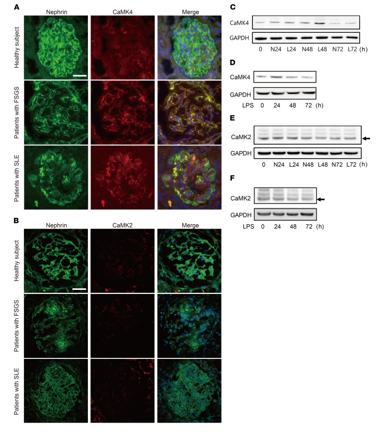
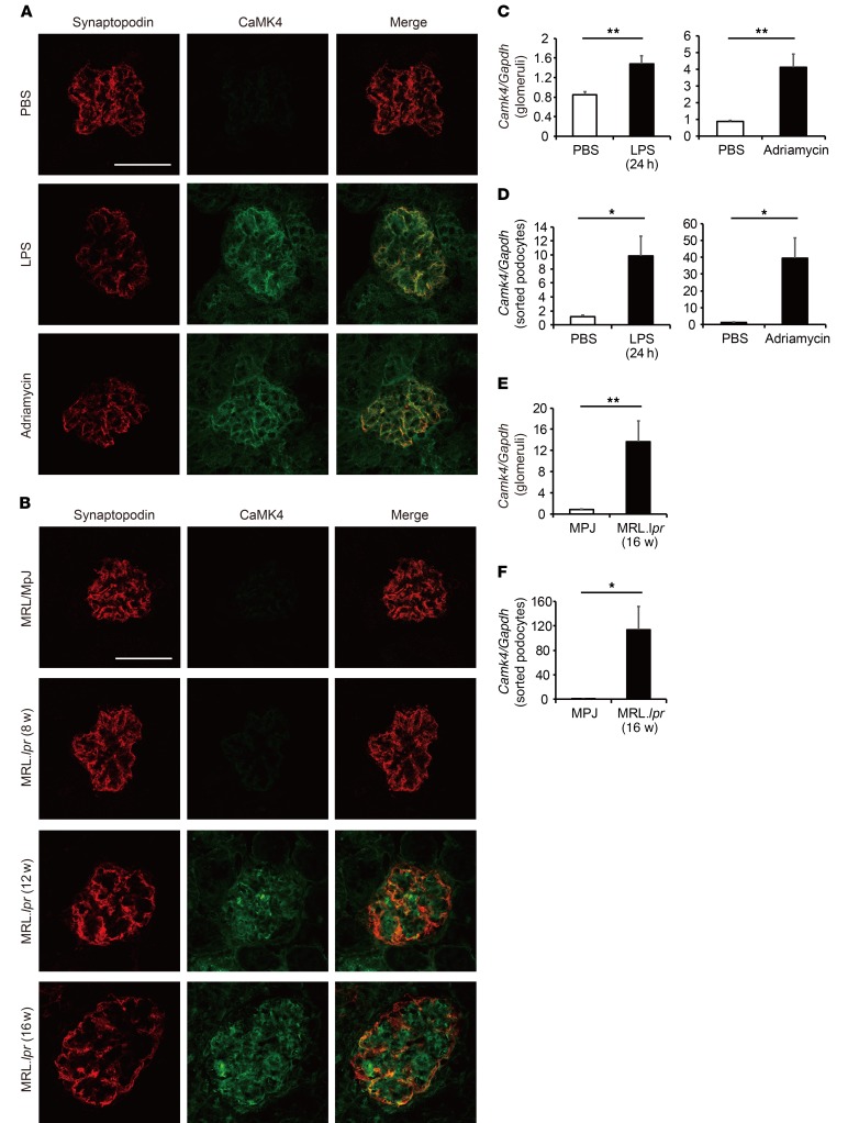
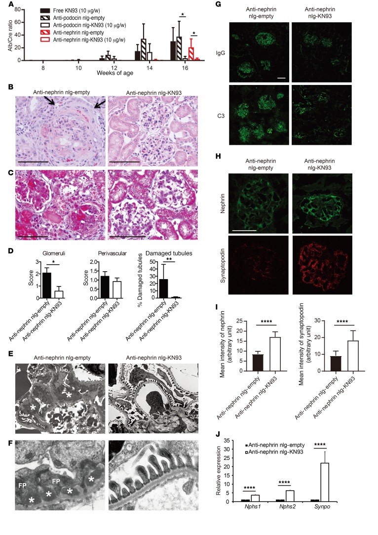
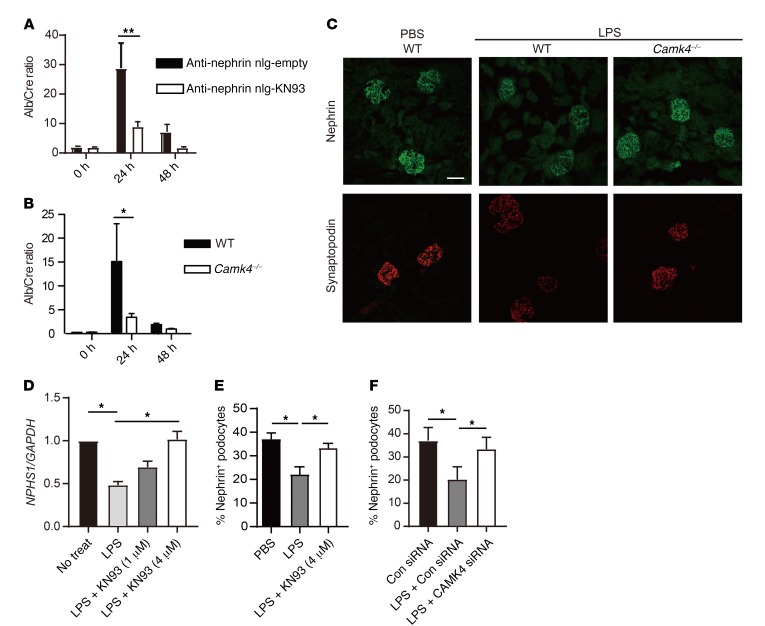
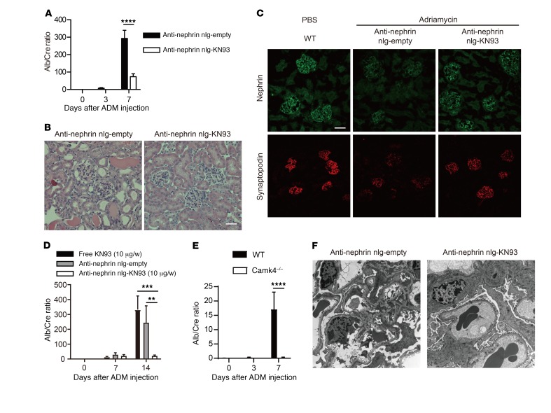
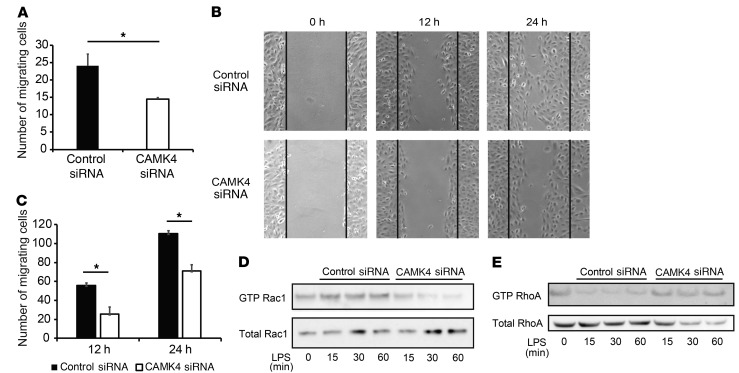
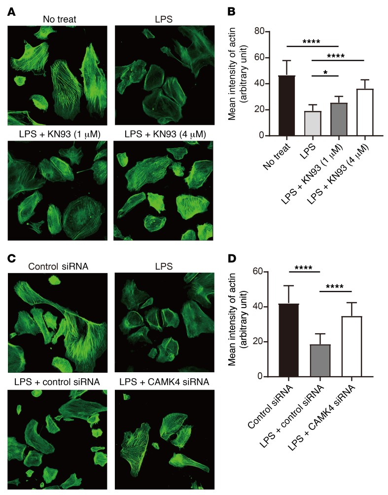
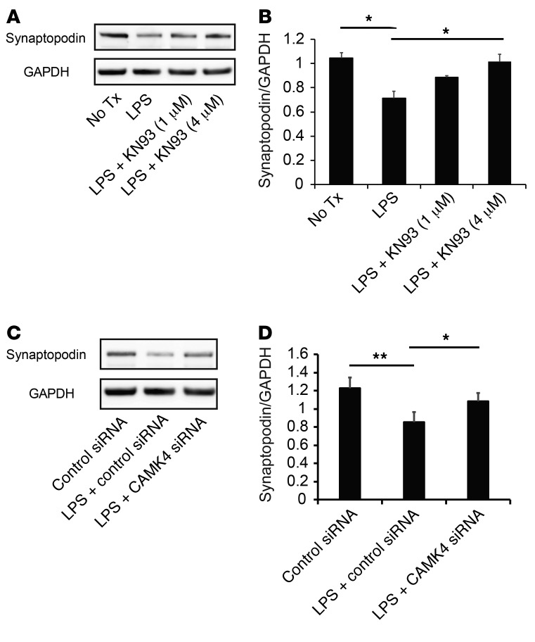
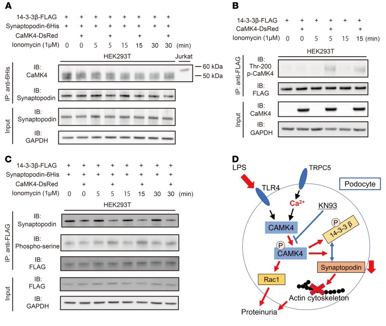
Podocyte malfunction occurs in autoimmune and nonautoimmune kidney disease. Calcium signaling is essential for podocyte injury, but the role of Ca2+/calmodulin-dependent kinase (CaMK) signaling in podocytes has not been fully explored. We report that podocytes from patients with lupus nephritis and focal segmental glomerulosclerosis and lupus-prone and lipopolysaccharide- or adriamycin-treated mice display increased expression of CaMK IV (CaMK4), but not CaMK2. Mechanistically, CaMK4 modulated podocyte motility by altering the expression of the GTPases Rac1 and RhoA and suppressed the expression of nephrin, synaptopodin, and actin fibers in podocytes. In addition, it phosphorylated the scaffold protein 14-3-3β, which resulted in the release and degradation of synaptopodin. Targeted delivery of a CaMK4 inhibitor to podocytes preserved their ultrastructure, averted immune complex deposition and crescent formation, and suppressed proteinuria in lupus-prone mice and proteinuria in mice exposed to lipopolysaccharide-induced podocyte injury by preserving nephrin/synaptopodin expression. In animals exposed to adriamycin, podocyte-specific delivery of a CaMK4 inhibitor prevented and reversed podocyte injury and renal disease. We conclude that CaMK4 is pivotal in immune and nonimmune podocyte injury and that its targeted cell-specific inhibition preserves podocyte structure and function and should have therapeutic value in lupus nephritis and podocytopathies, including focal segmental glomerulosclerosis.

Keywords: Autoimmunity; Calcium signaling; Calmodulin; Lupus; Nephrology.


Conflict of interest statement

Conflict of interest: The authors have declared that no conflict of interest exists.

Figures

Figure 1
Figure 1. CaMK4 is upregulated in podocytes from autoimmune/nonautoimmune kidney diseases.
(A) Representative images of immunofluorescence for nephrin or CaMK4 from patients with LN (n = 10) or FSGS (n = 10) or from a subject without renal disease. Green or red color represents nephrin or CaMK4, respectively, and merged figure shows the signals from nephrin, CaMK4, and DAPI staining. Scale bar: 50 μm. (B) Representative images of immunofluorescence for nephrin or CaMK2 from patients with LN (n = 10) or FSGS (n = 10) or from a subject without renal disease. Green or red color represents nephrin or CaMK2, respectively, and the merged figure shows the signals from nephrin, CaMK2, and DAPI staining. Scale bar: 50 μm. (C and D) Time course of CaMK4 expression in human podocytes after exposure to IgG from LN patients (L) or healthy donors (N) (C) or LPS (D). The data are representative of 3 independent experiments. (E and F) Time course of CaMK2 (arrow) expression in human podocytes after exposure to IgG from LN patients or healthy donors (E) or LPS (F). The data are representative of 3 independent experiments.
Figure 2
Figure 2. CaMK4 expression is increased in podocytes from lupus-prone and LPS- or adriamycin-treated mice.
(A) Representative images of immunofluorescence for synaptopodin or CaMK4 in PBS-, LPS-, and adriamycin-treated mice. Scale bar: 50 μm. (B) Representative images of immunofluorescence for synaptopodin or CaMK4 in MRL/MpJ and MRL.lpr mice (8, 12, 16 weeks old). Scale bar: 50 μm. (CF) Camk4 mRNA expression in glomeruli (C and E) or sorted podocytes by flow cytometry (D and F) from PBS-, LPS-, and adriamycin- treated mice and MRL/MpJ and MRL.lpr mice (16 weeks old). Results were normalized to the expression of GAPDH (n = 5 in each group). *P < 0.05; **P < 0.01, Student’s t test.
Figure 3
Figure 3. Podocyte-targeted delivery of KN93 suppresses kidney disease development in lupus-prone mice.
MRL.lpr mice were injected i.p. with free KN93 (10 μg/wk), anti-podocin or anti-nephrin antibody–coated KN93-loaded nlg (10 μg of KN93/week), or empty nlg at from 8 to 16 weeks of age (n = 5–7 mice in each group). (A) Urine albumin/creatinine (Alb/Cre) ratio. Urine samples were obtained biweekly, and albumin and creatinine levels were determined by ELISA. Error bars represent mean ± SEM. *P < 0.05, 2-way ANOVA with Bonferroni’s post test. (B and C) Representative images of glomeruli from 16-week- old MRL.lpr mice treated with anti-nephrin antibody–coated empty nlg or KN93-loaded nlg. PAS (B) and Masson’s trichrome staining (C) are shown. The arrows point to the crescent. Scale bars: 50 μm. (D) Mean histological scores in the kidneys of mice from the indicated treatment groups (n = 5 in each group). *P < 0.05; **P < 0.01, Student’s t test. (E and F) Electron microscopic images of a glomerulus from a mouse treated with anti-nephrin empty nlg (left) showing diffuse podocyte foot process (FP) effacement with slit diaphragm occlusion (arrows) and subepithelial dense deposits (asterisks) and a mouse treated with anti-nephrin KN93-loaded nlg (right), which shows podocytes with normal foot processes and slit diaphragms and no deposits in the basement membrane. Original magnification, ×8,000 (E); ×30,000 (F). Three glomeruli were evaluated in each of 3 mice in each experimental condition. (G) C3 and IgG deposition was significantly suppressed in the glomeruli of MRL.lpr mice treated with KN93 targeted to podocytes, as indicated. Kidney sections from MRL/MpJ (16 weeks of age) mice are shown as controls (n = 5 mice in each group). (H) Nephrin (green) and synaptopodin (red) expression detected by immunofluorescence. Scale bars: 50 μm. (I) The fluorescence intensity of nephrin and synaptopodin quantified in glomeruli from mice subjected to the indicated treatments. ****P < 0.0001, Student’s t test. (J) Nphs1, Nphs2, and Synpo expression in glomeruli from mice treated with anti-nephrin–coated empty or KN93-loaded nlg. Results were normalized to the expression of GAPDH (n = 5 in each group). ****P < 0.0001, Student’s t test.
Figure 4
Figure 4. Inhibition or genetic deletion of CaMK4 protects mice from LPS-induced podocyte injury.
Mice treated with KN93-loaded nlg targeted to podocytes and CaMK4-deficient mice develop proteinuria after exposure to LPS. Each B6 or B6 Camk4–/– mouse was injected i.p. with LPS on day 0. (A) Urine albumin/creatinine ratio of mice treated with anti-nephrin antibody–coated nlg either empty or loaded with KN93. Nlg were injected i.p. on day –1 (n = 10 mice in each group). (B) Urine albumin/creatinine ratio of B6 or B6 Camk4–/– mice (n = 6 in each group). Error bars represent mean ± SEM. *P < 0.05; **P< 0.01, 2-way ANOVA with Bonferroni’s post test. (C) Representative immunofluorescent images of nephrin (upper panels) and synaptopodin (lower panels) in the kidneys of mice injected with LPS or PBS. Scale bar: 50 μm. (D) Nphs1 expression in human podocytes after stimulation with LPS with or without KN93. Cells were treated with KN93 1 hour before stimulation. Results were normalized by the expression of GAPDH. Four independent experiments were performed. *P < 0.05, 1-way ANOVA with Tukey’s post test. (E and F) Mean percentage of nephrin-positive human podocytes evaluated by flow cytometry after stimulation with LPS for 72 hours with KN93 (E) or CAMK4 siRNA (F). Four independent experiments were performed. *P < 0.05, 1-way ANOVA with Tukey’s post test.
Figure 5
Figure 5. Podocyte-targeted delivery of KN93 prevents and reverses adriamycin-induced podocyte injury in mice.
Each mouse was injected i.v. with adriamycin (ADM) on day 0. (AC) Anti-nephrin antibody–coated empty or KN93-loaded nlg were injected i.p. into BALB/c mice on day –1 and day 3 (n = 7 in each group). (A) Mean urine albumin/creatinine ratio from mice subjected to the indicated treatment. Error bars represent mean ± SEM. ****P < 0.0001, 2-way ANOVA with Bonferroni’s post test (B) Representative images showing PAS staining of kidney from BALB/c mice treated with anti-nephrin antibody–coated empty nlg or KN93-loaded nlg. Scale bar: 50 μm. (C) Representative immunofluorescence images of nephrin and synaptopodin expression in glomeruli. Scale bar: 100 μm. (D) Free KN93 (10 μg/wk), anti-nephrin antibody–coated empty, or KN93-loaded nlg (10 μg of KN93/wk) were injected i.p. into BALB/c mice on day 7 (n = 5 in each group). **P < 0.01; ***P < 0.001, 2-way ANOVA with Bonferroni’s post test. (E) Mean urine albumin/creatinine ratio of B6 or B6 Camk4–/– mice treated with adriamycin. (n = 5 in each group; 2 independent experiments were performed). ****P < 0.0001, 2-way ANOVA with Bonferroni’s post-test. (F) Representative electron microscopy images of glomeruli from BALB/c mice at 7 days after exposure to adriamycin, treated with anti-nephrin antibody–coated empty nlg (left) or KN93-loaded nlg (right). Original magnification, ×8,000.
Figure 6
Figure 6. CaMK4 regulates podocyte migration.
(A) Result of Transwell migration experiments using human podocytes treated with control or CaMK4 siRNA. Error bars represent mean ± SEM (n = 3 independent experiments). *P < 0.05, Student’s t test. (B) Representative images of wound-healing assay 0, 12, and 24 hours after wounding. (C) Quantification of the results in B. Error bars represent mean ± SEM (n = 3 independent experiments). *P < 0.05, Student’s t test. (D and E) Western blotting analysis of GTP-Rac1 and total Rac1 expression (D) and GTP-RhoA and total RhoA (E) in human podocytes treated with control or CaMK4 siRNA for the indicated times. Three independent experiments were performed.
Figure 7
Figure 7. Inhibition or silencing of CaMK4 preserves actin structure of podocytes.
(A) Representative results of phalloidin staining of human podocytes after LPS treatment with or without KN93 (1 μM or 4 μM). (B) Quantification of the results in A (n = 20 cells; 3 independent experiments were performed). Error bars represent mean ± SEM. *P < 0.05; ****P < 0.0001, 1-way ANOVA with Tukey’s post test. (C) Representative results of phalloidin staining of human podocytes after LPS treatment with transfection of control or CaMK4 siRNA. (D) Quantification of the results in C (n = 20 cells; 3 independent experiments were performed). Error bars represent mean ± SEM. ***P < 0.0001, 1-way ANOVA with Tukey’s post test. Original magnification, ×200.
Figure 8
Figure 8. Inhibition or silencing of CaMK4 preserves synaptopodin expression.
(A) Western blotting analysis of synaptopodin expression in human podocytes 72 hours after exposure to LPS with or without KN93 (1 μM or 4 μM). Tx, treatment. (B) Quantification of the results in A. (C) Western blotting analysis of synaptopodin expression in human podocytes 72 hours after exposure to LPS with transfection of control or CaMK4 siRNA. (D) Quantification of the results in C. Error bars represent mean ± SEM (n = 3–5 independent experiments). *P < 0.05; **P < 0.01, 1-way ANOVA with Tukey’s post-test.
Figure 9
Figure 9. Activated CaMK4 disrupts the binding of synaptopodin to 14-3-3β.
Human 14-3-3β-FLAG, synaptopodin-6His, and CaMK4-DsRed plasmids are transfected into HEK293T cells, followed by immunoprecipitation with an anti-FLAG or anti-6His antibodies. (A) Immunoprecipitation with anti-6His antibody and immunoblot analysis of CaMK4 and synaptopodin. CaMK4 does not interact with His-tagged synaptopodin in cotransfected HEK293T cells. Jurkat cell lysate was used as a positive control of CaMK4. Data are representative of 2 independent experiments. (B) Phospho-CaMK4 precipitates with flag-tagged 14-3-3β after exposure to ionomycin from cotransfected HEK293T cells. Data are representative of 2 independent experiments. (C) Immunoprecipitation and immunoblot analysis of synaptopodin and phospho-serine expression. Cotransfected HEK293T cells were stimulated with ionomycin for the indicated times. Cell lysates were immunoprecipitated with anti-FLAG antibody and then analyzed by immunoblotting with anti-synaptopodin and anti–phospho-serine antibodies. Data are representative of 3 independent experiments. (D) Schematic model of the relationship between Ca2+/CaMK4 signaling and actin stability in podocytes.

Comment in

Similar articles

Cited by

References

    1. Breyer MD, Susztak K. The next generation of therapeutics for chronic kidney disease. Nat Rev Drug Discov. 2016;15(8):568–588. doi: 10.1038/nrd.2016.67. - DOI - PMC - PubMed
    1. D’Agati VD, Kaskel FJ, Falk RJ. Focal segmental glomerulosclerosis. N Engl J Med. 2011;365(25):2398–2411. doi: 10.1056/NEJMra1106556. - DOI - PubMed
    1. Scott RP, Quaggin SE. Review series: The cell biology of renal filtration. J Cell Biol. 2015;209(2):199–210. doi: 10.1083/jcb.201410017. - DOI - PMC - PubMed
    1. Welsh GI, Saleem MA. The podocyte cytoskeleton--key to a functioning glomerulus in health and disease. Nat Rev Nephrol. 2011;8(1):14–21. - PubMed
    1. Perico L, Conti S, Benigni A, Remuzzi G. Podocyte-actin dynamics in health and disease. Nat Rev Nephrol. 2016;12(11):692–710. doi: 10.1038/nrneph.2016.127. - DOI - PubMed

Publication types

MeSH terms

Substances