. 2018 Aug 1;128(8):3402-3412.
doi: 10.1172/JCI97192. Epub 2018 Jul 9.

The cardiac lymphatic system stimulates resolution of inflammation following myocardial infarction

Affiliations

The cardiac lymphatic system stimulates resolution of inflammation following myocardial infarction

Joaquim Miguel Vieira et al. J Clin Invest. .

Abstract

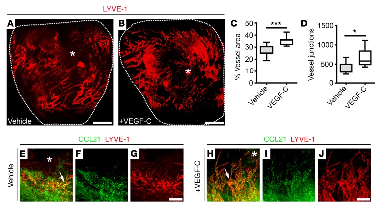
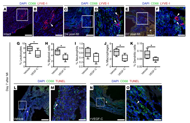
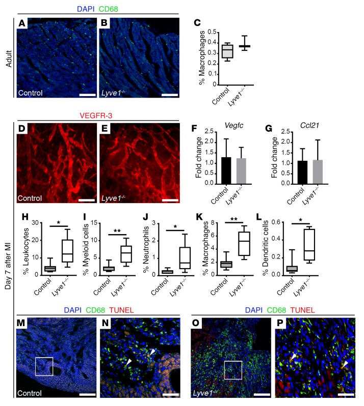
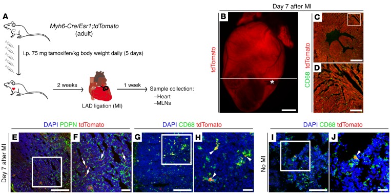
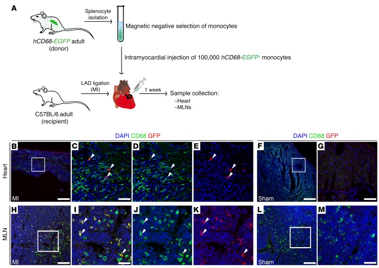
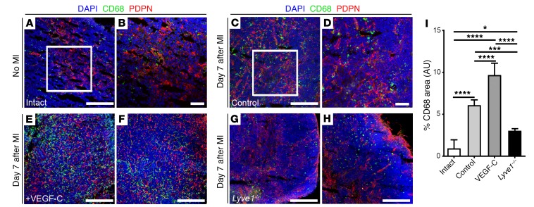
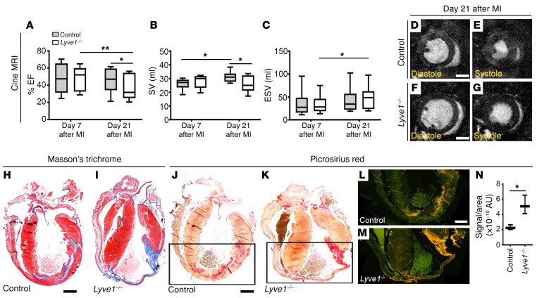
Myocardial infarction (MI) arising from obstruction of the coronary circulation engenders massive cardiomyocyte loss and replacement by non-contractile scar tissue, leading to pathological remodeling, dysfunction, and ultimately heart failure. This is presently a global health problem for which there is no effective cure. Following MI, the innate immune system directs the phagocytosis of dead cell debris in an effort to stimulate cell repopulation and tissue renewal. In the mammalian adult heart, however, the persistent influx of immune cells, coupled with the lack of an inherent regenerative capacity, results in cardiac fibrosis. Here, we reveal that stimulation of cardiac lymphangiogenesis with VEGF-C improves clearance of the acute inflammatory response after MI by trafficking immune cells to draining mediastinal lymph nodes (MLNs) in a process dependent on lymphatic vessel endothelial hyaluronan receptor 1 (LYVE-1). Deletion of Lyve1 in mice, preventing docking and transit of leukocytes through the lymphatic endothelium, results in exacerbation of chronic inflammation and long-term deterioration of cardiac function. Our findings support targeting of the lymphatic/immune cell axis as a therapeutic paradigm to promote immune modulation and heart repair.

Keywords: Cardiovascular disease; Inflammation; Vascular Biology.


Conflict of interest statement

Conflict of interest: PRR is cofounder of and equity holder in OxStem Cardio, an Oxford University spin-out that seeks to exploit therapeutic strategies stimulating endogenous repair in cardiovascular regenerative medicine.

Figures

Figure 1
Figure 1. VEGF-C treatment augments cardiac lymphangiogenesis after injury.
(A and B) Whole-mount immunostaining for LYVE-1 (red) to visualize the subepicardial lymphatic plexus of vehicle- and recombinant VEGF-C(C156S)–treated hearts on day 7 after MI. (C and D) Quantification of the lymphangiogenic response on day 7 after MI as percent LYVE-1+ lymphatic vessel area (C) and junction number (D). Data are presented as mean ± SEM; vehicle, n = 7 hearts; VEGF-C, n = 5 hearts. Significant differences were calculated using an unpaired, 2-tailed Student’s t test (*P ≤ 0.05, ***P ≤ 0.001). (EJ) Immunostaining for CCL21 (green) and LYVE-1 (red) in vehicle- (EG) and recombinant VEGF-C(C156S)–treated (HJ) whole adult hearts on day 7 after MI. White arrows highlight expression of the immune cell chemoattractant cue CCL21 by lymphatic capillaries after injury. White asterisks indicate the ligating suture. Scale bars: A and B, 1 mm; G and J, 20 μm.
Figure 2
Figure 2. VEGF-C–driven cardiac lymphangiogenesis increases clearance of immune cells after injury.
(AF) CD68 (green) and LYVE-1 (red) immunostaining of tissue sections derived from adult intact hearts (A and B) from hearts day 4 (C and D) and day 7 (E and F) after MI, documenting close association of macrophages to lymphatic vessels (white arrowheads). (B) Magnified view of box shown in A. (D) Magnified view of box shown in C. (F) Magnified view of box shown in E. DAPI (blue) labels cell nuclei. Asterisk in E denotes fibrotic scarring. (GK) Characterization of immune cell content in vehicle- and VEGF-C–treated hearts collected on day 7 after MI and analyzed by flow cytometry using antibodies against CD45 (pan-leukocyte marker), CD11b (CD45+CD11b+, myeloid cells), Ly6G (CD45+CD11b+Ly6G+, neutrophils), F4/80 (CD45+CD11b+Ly6GF4/80+, macrophages), and CD11c (CD45+CD11b+Ly6GF4/80CD11c+, dendritic cells). Animals received i.p. injections of vehicle (PBS) or recombinant VEGF-C(C156S) on days 0, 2, 4, and 6 after MI. Data are presented as mean ± SEM; vehicle, n = 5 hearts; VEGF-C, n = 6 hearts. Significant differences were calculated using an unpaired, 2-tailed Student’s t test (*P ≤ 0.05). (LO) CD68 (green) and TUNEL (red) immunostaining of tissue sections derived from vehicle- (L and M) and recombinant VEGF–C(C156S)–treated (N and O) hearts on day 7 after MI. (M) Magnified view of box shown in L. (O) Magnified view of box shown in N. Asterisks in L and N indicate fibrotic scarring; DAPI (blue) labels cell nuclei. White arrowheads mark rare macrophage cells undergoing apoptosis (CD68+TUNEL+). Scale bars: A, C, E, L, and N, 100 μm; B, D, F, M, and O, 20 μm.
Figure 3
Figure 3. LYVE-1 is required for immune cell clearance after MI.
(A and B) CD68 (green) immunostaining of sections derived from control and Lyve1–/– intact hearts, documenting the presence of resident macrophages throughout the myocardium of mutant hearts, compared with controls. DAPI (blue) labels cell nuclei. (C) Quantification of resident macrophages (CD45+CD11b+Ly6GF4/80+) in the intact adult heart by flow cytometry. Data are presented as mean ± SEM; control, n = 4 hearts; Lyve1–/–, n = 4 hearts. No significant differences were observed (unpaired, 2-tailed Student’s t test). (D and E) Whole-mount immunostaining for VEGFR-3 (red) revealing comparable superficial lymphatic networks with lymphangiogenic capillary tips in control and Lyve1–/– hearts on day 7 after MI. (F and G) Vegfc and Ccl21 expression analysis by qRT-PCR showing no differences in expression levels in control and Lyve1–/– hearts on day 7 after MI. Data are presented as mean ± SEM; control, n = 4 hearts; Lyve1–/–, n = 6 hearts. (HL) Characterization of the immune cell content in control and Lyve1–/– hearts collected on day 7 after MI and analyzed by flow cytometry using antibodies against CD45 (pan-leukocyte marker), CD11b (CD45+CD11b+, myeloid cells), Ly6G (CD45+CD11b+Ly6G+, neutrophils), F4/80 (CD45+CD11b+Ly6GF4/80+, macrophages), and CD11c (CD45+CD11b+Ly6GF4/80CD11c+, dendritic cells). Data are presented as mean ± SEM; control, n = 7 hearts; Lyve1–/–, n = 5 hearts. Significant differences were calculated using an unpaired, 2-tailed Student’s t test (*P ≤ 0.05, **P ≤ 0.01). (MP) CD68 (green) and TUNEL (red) immunostaining of sections derived from control (M and N) and Lyve1–/– (O and P) hearts on day 7 after MI. (N and P) Magnified views of boxes shown in M and O. DAPI (blue) labels cell nuclei. White arrowheads mark rare macrophages undergoing apoptosis (CD68+TUNEL+). Note increased CD68 expression in Lyve1–/– compared with controls. Scale bars: A, B, M, and O, 100 μm; D and E, 200 μm; N and P, 20 μm.
Figure 4
Figure 4. Cardiac immune cells are cleared to MLNs after injury.
(A) Schematic of tamoxifen-induced labeling of adult cardiomyocytes in Myh6-Cre/Esr1;tdTomato mice to probe phagocytic cell trafficking to MLNs via the cardiac lymphatic system on day 7 after injury. (BD) Visualization of endogenous tdTomato (red) fluorescence alone or in combination with CD68 (green) immunostaining (macrophage marker), documenting efficient labeling of cardiomyocytes in the adult heart. White asterisk marks the ligating suture; white line marks the plane of sectioning of the whole heart. Note that the section in C is derived from the heart in B, and D is a magnified view of white box in C. (E and F) PDPN (green) immunostaining and tdTomato fluorescence marking red-labeled particles in close association with PDPN-expressing lymphatic capillaries (white arrows) within MLNs of tamoxifen-induced Myh6-Cre/Esr1;tdTomato mice at 7 days after MI. (GJ) CD68 (green) immunostaining combined with tdTomato (red) fluorescence indicating that red particles are contained within CD68+ phagocytic cells (white arrowheads). The CD68+/tdTomato-labeled cell population is increased in MLNs after MI (compare G and H with I and J). (F, H, and J) Magnified views of white boxes in E, G, and I. DAPI (blue) labels cell nuclei. Scale bars: C, E, G, and I, 100 μm; B, 1 mm; D, 300 μm; F, 50 μm; H and J, 20 μm.
Figure 5
Figure 5. Adoptive transfer of splenic GFP+ monocytes confirms immune cell clearance to MLNs after MI.
(A) Schematic of the adoptive cell transfer approach using hCD68-EGFP transgenic mice as splenic GFP+ monocyte donor and recipient C57BL/6 adult mice receiving intramyocardial delivery of labeled monocytes at the time of LAD ligation, to assess immune cell trafficking to MLNs. (BG) GFP (red) and CD68 (green) immunostaining (macrophage marker) of tissue sections documenting engraftment of CD68+GFP+ monocytes within the injury area at 7 days after MI (white arrowheads). No GFP-labeled cells were detected in sham-operated animals (F and G). (CE) Magnified views of box shown in B. (G) Magnified view of box shown in F. DAPI (blue) labels cell nuclei. (HM) GFP (red) and CD68 (green) immunostaining of tissue sections derived from MLNs of MI (HK) and sham-operated (LM) animals, indicating the presence of cleared CD68+GFP+ phagocytic cells (white arrowheads) in MLNs after MI. (I, J, and K) Magnified views of box in H. (M) Magnified view of box in L. DAPI (blue) labels cell nuclei. Scale bars: 100 μm; except C, G, I, and M, 20 μm.
Figure 6
Figure 6. VEGF-C–driven cardiac lymphangiogenesis promotes clearance of immune cells to MLNs after injury.
(AH) CD68 (green) and PDPN (red) immunostaining of tissue sections derived from intact (no MI; A and B), control (C and D), recombinant VEGF-C(C156S)–treated (E and F), and Lyve1–/– (G and H) MLNs collected 7 days after MI. (B and D) Magnified views of white boxes in A and C. (E and F) Representative views of 2 different VEGF-C–treated MLNs. (G and H) Representative views of 2 different Lyve1–/– MLNs. Note the relative abundance of CD68+ macrophages in VEGF-C–treated MLNs, compared with Lyve1–/– (compare E and F with G and H). DAPI (blue) labels cell nuclei. (I) Quantification of macrophage proportion as percent CD68+ total staining area/total DAPI-labeled tissue area × 100. Data are presented as mean ± SEM; intact, n = 4 MLNs; control, n = 8 MLNs; VEGF-C, n = 4 MLNs; Lyve1–/–, n = 4 MLNs. Note that 1 MLN was analyzed per mouse. Significant differences were calculated using 1-way ANOVA followed by Tukey’s multiple comparisons test (*P ≤ 0.05 for intact vs. Lyve1–/–; ***P ≤ 0.001 for control vs. Lyve1–/–; and ****P ≤ 0.0001 for intact vs. control, control vs. VEGF-C, intact vs. VEGF-C, and VEGF-C vs. Lyve1–/–). Scale bars: 100 μm; except B and D, 50 μm.
Figure 7
Figure 7. Disruption of LYVE-1–dependent clearance of immune cells by lymphatics is detrimental to cardiac function after injury.
(AC) Longitudinal cine MRI analyses of infarcted control and Lyve1–/– hearts on days 7 and 21 after injury showing reduced EF (A), SV (B), and ESV (C) in mutants, compared with controls. Data are presented as mean ± SEM; control, n = 10 hearts; Lyve1–/– , n = 10 hearts. Significant differences were calculated using 2-way ANOVA with repeated measures (*P ≤ 0.05, **P ≤ 0.01). (DG) Representative 1-mm-thick mid-ventricular short-axis cine MRI frames for control (D and E) and Lyve1–/– (F and G) hearts in diastole (D and F) and systole (E and G) on day 21 after MI. (HN) Histological characterization of control and Lyve1–/– hearts on day 21 after MI using Masson’s trichrome (H and I) and Picrosirius red staining (JN), documenting excessive collagen deposition/fibrotic scarring (blue in H and I; red in J and K; yellow-orange birefringence in L and M) in mutant hearts. Note that Picrosirius staining was visualized under brightfield (J and K) and polarized light (L and M), leading to birefringence of the collagen fibers, to further characterize the type of fibers making up the scar, i.e. type I (thicker; yellow-orange) or type III (thin; green). (N) Quantification of fibrotic scarring as (Picrosirius) yellow-orange birefringence signal/area ratio. Data are presented as mean ± SEM; control, n = 5 hearts; Lyve1–/–, n = 5 hearts. Significant differences were calculated using an unpaired, 2-tailed Student’s t test (*P ≤ 0.05). Scale bars: 1 mm; except D and F, 2 mm.

Comment in

References

    1. Aspelund A, Robciuc MR, Karaman S, Makinen T, Alitalo K. Lymphatic system in cardiovascular medicine. Circ Res. 2016;118(3):515–530. doi: 10.1161/CIRCRESAHA.115.306544. - DOI - PubMed
    1. Card CM, Yu SS, Swartz MA. Emerging roles of lymphatic endothelium in regulating adaptive immunity. J Clin Invest. 2014;124(3):943–952. doi: 10.1172/JCI73316. - DOI - PMC - PubMed
    1. Kaipainen A, et al. Expression of the fms-like tyrosine kinase 4 gene becomes restricted to lymphatic endothelium during development. Proc Natl Acad Sci U S A. 1995;92(8):3566–3570. doi: 10.1073/pnas.92.8.3566. - DOI - PMC - PubMed
    1. Kim H, Kataru RP, Koh GY. Inflammation-associated lymphangiogenesis: a double-edged sword? J Clin Invest. 2014;124(3):936–942. doi: 10.1172/JCI71607. - DOI - PMC - PubMed
    1. Kataru RP, et al. Critical role of CD11b+ macrophages and VEGF in inflammatory lymphangiogenesis, antigen clearance, and inflammation resolution. Blood. 2009;113(22):5650–5659. doi: 10.1182/blood-2008-09-176776. - DOI - PubMed

Publication types

MeSH terms

Substances