Review
. 2018 Aug;14(8):482-495.
doi: 10.1038/s41582-018-0025-8.

Multidimensional communication in the microenvirons of glioblastoma

Affiliations
Review

Multidimensional communication in the microenvirons of glioblastoma

Marike L Broekman et al. Nat Rev Neurol. 2018 Aug.

Abstract

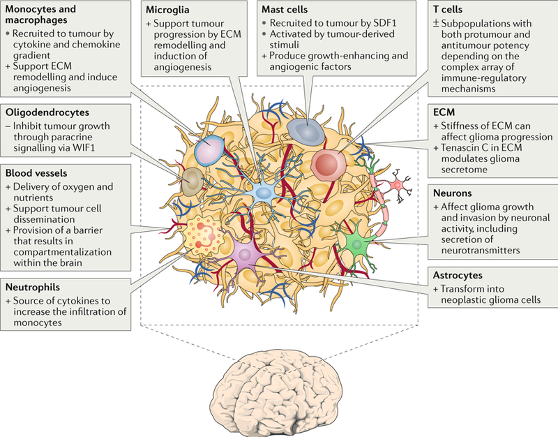
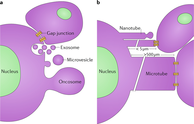
Glioblastomas are heterogeneous and invariably lethal tumours. They are characterized by genetic and epigenetic variations among tumour cells, which makes the development of therapies that eradicate all tumour cells challenging and currently impossible. An important component of glioblastoma growth is communication with and manipulation of other cells in the brain environs, which supports tumour progression and resistance to therapy. Glioblastoma cells recruit innate immune cells and change their phenotype to support tumour growth. Tumour cells also suppress adaptive immune responses, and our increasing understanding of how T cells access the brain and how the tumour thwarts the immune response offers new strategies for mobilizing an antitumour response. Tumours also subvert normal brain cells - including endothelial cells, neurons and astrocytes - to create a microenviron that favours tumour success. Overall, after glioblastoma-induced phenotypic modifications, normal cells cooperate with tumour cells to promote tumour proliferation, invasion of the brain, immune suppression and angiogenesis. This glioblastoma takeover of the brain involves multiple modes of communication, including soluble factors such as chemokines and cytokines, direct cell-cell contact, extracellular vesicles (including exosomes and microvesicles) and connecting nanotubes and microtubes. Understanding these multidimensional communications between the tumour and the cells in its environs could open new avenues for therapy.


Conflict of interest statement

Competing interests

The authors declare no competing interests.

Figures

Fig. 1 |
Fig. 1 |. Glioblastoma microenvironment.
The glioblastoma environ consists of tumour cells, extracellular matrix (ECM), blood vessels, innate immune cells (monocytes, macrophages, mast cells, microglia and neutrophils), T cells and non-tumorous neurons, astrocytes and oligodendrocytes. +, protumour function; −, antitumour function; ±, mixed protumour and antitumour functions; SDF1, stromal cell-derived factor 1; WIF1, WNT inhibitory factor 1.
Fig. 2 |
Fig. 2 |. Routes of communication between tumour cells and cells in their environs.
a | Gap junctions (24 nm in diameter) form across the adjacent membranes of cells that are in physical contact, enabling the passage of small molecules. Cells also release exosomes (50–100 μm) from multivesicular bodies that fuse with the plasma membrane. In addition, microvesicles (100–200 μm) and even large oncosomes (1–10 μm) bud off from the plasma membrane and can interact with and be taken up by other cells. b | Tunnelling nanotubes (50–200 nm in width and up to 5 μm in length) extend out from cells and can either bud off vesicles at their tips or form gap junctions with other cells. Microtubes extend out from tumour cells (1–2 μm in width and >500 μm in length) and can form gap junctions with other cells.
Fig. 3 |
Fig. 3 |. Interactions between glioma and TAMs.
Recruitment of tumour-associated macrophages or myeloid cells (TAMs), including blood monocytes and brain-resident microglia, is based on the gradient of chemokines and cytokines released by the glioblastoma cells. Once recruited, TAMs can be activated and differentiated under the influence of the secretome and extracellular vesicles (EVs) released by the tumour. The various recruited and activated TAMs can affect tumour growth by promoting angiogenesis through secretion of epidermal growth factor (EGF), vascular endothelial growth factor (VEGF), CXC-chemokine ligand 2 (CXCL2) and insulin-like growth factor-binding protein 1 (IGFBP1). This process is further promoted by the release of tumour-derived VEGF and EGF. Invasion and growth of the tumour are accomplished by remodelling the extracellular matrix (ECM) surrounding the tumour. For example, versican and EVs from the tumour induce the release of matrix metalloproteinase 14 (MMP14) by microglia. The release will facilitate the cleavage of tumour-derived pro-MMP2 following extracellular degradation by the active enzyme MMP2. CCL2, CC-chemokine ligand 2 (also known as MCP1); CSF1, macrophage colony-stimulating factor 1; GDNF, glial cell line-derived neurotrophic factor; HGF, hepatocyte growth factor; SDF1, stromal cell-derived factor 1; TNF, tumour necrosis factor.
Fig. 4 |
Fig. 4 |. T lymphocytes in the glioblastoma environment.
a | A hypothetical scenario for the induction of anti-glioblastoma T cell responses. Tumour antigens travel with interstitial fluid along the perivascular glymphatic system to the brain surface (step 1), where they enter the cerebrospinal fluid or are taken up by local antigen-presenting cells (APCs). A fraction of cerebrospinal fluid enters dural lymphatic vessels, potentially along with migratory APCs (step 2), and by this route tumour antigen reaches the deep cervical lymph nodes (dcLNs). There, resident or migratory APCs interact with and activate tumour antigen-specific naive T cells (step 3). b | Potential pathways by which activated or memory T cells reach the CNS and glioblastoma tumours. T cells expressing CC-chemokine receptor 6 (CCR6; and possibly other chemokine receptors) enter the cerebrospinal fluid in ventricles via the choroid plexus (step 4) and percolate along the brain surface. Upon re-encounter with their cognate tumour antigen (step 5), they initiate a local inflammatory reaction that activates the local microvasculature to recruit additional T cells in a CCR6-independent and potentially CXC-chemokine receptor 3 (CXCR3)-dependent fashion (step 6), facilitating their accumulation in the glioblastoma environment. c | In and around the glioblastoma, effector T cells, including cytotoxic T lymphocytes (CTLs), are exposed to a network of immune-regulatory mechanisms that promote their differentiation into a dysfunctional state. CTLA4, cytotoxic T lymphocyte protein 4; FasL, Fas antigen ligand; IDO, indoleamine 2,3-dioxygenase; MDSC, myeloid-derived suppressor cell; PDL1, programmed cell death 1 ligand 1; TAM, tumour-associated macrophages or myeloid cells; TGFβ, transforming growth factor-β; Treg cell, regulatory T cell; VEGFA, vascular endothelial growth factor A.

References

    1. Stupp R et al. Effects of radiotherapy with concomitant and adjuvant temozolomide versus radiotherapy alone on survival in glioblastoma in a randomised phase III study: 5-year analysis of the EORTC-NCIC trial. Lancet Oncol 10, 459–466 (2009). - PubMed
    1. Jhaveri N, Chen TC & Hofman FM Tumor vasculature and glioma stem cells: contributions to glioma progression. Cancer Lett 380, 545–551 (2016). - PubMed
    1. See AP, Parker JJ & Waziri A The role of regulatory T cells and microglia in glioblastoma-associated immunosuppression. J. Neurooncol 23, 405–412 (2015). - PubMed
    1. Roesch S, Rapp C, Dettling S & Herold-Mende C When immune cells turn bad-tumor-associated microglia/macrophages in glioma. Int. J. Mol. Sci 19, E436 (2018). - PMC - PubMed
    1. Okolie O et al. Reactive astrocytes potentiate tumor aggressiveness in a murine glioma resection and recurrence model. Neuro Oncol 18, 1622–1633 (2016). - PMC - PubMed

Publication types

MeSH terms