. 2019 Jan;33(1):358-372.
doi: 10.1096/fj.201800534R. Epub 2018 Jul 9.

High-depth transcriptomic profiling reveals the temporal gene signature of human mesenchymal stem cells during chondrogenesis

Affiliations

High-depth transcriptomic profiling reveals the temporal gene signature of human mesenchymal stem cells during chondrogenesis

Nguyen P T Huynh et al. FASEB J. 2019 Jan.

Abstract

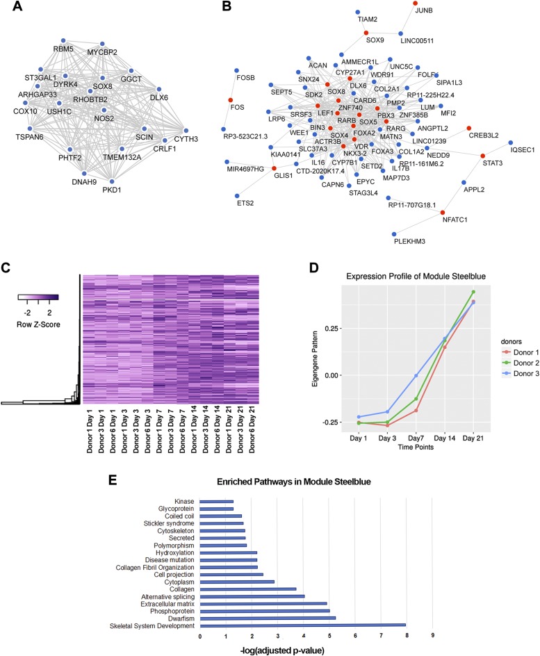
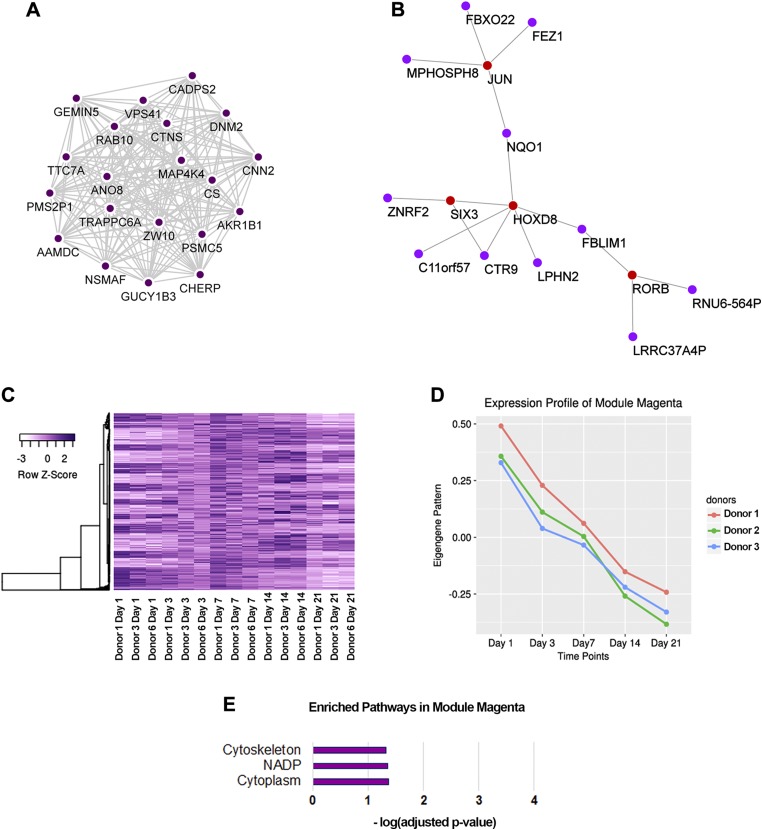
Mesenchymal stem/stromal cells (MSCs) provide an attractive cell source for cartilage repair and cell therapy; however, the underlying molecular pathways that drive chondrogenesis of these populations of adult stem cells remain poorly understood. We generated a rich data set of high-throughput RNA sequencing of human MSCs throughout chondrogenesis at 6 different time points. Our data consisted of 18 libraries with 3 individual donors as biologic replicates, with each library possessing a sequencing depth of 100 million reads. Computational analyses with differential gene expression, gene ontology, and weighted gene correlation network analysis identified dynamic changes in multiple biologic pathways and, most importantly, a chondrogenic gene subset, whose functional characterization promises to further harness the potential of MSCs for cartilage tissue engineering. Furthermore, we created a graphic user interface encyclopedia built with the goal of producing an open resource of transcriptomic regulation for additional data mining and pathway analysis of the process of MSC chondrogenesis.-Huynh, N. P. T., Zhang, B., Guilak, F. High-depth transcriptomic profiling reveals the temporal gene signature of human mesenchymal stem cells during chondrogenesis.

Keywords: RNA-Seq; chondrocyte; lncRNA; miRNA; pericyte.


Conflict of interest statement

The authors thank the Duke University Genomics Core Facility, the Washington University in St. Louis Pathology Core Labs, and the Washington University Center of Regenerative Medicine for their resources and support. The authors also wish to thank S. Oswald (Washington University in St. Louis) for assistance with technical writing. This work was supported by the Arthritis Foundation, U.S. National Institutes of Health (NIH) Grants AR50245, AR48852, AG15768, AR48182, and AR067467, the Nancy Taylor Foundation for Chronic Diseases, the Collaborative Research Center of the AO Foundation, Davos, Switzerland, and the Washington University Musculoskeletal Research Center (NIH P30 AR057235). B.Z. was supported by NIH grant DA027995 and the Goldman Sachs Philanthropy Fund. The authors declare no conflicts of interest.

Figures

Figure 1
Figure 1
Distribution of MSC surface markers. A) Experimental design. MSCs were formed into pellets after passage 4 and cultured under chondrogenic conditions for up to 21 d. Pellets were collected at d 0, 1, 3, 7, 14, and 21. B) Representative histograms from 3 donors. Control samples stained with isotypes are depicted in red; samples stained with surface markers are depicted in blue.
Figure 2
Figure 2
MSC chondrogenesis in pellet culture. A) Biochemical assays measuring GAG and DNA over time course of analysis. There was increase in GAG accumulation from d 3 to later time points (left and right), while DNA levels remained similar across days (middle). Mean ± sd. n = 3 independent specimens per donor (3 donors as biologic replicates). Two-way ANOVA followed by Tukey post hoc test (α = 0.05). Groups of different letters are statistically different from one another. B) Safranin O/Fast Green staining on pellets from 3 donors at different time points. CE) Immunohistochemistry staining on pellets from 3 donors at different time points: collagen type II (C); collagen type I (D); collagen type X (E). Scale bars, 500 µm.
Figure 3
Figure 3
Differential expression (DE) analysis. A) PCA of 18 MSC-derived pellet samples. Samples were closely related by days, and there was minor donor-to-donor variation. B) PCA of MSC-derived pellet samples vs. human cartilage at different developmental stages. C) Expression patterns of MSC markers and markers of potential lineages. Gene expression was represented as log2 fold change compared to d 0 samples. D) K-means clustering grouped DE genes into 4 patterns. Gene expression was represented as log2 fold change compared to d 0 samples. E) Bar graphs of enriched pathways from gene ontology analysis. Datasets for human cartilage were retrieved using GEO accession number GSE106292 (79).
Figure 4
Figure 4
Modules and their relationships to biochemical changes. A) Dendrogram of RNA-Seq samples and corresponding changes in traits. Lowest values are depicted in white; highest values are depicted in red; missing values are depicted in gray. B) Eigengene adjacency heat map with blocks of metamodules. C) Modules whose eigengenes are highly correlated with changes in GAG per DNA and their corresponding Pearson correlation values.
Figure 5
Figure 5
Characteristics of module steel blue. A) Coexpression network of 20 top hubs. B) Coexpression network of involved TFs (red) and their top 3 connected genes (steel blue). C) Heat map of steel blue modular gene expression. D) Eigengene vector of module steel blue. E) Enriched Gene Ontology terms and pathways from modular genes with gene ontology analysis.
Figure 6
Figure 6
Characteristics of module magenta. A) Coexpression network of 20 top hubs. B) Coexpression network of involved TFs (red) and their top 3 connected genes (magenta). C) Heat map of magenta modular gene expression. D) Eigengene vector of module magenta. E) Enriched Gene Ontology terms and pathways from modular genes with gene ontology analysis.

References

    1. Nguyen U. S., Zhang Y., Zhu Y., Niu J., Zhang B., Felson D. T. (2011) Increasing prevalence of knee pain and symptomatic knee osteoarthritis: survey and cohort data. Ann. Intern. Med. 155, 725–732 10.7326/0003-4819-155-11-201112060-00004 - DOI - PMC - PubMed
    1. Ahmed T. A., Hincke M. T. (2014) Mesenchymal stem cell–based tissue engineering strategies for repair of articular cartilage. Histol. Histopathol. 29, 669–689 - PubMed
    1. Caplan A. I. (1991) Mesenchymal stem cells. J. Orthop. Res. 9, 641–650 10.1002/jor.1100090504 - DOI - PubMed
    1. Caplan A. I. (2008) All MSCs are pericytes? Cell Stem Cell 3, 229–230 10.1016/j.stem.2008.08.008 - DOI - PubMed
    1. Crisan M., Yap S., Casteilla L., Chen C. W., Corselli M., Park T. S., Andriolo G., Sun B., Zheng B., Zhang L., Norotte C., Teng P. N., Traas J., Schugar R., Deasy B. M., Badylak S., Buhring H. J., Giacobino J. P., Lazzari L., Huard J., Péault B. (2008) A perivascular origin for mesenchymal stem cells in multiple human organs. Cell Stem Cell 3, 301–313 10.1016/j.stem.2008.07.003 - DOI - PubMed

Publication types