. 2018 Nov 2;46(19):e113.
doi: 10.1093/nar/gky596.

MetClo: methylase-assisted hierarchical DNA assembly using a single type IIS restriction enzyme

Affiliations

MetClo: methylase-assisted hierarchical DNA assembly using a single type IIS restriction enzyme

Da Lin et al. Nucleic Acids Res. .

Abstract

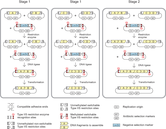
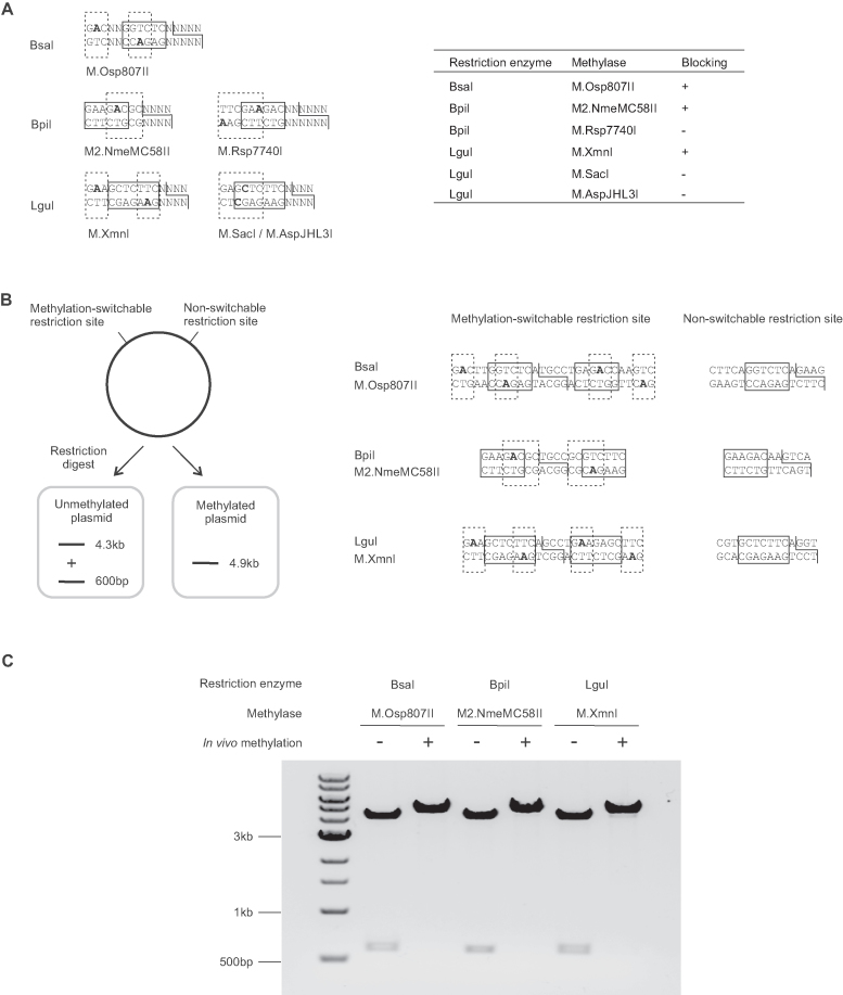
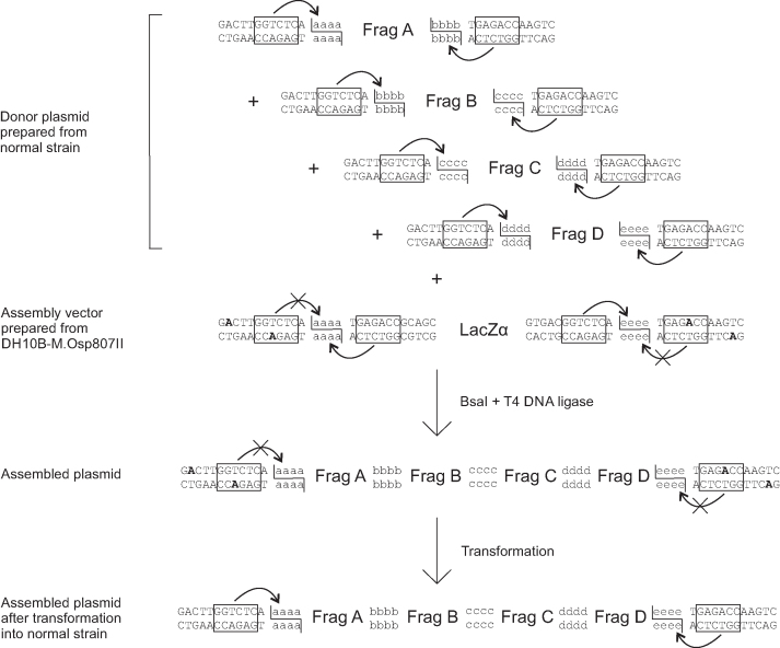
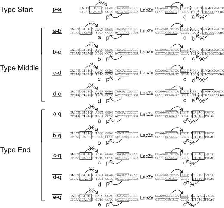
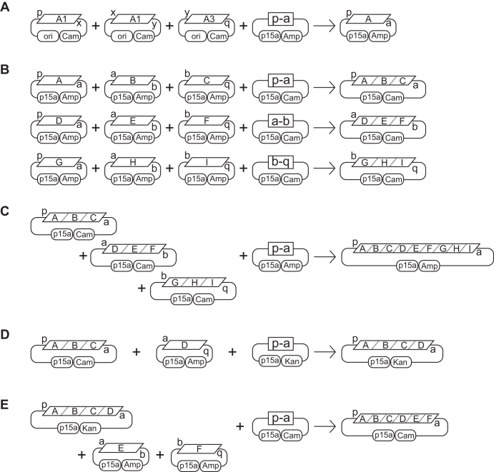
Efficient DNA assembly is of great value in biological research and biotechnology. Type IIS restriction enzyme-based assembly systems allow assembly of multiple DNA fragments in a one-pot reaction. However, large DNA fragments can only be assembled by alternating use of two or more type IIS restriction enzymes in a multi-step approach. Here, we present MetClo, a DNA assembly method that uses only a single type IIS restriction enzyme for hierarchical DNA assembly. The method is based on in vivo methylation-mediated on/off switching of type IIS restriction enzyme recognition sites that overlap with site-specific methylase recognition sequences. We have developed practical MetClo systems for the type IIS enzymes BsaI, BpiI and LguI, and demonstrated hierarchical assembly of large DNA fragments up to 218 kb. The MetClo approach substantially reduces the need to remove internal restriction sites from components to be assembled. The use of a single type IIS enzyme throughout the different stages of DNA assembly allows novel and powerful design schemes for rapid large-scale hierarchical DNA assembly. The BsaI-based MetClo system is backward-compatible with component libraries of most of the existing type IIS restriction enzyme-based assembly systems, and has potential to become a standard for modular DNA assembly.


Figures

Figure 1.
Figure 1.
DNA assembly using MetClo. The figure illustrates the assembly of four DNA fragments (Fragments A, B, C and D, shown in yellow) into a single DNA fragment (Fragment ABCD) through two stages of MetClo using a single type IIS restriction enzyme. Stage 1 and Stage 2 are effectively the same process with progressively larger inserts. In Stage 1, the insert donor plasmids contain insert fragments (Fragments A, B, C or D) flanked by methylation-switchable type IIS restriction sites that generate compatible adhesive ends. Preparation of insert plasmids in normal E. coli strains, which lack the appropriate switch methylase, results in insert fragments that can be cut out by the type IIS restriction enzyme. The assembly vector contains the negative selection marker LacZα flanked by a purposely designed head-to-head configuration of type IIS restriction sites. The inner pair of restriction sites are designed to be non-switchable, but the outer pair are methylation-switchable, because the restriction enzyme recognition site overlaps with a recognition site for the switch methylase. Preparation of the assembly vector in E. coli strains expressing the switch methylase leads to selective methylation of the outer methylation-switchable restriction sites, and subsequent digestion with the type IIS restriction enzyme generates a vector backbone that retains the outer pair of methylated type IIS restriction sites. Ligation of the insert fragments into the vector backbone generates the correctly assembled plasmid, which is not cut by the type IIS restriction enzyme because these sites are still methylated. All of the other unwanted fragments including the vector backbone of the insert plasmid and the LacZα fragment from the assembly vector contain unmethylated type IIS restriction sites, which are cut by the restriction enzyme. This design allows a one-pot assembly reaction that combines the restriction and ligation step because the reaction favors generation of the correctly assembled plasmid. Transformation of the assembly reaction into a normal E. coli strain, which lacks the appropriate switch methylase, effectively results in demethylation of the methylated type IIS restriction sites flanking the assembled fragment in the assembled plasmid. In Stage 2, the assembled inserts (Fragments AB and CD) can then be used for a subsequent round of MetClo assembly into a larger fragment (Fragment ABCD).
Figure 2.
Figure 2.
Identification of suitable methylases for methylation-switching. (A) Initial screening of functional methylases for selective blocking of overlapping methylation/restriction sites. The diagrams show the design of overlapping sites for screening of methylase activity. The table shows the screening result using methylases expressed in vivo from an F-ori based low copy number vector. Restriction enzyme recognition sites are boxed in solid lines. The adhesive ends generated by the restriction enzyme are shown by solid lines. Methylase recognition sites are boxed in dashed lines, and methylated bases are in bold font. All the listed methylases modify N6-adenine, except M.SacI and M.AspJHL3I, which modify C5-cytosine and N4-cytosine respectively. (B) Experimental designs to test blocking of methylation-switchable type IIS restriction enzyme sites by in vivo methylation. For each methylase/restriction enzyme combination tested, the test plasmid contains a head-to-head potentially methylation-switchable restriction site and a non-methylatable restriction site. Restriction digestion of test plasmid prepared from a normal E. coli strain would result in cutting at both sites and the release of a 600 bp fragment from the 4.3 kb vector backbone. Restriction digestion of test plasmid prepared from a strain expressing the switch methylase would result in a single 4.9 kb band, due to blocking of the methylation-switchable restriction sites by in vivo methylation. The restriction sites of the test plasmids for each restriction enzyme are shown, with the restriction site boxed in solid line, the methylase recognition site boxed in dashed line, and the methylated bases in bold. The head-to-head arrangement of overlapping methylation/restriction site allows the same assay to be used to detect any residual single strand nicking activity of the restriction enzyme towards the methylated restriction site. (C) Agarose gel electrophoresis analysis of the test plasmids for each methylation-switchable restriction site after preparation of the plasmids in a normal strain (–) or in a strain expressing the appropriate DNA methylase (+) and digested with the corresponding type IIS restriction enzymes. The combinations tested were BsaI with M.Osp807II methylase using test plasmid pMOP_BsaINC, BpiI with M2.NmeMC58II methylase using test plasmid pMOP_BpiINC, and LguI with M.XmnI methylase using test plasmid pMOP_LguINC. Test conditions were 60 fmol test plasmid digested using 5 U BsaI or BpiI, or 2.5 U LguI in 10 μl reactions at 37°C for 1 h. The results show that in vivo methylation by each of the methylases successfully blocked the restriction site for the corresponding type IIS restriction enzyme when the methylase recognition site overlapped the restriction enzyme site. The data shown represents results from three independent experiments.
Figure 3.
Figure 3.
BsaI-M.Osp807II based MetClo system. The donor plasmids contain DNA fragments to be assembled (Fragments A–D) flanked by BsaI sites that generate compatible adhesive ends (schematically labelled aaaa-eeee). The BsaI sites overlap with the M.Osp807II methylase recognition sequence. Donor plasmids were prepared from a normal strain that does not express the M.Osp807II switch methylase. As a result, the BsaI sites are not methylated and so the insert DNA fragments can be released by BsaI digestion. The recipient assembly vector contains a LacZα selection marker flanked by head-to-head BsaI sites. The outer pair of BsaI sites closer to the vector backbone overlap with an M.Osp807II methylation sequence and so are methylation-switchable, whereas the inner pair of BsaI sites are not. Preparation of the assembly vector in the M.Osp807II switch methylase-expressing DH10B strain results in selective blocking of the outer pair of BsaI sites. The LacZα fragment can be released by BsaI through cutting at inner pair of BsaI sites. Following a one-pot reaction using BsaI and T4 DNA ligase, ligation among compatible adhesive ends results in ordered assembly of DNA fragments into the assembly vector backbone. The assembled fragment in the assembled plasmid is flanked by methylated BsaI sites, which are not cut by BsaI. Following transformation into a normal strain that does not express the M.Osp807II switch methylase, methylation of the flanking restriction sites is lost, and the assembled fragment can be released by BsaI for the next stage assembly.
Figure 4.
Figure 4.
Design of a standard MetClo vector set. Three types of adaptor sequences were designed for the standard MetClo vector set. The vectors contain two head-to-head BsaI sites (boxed) flanking the negative selection marker LacZα. The pair of unmethylated BsaI sites closest to the negative selection marker can be used to release LacZα and generates adhesive ends ‘p’ and ‘q’. This allows any fragments that start with adhesive end ‘p’ and end with adhesive end ‘q’ to be cloned into any of the three types of adaptor sequences (types ‘Start’, ‘Middle’, ‘End’). The outer pair of BsaI sites closest to the vector backbone overlap with the M.Osp807II recognition sequence and are both methylated at the adenine bases highlighted in bold when the vector is prepared in M.Osp807II-expressing E. coli strain. A DNA fragment assembled into these vectors, following transformation into a normal E. coli strain which lacks this switch methylase activity, can be released from the assembled plasmid using BsaI, which can now recognize this unmethylated BsaI site. The released fragment will carry different adhesive ends depending on the type of the vector used. Assembly into a type ‘Start’ vector will generate a fragment flanked by adaptors ‘p-a’; assembly into a type ‘Middle’ vector will generate a fragment flanked by ‘a-b’, ‘b-c’, ‘c-d’ or ‘d-e’; assembly into a type ‘End’ vector will generate a fragment flanked by ‘a-q’, ‘b-q’, ‘c-q’, ‘d-q’ or ‘e-q’. The design of these LacZα selection cassettes flanked by unique adaptors are represented in the figure by the letter codes for the outside adaptor sequences.
Figure 5.
Figure 5.
Schemes for DNA assembly using standard MetClo vector set. The MetClo vector set supports both multiplicative and linear additive topology for DNA assembly. (A) As an example, a hypothetical transcription unit ‘A’ can be assembled from a promoter fragment ‘A1’ that starts with adaptor sequence ‘p’ and ends with adaptor sequence ‘x’, a coding region fragment ‘A2’ that starts and ends with adaptor sequences ‘x’ and ‘y’, and a transcription terminator fragment ‘A3’ that starts and ends with adaptor sequences ‘y’ and ‘q’. The internal adaptor sequences ‘x’ and ‘y’ used to link the DNA parts in order at this stage are arbitrary adaptor sequences not specified by the MetClo vector set. Any compatible DNA parts can be assembled into the vector as long as the first part start with adaptor sequence ‘p’ and the last part end with adaptor sequence ‘q’. MetClo assembly of these parts into MetClo vector ‘p-a’ generates assembled transcription unit ‘A’ flanked by adaptor sequence ‘p’ and ‘a’. (B) With a multiplicative assembly scheme, assembled transcription units can be assembled three per group in different MetClo vectors into larger fragments that contain three transcription units ‘ABC’, ‘DEF’ or ‘GHI’. (C) These three fragments, ‘ABC’, ‘DEF’ and ‘GHI’, can then be assembled into a single fragment containing 9 transcription units. (D) Alternatively, additional transcription units can be added to an existing fragment containing multiple transcription units using a linear addition assembly scheme. Addition of transcription unit ‘D’ flanked by adaptors ‘a’ and ‘q’ to fragment ‘ABC’ flanked by adaptors ‘p’ and ‘a’ using assembly vector ‘p-a’ generates an assembled fragment ‘ABCD’ which is still flanked by adaptors ‘p’ and ‘a’. (E) Further addition of fragments ‘E’ and ‘F’ can be undertaken to generate a larger fragment ‘ABCDEF’.

References

    1. Casini A., Storch M., Baldwin G.S., Ellis T.. Bricks and blueprints: methods and standards for DNA assembly. Nat. Rev. Mol. Cell Biol. 2015; 16:568–576. - PubMed
    1. Juhas M., Ajioka J.W.. High molecular weight DNA assembly in vivo for synthetic biology applications. Crit. Rev. Biotechnol. 2017; 37:277–286. - PubMed
    1. Gibson D.G., Young L., Chuang R.Y., Venter J.C., Hutchison C.A. 3rd, Smith H.O.. Enzymatic assembly of DNA molecules up to several hundred kilobases. Nat. Methods. 2009; 6:343–345. - PubMed
    1. Shao Z., Zhao H., Zhao H.. DNA assembler, an in vivo genetic method for rapid construction of biochemical pathways. Nucleic Acids Res. 2009; 37:e16. - PMC - PubMed
    1. Itaya M., Fujita K., Kuroki A., Tsuge K.. Bottom-up genome assembly using the Bacillus subtilis genome vector. Nat. Methods. 2008; 5:41–43. - PubMed

Publication types

MeSH terms