. 2018 Sep:18:54-64.
doi: 10.1016/j.redox.2018.06.009. Epub 2018 Jun 25.

Exosomal transfer of mitochondria from airway myeloid-derived regulatory cells to T cells

Affiliations

Exosomal transfer of mitochondria from airway myeloid-derived regulatory cells to T cells

Kenneth P Hough et al. Redox Biol. 2018 Sep.

Abstract

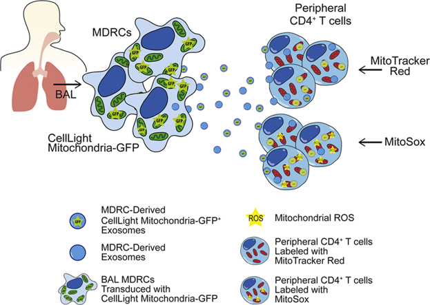
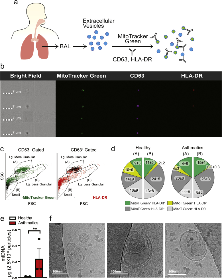
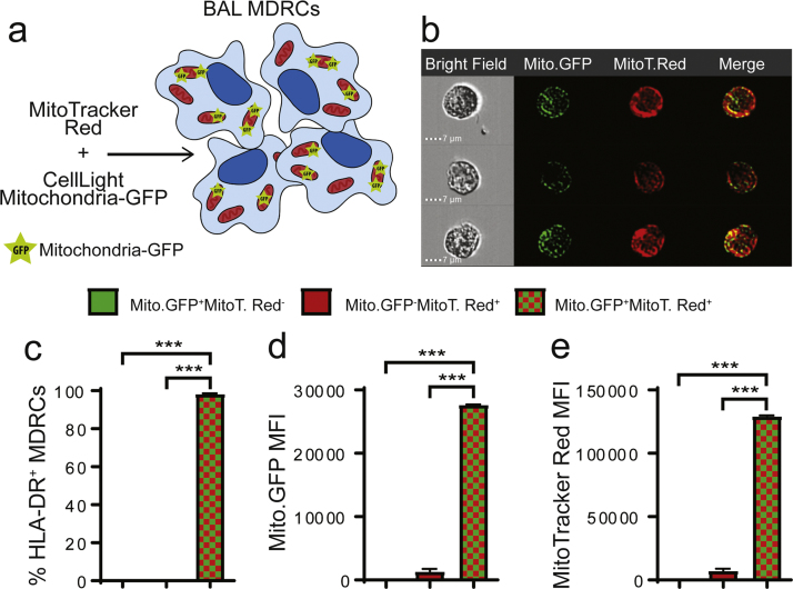
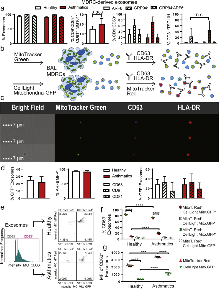
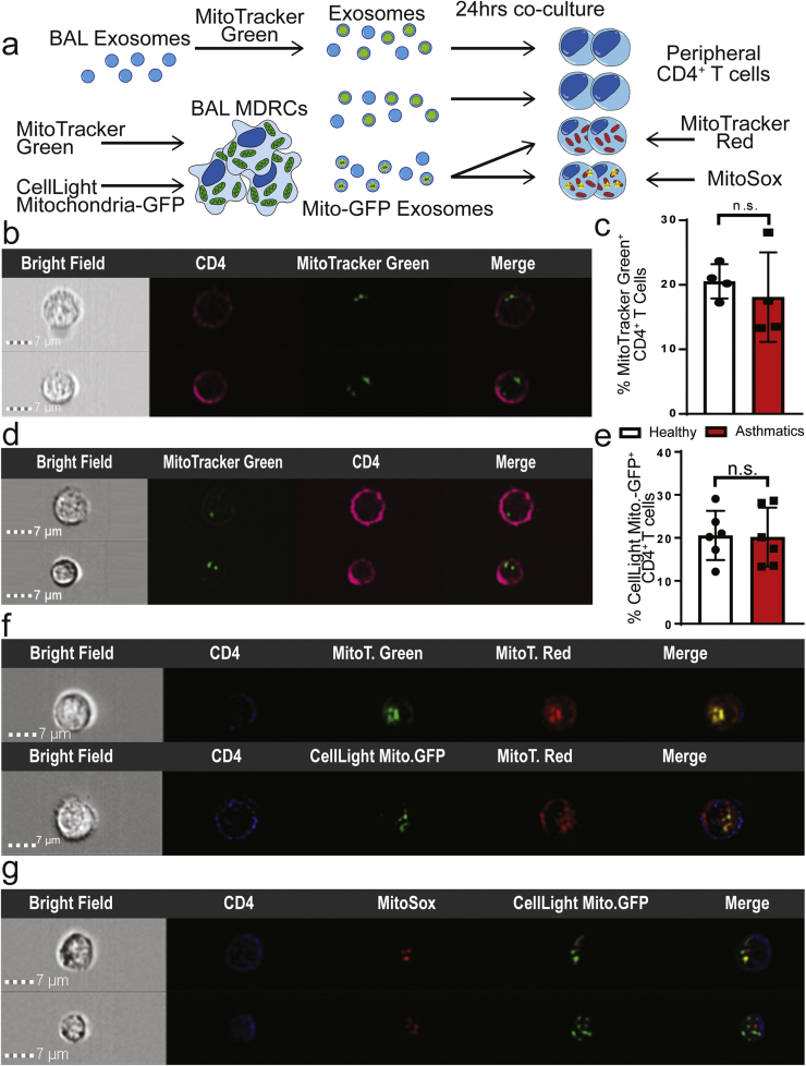
Chronic inflammation involving both innate and adaptive immune cells is implicated in the pathogenesis of asthma. Intercellular communication is essential for driving and resolving inflammatory responses in asthma. Emerging studies suggest that extracellular vesicles (EVs) including exosomes facilitate this process. In this report, we have used a range of approaches to show that EVs contain markers of mitochondria derived from donor cells which are capable of sustaining a membrane potential. Further, we propose that these participate in intercellular communication within the airways of human subjects with asthma. Bronchoalveolar lavage fluid of both healthy volunteers and asthmatics contain EVs with encapsulated mitochondria; however, the % HLA-DR+ EVs containing mitochondria and the levels of mitochondrial DNA within EVs were significantly higher in asthmatics. Furthermore, mitochondria are present in exosomes derived from the pro-inflammatory HLA-DR+ subsets of airway myeloid-derived regulatory cells (MDRCs), which are known regulators of T cell responses in asthma. Exosomes tagged with MitoTracker Green, or derived from MDRCs transduced with CellLight Mitochondrial GFP were found in recipient peripheral T cells using a co-culture system, supporting direct exosome-mediated cell-cell transfer. Importantly, exosomally transferred mitochondria co-localize with the mitochondrial network and generate reactive oxygen species within recipient T cells. These findings support a potential novel mechanism of cell-cell communication involving exosomal transfer of mitochondria and the bioenergetic and/or redox regulation of target cells.

Keywords: Asthma; Derived Regulatory Cells; Exosomes; Mitochondria; Myeloid-derived.


Figures

fx1
Graphical abstract
Fig. 1
Fig. 1
Characterization of extracellular vesicles (EVs) from the bronchoalveolar lavage (BAL) fluid of healthy controls and asthmatic subjects. (a) Schematic diagram of characterization of BAL EVs by labeling with MitoTracker-Green (MitoT-Green) and anti-CD63 and HLA-DR antibodies (b) ImageStream flow cytometry images showing positive staining of MitoT-Green in airway EVs, along with CD63 and HLA-DR (n = 3 healthy, n = 4 asthmatics; 3 technical replicates each) (c) Representative plot of particle heterogeneity. Granularity is represented on the y-axis and the x-axis represents size (n = 3 healthy, n = 4 asthmatics; 3 technical replicates each) (d) Quantitation of the proportionality of EVs that are positive for MitoT-Green and HLA-DR. The majority of the MitoT-Green signal was found in small and large granular particles. Statistical significance was observed for the following comparison: MitoTGreen+HLA-DR+ Healthy (A+B) vs Asthmatics (A+B) p-value < 0.05; and MitoTGreen+HLA-DR- Asthmatics (A) vs Asthmatics (B) p-value < 0.05 (unpaired Student's T-test) (e) Quantitation of mitochondrial DNA in pooled BAL EVs (total n = 11/group, with 3 independent experiments (** p < 0.01; unpaired Student's T-test) (f) Representative Cryo-Electron Microscopy images of airway EVs (n = 3 independent experiments) showing heterogeneous morphology. Airway EVs were multi-vesicular with electron dense cargo.
Fig. 2
Fig. 2
BAL MDRCs were transduced to express CellLight Mitochondria-GFP. Mitochondria-GFP (Mito-GFP) had proper subcellular localization as seen by the MitoTracker Red (MitoT-Red) overlay. (a) Schematic for validation of transduction and subcellular localization of Mito-GFP (b) ImageStream flow cytometry images illustrating Mito-GFP expression and co-localization with MitoT-Red. (c) Quantitation of % HLA-DR+ MDRCs that are positive for Mito-GFP, MitoT-Red, or both (*** p < 0.001; unpaired Student's T-test) (d-e) Quantitation of mean fluorescence intensity (MFI) of either Mito-GFP or MitoT-Red signal in HLA-DR+ MDRCs gated for either Mito-GFP+, MitoT-Red+, or Mito-GFP+MitoT-Red+ (*** p < 0.001; unpaired Student's T-test) (n = 3 subjects, 3 technical replicates).
Fig. 3
Fig. 3
MDRC-derived exosomes contain polarized mitochondria. (a) Characterization of MDRC-derived exosomes after purification. Quantitation of MDRC-derived EVs that were negative for non-exosomal markers ARF6 and GRP94. Quantitation of percent MDRC-derived exosomes with Tetraspanins (CD63, CD9, CD81) and Tsg101, an ESCRT pathway molecule (no statistical significance observed; Student's T-test) (n = 3 healthy, n = 3 asthmatics; 3 technical replicates each) (b) Schematic for characterization and validation of Mito-GFP+exosomes derived from MDRCs. MDRC mitochondria were marked with MitoT-Green or Mito-GFP. MDRC-derived exosomes marked with Mito-GFP were labeled with MitoT-Red (c) ImageStream flow cytometry images of MDRC-derived exosomes positive for MitoT-Green, CD63, and HLA-DR (n = 3 healthy, n = 3 asthmatics; 3 technical replicates each) (d) Characterization of MDRC-derived Mito-GFP+ exosomes. BAL MDRCs were isolated by FACS sorting from n = 3 healthy and n = 3 asthmatic subjects. Purified MDRCs were transduced with Mito-GFP and exosomes isolated from cell culture supernatant at 48 h. Histogram showing quantitation of percentages of CD63+Mito-GFP+ MDRC-derived exosomes, ARF6-Mito-GFP+ MDRC-derived exosomes and individual percentages of CD63+, CD9+ and CD81+ MDRC-derived exosomes (no statistical significance observed; Student's T-test). (e) Histogram and scatter plots of exosomes isolated from MDRCs transduced with Mito-GFP and then labeled with MitoT-Red (n = 3 healthy, n = 3 asthmatics; 3 technical replicates each) (f) Quantitation of the MDRC-derived Mito-GFP+ exosomes labeled ex vivo with MitoT-Red. A significant fraction of exosomes generated were positive for both Mito-GFP and MitoT-Red. (**** p < 0.0001; Two-way ANOVA) (g) Quantitation of the mean fluorescent intensity (MFI) of the MDRC-derived Mito-GFP+ exosomes labeled with MitoT-Red ex vivo. MDRC-derived Mito-GFP+ exosomes from asthmatics had significantly higher MFI of MitoT- Red and Mito-GFP than those derived from MDRCs of healthy subjects (*** p < 0.001, **** p < 0.0001; Two-way ANOVA).
Fig. 4
Fig. 4
Exosomal transfer of mitochondria by MDRCs to T cells. Transferred exosomal mitochondria merge with T cell mitochondrial network. (a) Schematic for experimental approach to validate exosomal transfer of mitochondria in co-cultures of exosomes and peripheral CD4+ T cells. (b) ImageStream flow cytometry images showing MitoT-Green+ BAL-derived exosomes within peripheral autologous CD4+ T cells (c) Quantitation of % CD4+ T cells in which MitoT- Green+ airway exosomes were transferred following co-culture (no statistical significance observed; Student's T-test) (b & c: n = 4 healthy, n = 4asthmatics, 3 technical replicates each) (d) ImageStream flow cytometry images of autologous peripheral CD4+ T cells following transfer of MDRC-derived MitoT-Green+ exosomes (n = 4 healthy, n = 3 asthmatics; 3 technical replicates each) (e) Quantitation of % CD4+ T cells in co-cultures with transferred MDRC-derived Mito-GFP+ exosomes (no statistical significance observed; Student's T-test) (n = 6 healthy, n = 6 asthmatics; 3 technical replicates each) (f) ImageStream flow cytometry images showing overlap of host T cell MitoT-Red signal with transferred Mito-GFP from MDRC-derived exosomes, validating merging of these transferred exosomes with the mitochondrial network of the host cells (f & g: n = 3 healthy, n = 3 asthmatics; 3 technical replicates each). (g) ImageStream Flow cytometry images of MitoSox labeling indicating ROS in peripheral autologous CD4+ T cells that partially overlaps with transferred MDRC-derived Mito-GFP+ exosomes.

Comment in

References

    1. Anderson G.P. Endotyping asthma: new insights into key pathogenic mechanisms in a complex, heterogeneous disease. Lancet. 2008;372:1107–1119. - PubMed
    1. Boushey H.A., Fahy J.V. Basic mechanisms of asthma. Environ. Health Perspect. 1995;103(Suppl 6):229–233. - PMC - PubMed
    1. Barnes P.J. Immunology of asthma and chronic obstructive pulmonary disease. Nat. Rev. Immunol. 2008;8:183–192. - PubMed
    1. Hammad H., Lambrecht B.N. Dendritic cells and epithelial cells: linking innate and adaptive immunity in asthma. Nat. Rev. Immunol. 2008;8:193–204. - PubMed
    1. Ray A., Khare A., Krishnamoorthy N., Qi Z., Ray P. Regulatory T cells in many flavors control asthma. Mucosal Immunol. 2010;3:216–229. - PMC - PubMed

Publication types