Review
. 2018 Jul 6;7(7):71.
doi: 10.3390/cells7070071.

Centrosome Remodelling in Evolution

Affiliations
Review

Centrosome Remodelling in Evolution

Daisuke Ito et al. Cells. .

Abstract

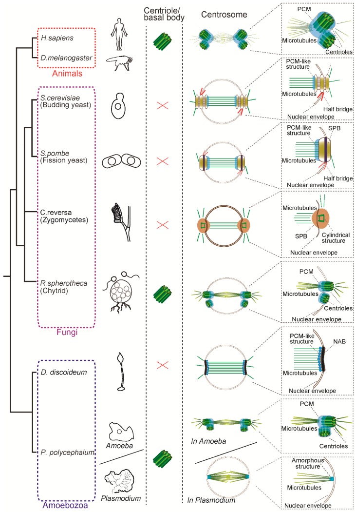
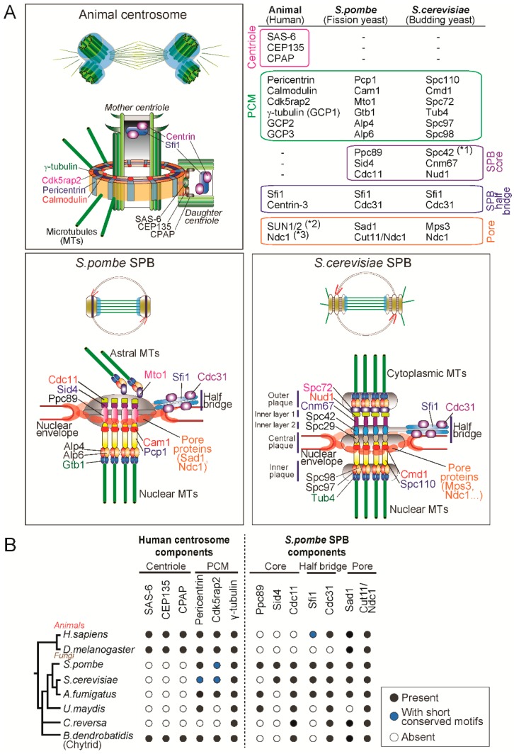
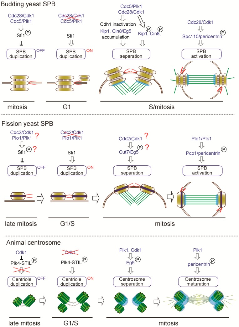
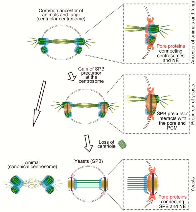
The centrosome is the major microtubule organizing centre (MTOC) in animal cells. The canonical centrosome is composed of two centrioles surrounded by a pericentriolar matrix (PCM). In contrast, yeasts and amoebozoa have lost centrioles and possess acentriolar centrosomes—called the spindle pole body (SPB) and the nucleus-associated body (NAB), respectively. Despite the difference in their structures, centriolar centrosomes and SPBs not only share components but also common biogenesis regulators. In this review, we focus on the SPB and speculate how its structures evolved from the ancestral centrosome. Phylogenetic distribution of molecular components suggests that yeasts gained specific SPB components upon loss of centrioles but maintained PCM components associated with the structure. It is possible that the PCM structure remained even after centrosome remodelling due to its indispensable function to nucleate microtubules. We propose that the yeast SPB has been formed by a step-wise process; (1) an SPB-like precursor structure appeared on the ancestral centriolar centrosome; (2) it interacted with the PCM and the nuclear envelope; and (3) it replaced the roles of centrioles. Acentriolar centrosomes should continue to be a great model to understand how centrosomes evolved and how centrosome biogenesis is regulated.

Keywords: PCM; SPB; centriole; centrosome; evolution; spindle pole body.


Conflict of interest statement

The authors declare no conflict of interest.

Figures

Figure 1
Figure 1
Structure of the centrosome in mitosis in animals, fungi and amoebozoa. Animals and basal fungi (chytrids) have a centriole/basal body and a canonical centrosome composed of a pair of centrioles surrounded by pericentriolar matrix (PCM), which anchors and nucleates microtubules. In contrast, the yeasts such as S. cerevisiae and S. pombe have a centriole-less centrosome called the spindle pole body (SPB). Another fungus C. reversa has a distinct SPB containing a cylindrical structure reminiscent of a centriole. In the Amoebozoa, while D. discoideum has a centriole-less nucleus-associated body (NAB), P. polycephalum has two modes of centrosomes: animal-like centriolar centrosome in the amoeba phase and an amorphous structure devoid of centriole at the pole in the plasmodium phase. Note that size of the centrosomes and spindle are not depicted to scale in this figure.
Figure 2
Figure 2
The molecular components of animal centrosomes and yeast SPBs. (A) Mapping of molecular components in the animal centrosome and budding and fission yeast SPBs. (*1. Spc42 seems functionally analogous to Ppc89 [61], *2,3. The protein is conserved but not known to be implicated in centrosome functions.) The illustrated SPB structure was adapted and modified from [13]. (B) Phylogenetic distribution of centrosome components in animals and fungi. We searched for orthologues of components of the human centrosome localizing to centrioles and PCM and the fission yeast SPB components by using reciprocal pairwise sequence-based (BLASTP and phmmer) and domain-based (hmmsearch) methods [87,88]. Black circles represent the presence of orthologues; blue circles indicate that previous studies showed the presence of a protein with short conserved motifs [47,56,60] although we failed to identify it by the above-mentioned computational methods; white circles indicate no detectable orthologue ([89] and our unpublished results).
Figure 3
Figure 3
Common regulators of centrosome biogenesis and function. Similar regulators are involved in centrosome biogenesis: (1) duplication; (2) separation and (3) maturation/activation in yeasts and animals.
Figure 4
Figure 4
Speculated scenario of centrosome evolution. The common ancestor of fungi and animals is likely to have had a canonical centrosome. Presumably, it might be like the extant chytrid centrosome in which the centriole pair is anchored in the NE to undergo closed mitosis. Eventually, the newly invented SPB precursor was formed closed to the centriolar centrosome and started to interact with the pore and PCM. When the centriole was lost, the SPB structure replaced the role of centrioles and maintained the same function as MTOC.

References

    1. Scheer U. Historical roots of centrosome research: Discovery of Boveri’s microscope slides in Wurzburg. Philos. Trans. R. Soc. Lond. B Biol. Sci. 2014;369:20130469. doi: 10.1098/rstb.2013.0469. - DOI - PMC - PubMed
    1. Bettencourt-dias M., Glover D.M. Centrosome biogenesis and function: Centrosomics brings new understanding. Nat. Rev. Mol. Cell Biol. 2007;8:451–463. doi: 10.1038/nrm2180. - DOI - PubMed
    1. Carvalho-Santos Z., Azimzadeh J., Pereira-Leal J.B., Bettencourt-Dias M. Evolution: Tracing the origins of centrioles, cilia and flagella. J. Cell Biol. 2011;194:165–175. doi: 10.1083/jcb.201011152. - DOI - PMC - PubMed
    1. Azimzadeh J. Exploring the evolutionary history of centrosomes. Philos. Trans. R. Soc. Lond. B Biol. Sci. 2014;369:20130453. doi: 10.1098/rstb.2013.0453. - DOI - PMC - PubMed
    1. Azimzadeh J., Bornens M. Centrosomes in Development and Disease. Wiley-Blackwell; Hoboken, NJ, USA: 2005. The Centrosome in Evolution; pp. 93–122.

LinkOut - more resources