. 2018 Aug 31;293(35):13662-13672.
doi: 10.1074/jbc.RA118.004123. Epub 2018 Jul 9.

Conserved motifs on the cytoplasmic face of the protein translocation channel are critical for the transition between resting and active conformations

Affiliations

Conserved motifs on the cytoplasmic face of the protein translocation channel are critical for the transition between resting and active conformations

Elisabet C Mandon et al. J Biol Chem. .

Abstract

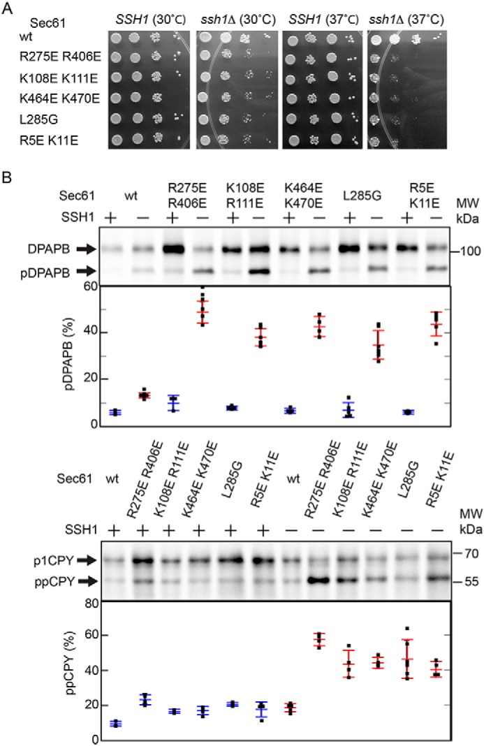
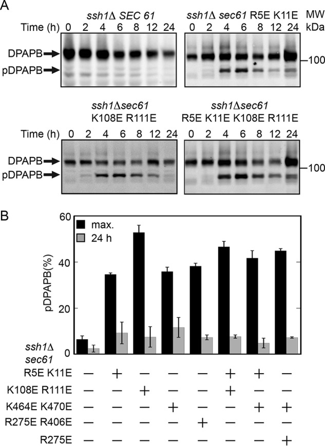
The Sec61 complex is the primary cotranslational protein translocation channel in yeast (Saccharomyces cerevisiae). The structural transition between the closed inactive conformation of the Sec61 complex and its open and active conformation is thought to be promoted by binding of the ribosome nascent-chain complex to the cytoplasmic surface of the Sec61 complex. Here, we have analyzed new yeast Sec61 mutants that selectively interfere with cotranslational translocation across the endoplasmic reticulum. We found that a single substitution at the junction between transmembrane segment TM7 and the L6/7 loop interferes with cotranslational translocation by uncoupling ribosome binding to the L6/7 loop from the separation of the lateral gate transmembrane spans. Substitutions replacing basic residues with acidic residues in the C-terminal tail of Sec61 had an unanticipated impact upon binding of ribosomes to the Sec61 complex. We found that similar charge-reversal mutations in the N-terminal tail and in cytoplasmic loop L2/3 did not alter ribosome binding but interfered with translocation channel gating. These findings indicated that these segments are important for the structural transition between the inactive and active conformations of the Sec61 complex. In summary our results have identified additional cytosolic segments of the Sec61 complex important for promoting the structural transition between the closed and open conformations of the complex. We conclude that positively charged residues in multiple cytosolic segments, as well as bulky hydrophobic residues in the L6/7-TM7 junction, are required for cotranslational translocation or integration of membrane proteins by the Sec61 complex.

Keywords: Sec61; endoplasmic reticulum (ER); membrane protein; protein synthesis; protein translocation; ribosome; translocation channel.


Conflict of interest statement

The authors declare that they have no conflicts of interest with the contents of this article

Figures

Figure 1.
Figure 1.
Location of yeast Sec61p residues selected for mutagenesis. A and B, the M. jannaschii SecYEβ channel (A) and lateral gate views (B) are color coded as follows: SecY (cyan), Secβ (yellow), SecE (orange), previously described mutants (magenta), new charge-reversal mutations (blue), and the L285G mutant (red). Conserved residues (Asn-302 and Gln-129) in the lateral gate polar cluster (36) that are important for channel gating are shown as magenta spheres. Sec61 residues selected for mutagenesis are mapped onto SecY based upon sequence alignment. The Sec61 R5E mutation is mapped onto M1 of M. jannashii SecY because S. cerevisiae Sec61 has a six-residue N-terminal extension relative to SecY. Residue numbers in all panels correspond to S. cerevisiae Sec61. Panels A and B were made using PYMOL v1.3 software and PDB ID 1RHZ. C, sequence logos for the N terminus, L2/3 and L6/TM7 junctions, and the C terminus of Sec61 were constructed by alignment of 125 diverse eukaryotic Sec61 sequences. Residues are color coded by side chain property; letter height is proportional to frequency. The M. jannaschii and S. cerevisiae sequences flank the logo. Arrowheads beneath the sequence logo designate residues selected for mutagenesis. Sequence logos were made using the website http://weblogo.berkeley.edu/logo.cgi.5
Figure 2.
Figure 2.
Ssh1p suppresses growth rate and protein translocation defects of the Sec61 cytoplasmic loop mutants. A, yeast strains (SSH1 or ssh1Δ) that express WT or mutant alleles of Sec61p were maintained in SEG media prior to spotting onto YPAD plates to evaluate growth at 30 or 37 °C for 2 days. Please note that the spotting order for the second and third dilutions was inverted on the SSH1 plate cultured at 37 °C. B, translocation assays of sec61 mutants in SSH1 or ssh1Δ strains. Yeast strains were shifted from SEG media into SD media and cultured for 4 h prior to pulse labeling. Integration of DPAPB and translocation of CPY was assayed by 7-min pulse labeling of WT and mutant yeast cells. CPY and DPAPB were immunoprecipitated from pulse-labeled cell extracts using CPY and DPAPB specific antisera. The glycosylated ER forms of CPY (p1) and DPAPB were resolved from nontranslocated precursors (ppCPY and pDPAPB) by SDS-PAGE. The percent integration (DPAPB) or translocation (CPY) is the average of two to eight determinations, one of which is shown here. Blue (SSH1) and red (ssh1Δ) bars represent mean and S.D. with individual data points plotted as black squares.
Figure 3.
Figure 3.
Transient accumulation of pDPAPB in sec61 mutants cultured in S. D. media. Yeast strains were transferred from SEG media into liquid SD media and cultured at 30 °C for 0–24 h. A, total cell lysates from selected strains were resolved by SDS-PAGE to allow protein immunoblot detection of pDPAPB and DPAPB. B, DPAPB immunoblots including those displayed in panel A were quantified by densitometry to determine the maximal percent accumulation of pDPAPB (4 or 6 h time point) and the percent pDPAPB remaining at the 24 h time point. The quantified values are the average of two determinations. Error bars designate individual data points.
Figure 4.
Figure 4.
Interaction between sec61 L285G and lateral gate polar cluster mutations. A, yeast strains that express WT or mutant alleles of Sec61p in the ssh1Δ background were maintained in SEG media prior to spotting onto YPAD plates to evaluate growth at 30 or 37 °C for 3 days. B, translocation assays of sec61 mutants in ssh1Δ strains. Yeast strains were shifted from SEG media into SD media and cultured for 4 h prior to pulse labeling. Integration of DPAPB and translocation of CPY was assayed as in Fig. 2. The percent integration of DPAPB (upper panel) and translocation of CPY (lower panel) is the average of two to nine determinations. Horizontal bars designate mean and S.D.s, with individual data points plotted as black squares.
Figure 5.
Figure 5.
Translocon gating assays of sec61 mutants. A, Dap2 reporters consist of i) the N-terminal cytoplasmic domain of Dap2p (green, Dap2p1–29) followed by the TM span (black, Dap2p30–45), ii) 49- to 265-residue spacer segments (blue) derived from Dap2p, iii) a Ub domain (red), iv) a 42-residue linker (cyan) with a processing site (arrowhead) for a Ub-specific protease, and v) a Ura3 reporter domain followed by a triple-HA epitope tag (yellow). Sites for N-linked glycosylation are indicated. B, cleavage of the Dap2 reporter defines the in vivo kinetics of translocon gating in sec61 mutants. A delay in RNC docking or in channel gating allows folding of Ub in the cytosol and cleavage of the reporter to generate Ura3-HA. The color code for Dap2 segments is as defined in panel A. C, in vivo cleavage of the Dap2 reporter in SEC61ssh1Δ and sec61ssh1Δ mutant strains after 24 h of growth at 30 °C in SD media was evaluated by pulse labeling. Labels designate the intact glycosylated (e.g. g49) and cleaved (U-HA) reporter domains. Downward-pointing arrowheads in assays of mutant strains designate nonglycosylated intact reporters that correspond to cytosolic Dap2 reporter aggregates (10, 34). D and E, spacer-length dependence of Dap2 reporter cleavage (percent cytosolic Ura3-HA) for Sec61 WT (D and E, black squares), sec61 R5E K11E (D, blue circles), sec61 K108E R111E (D, red circles), sec61 K464E K470E (D, cyan squares), sec61 L285G (E, cyan circles), and sec61 L285G Q129N (E, blue squares). Data points in panels D and E are averages of two experiments one of which is shown in panel C, with error bars designating individual data points. Error bars that are not visible are smaller than the data symbol.
Figure 6.
Figure 6.
Ribosome-binding assays of sec61 mutants. Purified WT and mutant Sec61 heterotrimers in detergent solution were incubated in the presence or absence of yeast 80S ribosomes prior to centrifugation to separate free Sec61 complexes in the supernatant fraction (S) from ribosome-bound Sec61 complexes in the pellet (P) fraction. Supernatant and pellet fractions were resolved by SDS-PAGE for protein immunoblot analysis using anti-FLAG to detect His6-FLAG-Sbh1p. Each Sec61 preparation was assayed in two or more experiments, one of which is shown here. The vertical lines designate samples from the same experiment that were electrophoresed on separate gels.

References

    1. Ng D. T. W., Brown J. D., and Walter P. (1996) Signal sequences specify the targeting route to the endoplasmic reticulum. J. Cell Biol. 134, 269–278 10.1083/jcb.134.2.269 - DOI - PMC - PubMed
    1. Ast T., Cohen G., and Schuldiner M. (2013) A network of cytosolic factors targets SRP-independent proteins to the endoplasmic reticulum. Cell 152, 1134–1145 10.1016/j.cell.2013.02.003 - DOI - PubMed
    1. Walter P., and Johnson A. E. (1994) Signal sequence recognition and protein targeting to the endoplasmic reticulum membrane. Ann. Rev. Cell Biol. 10, 87–119 10.1146/annurev.cb.10.110194.000511 - DOI - PubMed
    1. Halic M., Becker T., Pool M. R., Spahn C. M., Grassucci R. A., Frank J., and Beckmann R. (2004) Structure of the signal recognition particle interacting with the elongation-arrested ribosome. Nature 427, 808–814 10.1038/nature02342 - DOI - PubMed
    1. Song W., Raden D., Mandon E. C., and Gilmore R. (2000) Role of Sec61α in the regulated transfer of the ribosome-nascent chain complex from the signal recognition particle to the translocation channel. Cell 100, 333–343 10.1016/S0092-8674(00)80669-8 - DOI - PubMed

Publication types

MeSH terms

Substances

Associated data

LinkOut - more resources