

Large-Scale Single-Cell RNA-Seq Reveals Molecular Signatures of Heterogeneous Populations of Human Induced Pluripotent Stem Cell-Derived Endothelial Cells
- PMID: 29986945
- PMCID: PMC6202208
- DOI: 10.1161/CIRCRESAHA.118.312913
Large-Scale Single-Cell RNA-Seq Reveals Molecular Signatures of Heterogeneous Populations of Human Induced Pluripotent Stem Cell-Derived Endothelial Cells
Abstract
Rationale: Human-induced pluripotent stem cell-derived endothelial cells (iPSC-ECs) have risen as a useful tool in cardiovascular research, offering a wide gamut of translational and clinical applications. However, inefficiency of the currently available iPSC-EC differentiation protocol and underlying heterogeneity of derived iPSC-ECs remain as major limitations of iPSC-EC technology.
Objective: Here, we performed droplet-based single-cell RNA sequencing (scRNA-seq) of the human iPSCs after iPSC-EC differentiation. Droplet-based scRNA-seq enables analysis of thousands of cells in parallel, allowing comprehensive analysis of transcriptional heterogeneity.
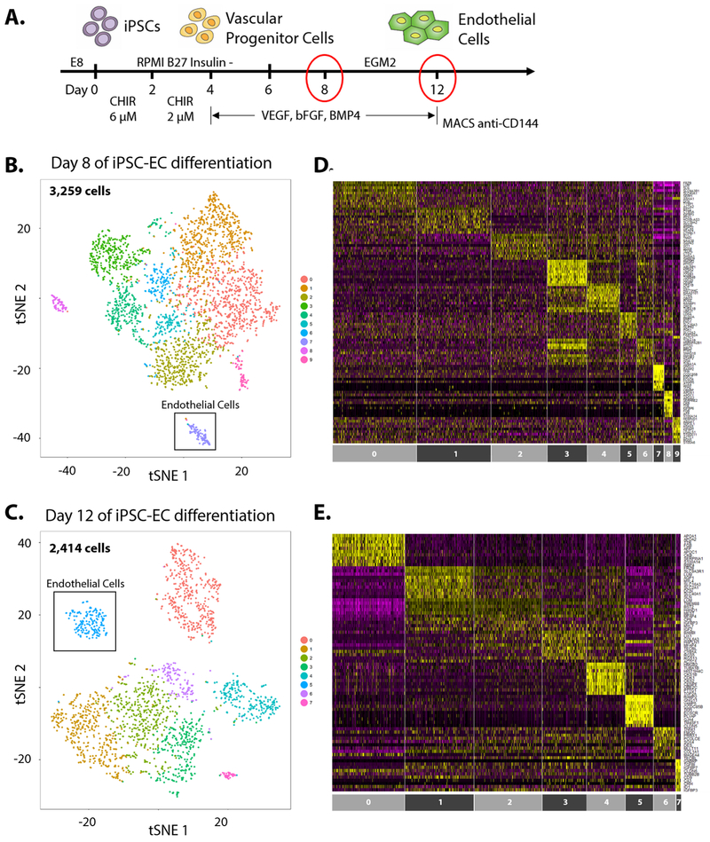
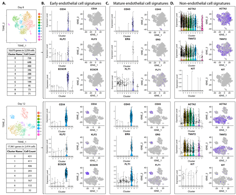
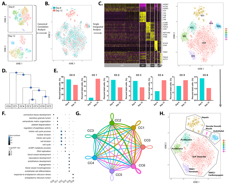
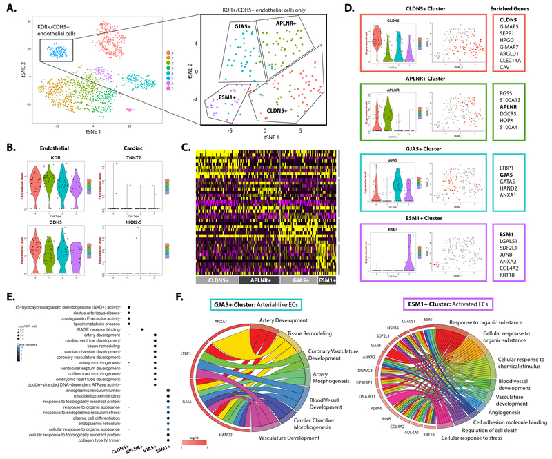
Methods and results: Bona fide iPSC-EC cluster was identified by scRNA-seq, which expressed high levels of endothelial-specific genes. iPSC-ECs, sorted by CD144 antibody-conjugated magnetic sorting, exhibited standard endothelial morphology and function including tube formation, response to inflammatory signals, and production of NO. Nonendothelial cell populations resulting from the differentiation protocol were identified, which included immature cardiomyocytes, hepatic-like cells, and vascular smooth muscle cells. Furthermore, scRNA-seq analysis of purified iPSC-ECs revealed transcriptional heterogeneity with 4 major subpopulations, marked by robust enrichment of CLDN5, APLNR, GJA5, and ESM1 genes, respectively.
Conclusions: Massively parallel, droplet-based scRNA-seq allowed meticulous analysis of thousands of human iPSCs subjected to iPSC-EC differentiation. Results showed inefficiency of the differentiation technique, which can be improved with further studies based on identification of molecular signatures that inhibit expansion of nonendothelial cell types. Subtypes of bona fide human iPSC-ECs were also identified, allowing us to sort for iPSC-ECs with specific biological function and identity.
Keywords: computational biology; endothelial cells; induced pluripotent stem cells; myocytes, cardiac; stem cells.
Figures
References
-
- Benjamin EJ, Virani SS, Callaway CW, Chang AR, Cheng S, Chiuve SE, Cushman M, Delling FN, Deo R, de Ferranti SD, Ferguson JF, Fornage M, Gillespie C, Isasi CR, Jiménez MC, Jordan LC, Judd SE, Lackland D, Lichtman JH, Lisabeth L, Liu S, Longenecker CT, Lutsey PL, Matchar DB, Matsushita K, Mussolino ME, Nasir K, O’Flaherty M, Palaniappan LP, Pandey DK, Reeves MJ, Ritchey MD, Rodriguez CJ, Roth GA, Rosamond WD, Sampson UKA, Satou GM, Shah SH, Spartano NL, Tirschwell DL, Tsao CW, Voeks JH, Willey JZ, Wilkins JT, Wu JH, Alger HM, Wong SS, Muntner P. Heart Disease and Stroke Statistics-2018 Update: A Report From the American Heart Association. Circulation. 2018; - PubMed
-
- Cai H, Harrison DG. Endothelial dysfunction in cardiovascular diseases: the role of oxidant stress. Circ Res. 2000;87:840–844. - PubMed
-
- Yoshida Y, Yamanaka S. Induced pluripotent stem cells 10 years later: For cardiac applications. Circ Res. 2017;120:1958–1968. - PubMed
Publication types
MeSH terms
Substances
Grants and funding
- R01 HL109512/HL/NHLBI NIH HHS/United States
- R01 HL130020/HL/NHLBI NIH HHS/United States
- R01 HL128503/HL/NHLBI NIH HHS/United States
- R01 HL123968/HL/NHLBI NIH HHS/United States
- R01 HL139478/HL/NHLBI NIH HHS/United States
- R33 HL120757/HL/NHLBI NIH HHS/United States
- K01 HL135455/HL/NHLBI NIH HHS/United States
- R01 HL134817/HL/NHLBI NIH HHS/United States
- P30 DK116074/DK/NIDDK NIH HHS/United States
- 13SDG17340025/AHA/American Heart Association-American Stroke Association/United States
- R01 HL132875/HL/NHLBI NIH HHS/United States
- R01 HL113006/HL/NHLBI NIH HHS/United States
- T32 EB009035/EB/NIBIB NIH HHS/United States
- F32 HL134221/HL/NHLBI NIH HHS/United States
- R01 HL128170/HL/NHLBI NIH HHS/United States
LinkOut - more resources
Full Text Sources
Other Literature Sources
Molecular Biology Databases
