. 2018 Jul 24;115(30):E7063-E7072.
doi: 10.1073/pnas.1805862115. Epub 2018 Jul 9.

Protein moonlighting elucidates the essential human pathway catalyzing lipoic acid assembly on its cognate enzymes

Affiliations

Protein moonlighting elucidates the essential human pathway catalyzing lipoic acid assembly on its cognate enzymes

Xinyun Cao et al. Proc Natl Acad Sci U S A. .

Abstract

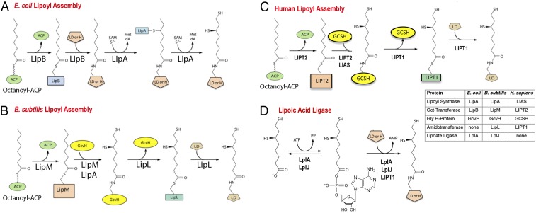
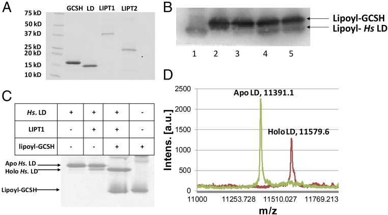
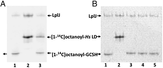
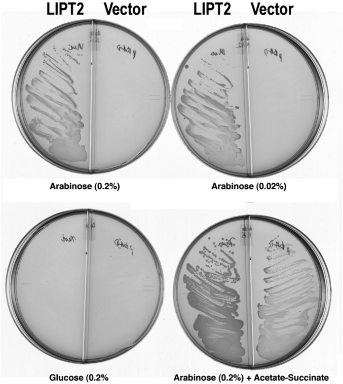
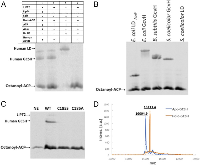
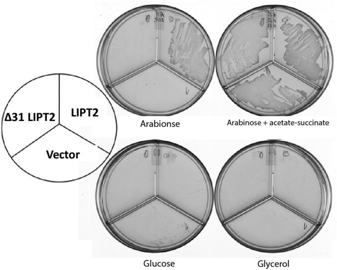
The lack of attachment of lipoic acid to its cognate enzyme proteins results in devastating human metabolic disorders. These mitochondrial disorders are evident soon after birth and generally result in early death. The mutations causing specific defects in lipoyl assembly map in three genes, LIAS, LIPT1, and LIPT2 Although physiological roles have been proposed for the encoded proteins, only the LIPT1 protein had been studied at the enzyme level. LIPT1 was reported to catalyze only the second partial reaction of the classical lipoate ligase mechanism. We report that the physiologically relevant LIPT1 enzyme activity is transfer of lipoyl moieties from the H protein of the glycine cleavage system to the E2 subunits of the 2-oxoacid dehydrogenases required for respiration (e.g., pyruvate dehydrogenase) and amino acid degradation. We also report that LIPT2 encodes an octanoyl transferase that initiates lipoyl group assembly. The human pathway is now biochemically defined.

Keywords: 2-oxoacid dehydrogenases; glycine cleavage system; inborn errors; lipoic acid; mitochondrial disorder.


Conflict of interest statement

The authors declare no conflict of interest.

Figures

Fig. 1.
Fig. 1.
Pathways of lipoyl assembly on cognate proteins. (A) The simplest assembly pathway is that of E. coli where only two enzymes are required (4). LipB transfers an octanoyl moiety from octanoyl-ACP to each of the cognate protein substrates. The LipA radical S-adenosyl-l-methionine enzyme then inserts two sulfur atoms to produce dihydrolipoyl moieties. (B) A more complex pathway is found in Firmicute bacteria such as Bacillus subtilis (10, 12, 68) and Staphyloccus aureus (11). In this pathway, lipoyl moieties are assembled on the GcvH protein of the glycine cleavage system and then transferred to the lipoyl domains (LDs) of the 2-oxoacid dehydrogenases. This pathway requires a lipoyl amidotransferase called LipL and a distinct octanoyl transferase called LipM. LipA catalyzes sulfur insertion as in A. (C) The pathway of lipoyl assembly in humans as elucidated in the present work. The pathway parallels the bacterial pathway of B. The differing nomenclatures of the human and bacterial lipoyl assembly proteins and enzymes are given in the Inset. Note that the LIPT1 acyl-enzyme intermediate is hypothetical. (D) The lipoate ligase reaction catalyzed by the lipoate salvage enzymes, E. coli LplA and B. subtilis LplJ (4). Acyl adenylate is a stably bound intermediate in the reaction. Human and bovine LIPT1s can catalyze only the second partial ligase reaction, transfer from the adenylate to the acceptor protein (17, 18). All three enzymes are active with octanoate in place of lipoate.
Fig. 2.
Fig. 2.
(A) Flow chart of the reconstitution of LIPT1lipoyl transfer from lipoyl-GCSH to a human LD acceptor in the E. coli ∆lipB strain, XC.127. The cultures were grown in glycerol minimal medium with differing supplements as shown in the figure. Supplementation with succinate and acetate bypasses the need for 2-oxoacid dehydrogenase lipoylation (4). Strain XC.127 transformed with the plasmid encoding the His6-LD acceptor protein was additionally transformed with the GCSH plasmid plus the LIPT1 plasmid, the GCSH plasmid alone, or the LIPT1 plasmid alone. The resulting cultures were induced with IPTG in the presence of lipoate to allow the host LplA ligase to synthesize lipoyl-GCSH (if present). The cells of each culture were then collected by centrifugation and washed to remove lipoate and IPTG. After resuspension in glycerol minimal medium containing acetate and succinate, arabinose was added to the three cultures to induce expression of LIPT1 and His6-LD. The cultures were incubated to allow further growth and accumulation of the His6-LD. The cells were then collected and lysed, and the His6-LD of each culture was purified by Ni2+ chelate chromatography. The purified samples were then submitted for mass-spectrometric analysis and the proteins expressed in each sample are summarized in B, where + or − denotes expression. The electrospray mass-spectrometric scans for each culture are given in CE. (C) Mass-spectrometric analysis of the His6-LD acceptor accumulated in the absence of the LIPT1 plasmid. The LD remained in the apo form (13,796.2 Da). Note that the apo LD mass was 18 Da less than that calculated (13,814.3 Da), consistent with either dehydration (−18 Da) or deamidation (−17 Da) during mass spectrometry (Protein Prospector, prospector.ucsf.edu). Dehydration seems more probable since almost a third of the protein is composed of residues (S, T, E, and D) known to undergo water loss. (D) Mass-spectrometric analysis of the His6-LD acceptor accumulated in the absence of the GCSH plasmid. The LD remained in the apo form (m/z 13,797.2 Da). (E) Mass-spectrometric scan of the His6-LD accumulated when both LIPT1 and GCSH were expressed. The mass of the lipoylated LD form (13,983.1 Da) agrees well with the calculated value (14,001.5 Da). The change in mass upon modification (calculated for lipoyl modification, 188 Da; observed, 187 Da) is within the accuracy of the instrument utilized. a.u., arbitrary units; Intens., intensity.
Fig. 3.
Fig. 3.
LIPT1 catalyzes transfer of lipoyl moieties from GCSH to an LD derived from human pyruvate dehydrogenase. (A) Purification of the lipoate assembly proteins. The proteins were purified as described in Experimental Procedures and analyzed by SDS/PAGE on 4–20% polyacrylamide gels. The molecular weights of the Bio-Rad broad-range protein standards are indicated. (B) Western blot analysis of SDS/PAGE assay of LIPT1 lipoyl amidotransferase activity using anti-lipoyl antibody and B. subtilis LplJ-generated lipoyl-GCSH as the substrate. Lipoyl-GCSH was synthesized with LplJ plus ATP and lipoic acid and then purified using anion exchange chromatography to remove LplJ and residual ATP. Lanes: 1, standard lipoyl-LD prepared by LplJ modification of human LD (Hs apo-LD); 2, lipoyl-GCSH; 3, lipoyl-GCSH plus the human LD; 4 and 5, lipoyl-GCSH incubated with LIPT1 and LD. (C) LIPT1 lipoyl amidotransferase activity analyzed by urea gel electrophoresis. Loss of the positive charge of the modified lysine ϵ-amino group of the LD results in faster migration of the modified form on these gels. The gel was stained with Coomassie Blue. (D) Electrospray mass-spectrometric analysis of lipoylated human LD and the remaining apo LD from the reaction of gel (B, lane 4). The calculated difference in mass (delta mass) between the apo and lipoyl forms was 188, whereas the observed delta mass was 188.5. Note that, in A and B, trace levels of LD lipoylation were seen in the absence of GCSH, which is attributed to LIPT1-bound lipoyl-AMP that survives purification and crystallization (28). The traces of lipoylation that appears without LIPT1 in B seems likely to be due the lipoate assembly pathway of the wild-type E. coli strain used for protein production.
Fig. 4.
Fig. 4.
Ability of LIPT1 to transfer octanoyl moieties from GCSH to an LD derived from human pyruvate dehydrogenase. (A) Autoradiograms of urea–PAGE gels of assays testing LipT1-catalyzed octanoyl amidotransfer from purified [1-14C]octanoyl-GCSH to the lipoyl domain (LD). Lanes: 1, [1-14C]octanoyl-GCSH synthesized using B. subtilis LplJ plus ATP and [1-14C]octanoic acid; 2, [1-14C]octanoyl-LD standard synthesized with LD as in lane 1; 3, LD added to the [1-14C]octanoyl-GCSH synthesis reaction. The [1-14C]octanoyl-labeled LD band indicates that residual ATP remaining from the [1-14C]octanoyl-GCSH synthetic reaction was used by LplJ to modify the LD. (B) Octanoyl amidotransferase urea–PAGE gel assays performed in the presence of an ATP trap (hexokinase plus d-glucose) to prevent LplJ modification of the LD. Lanes: 1 and 2 are a repetition of the experiment of lanes 1 and 2 of the gel in A, respectively; 3, LD added to the [1-14C]octanoyl-GCSH synthesis reaction in the presence of the ATP trap (2 units of hexokinase and 10 mM d-glucose). The lack of LD labeling indicates that the trap eliminated the ATP remaining from the [1-14C]octanoyl-GCSH synthesis reaction; 4 and 5, [1-14C]octanoyl-GCSH with addition of LIPT1, the ATP trap, and the LD acceptor.
Fig. 5.
Fig. 5.
Alignment and phylogeny of LIPT2. (A) Alignment of LIPT2 with the enzymatically characterized LipB and LipM proteins. Unweighted sequence alignments were performed using T-Coffee (69) at the European Bioinformatics Institute website (https://www.ebi.ac.uk) using the default settings and displayed using Jalview. The sequence name indicates the enzyme type, the Uniprot code indicates the organism of origin, and the numbers indicate the amino acid residues displayed. Positions having 50% or greater identity are highlighted in blue. The catalytic cysteine residues of the LIPT2, LipB, and LipM are boxed and highlighted in black, as is the conserved lysine residue. The leucine-to-arginine mutations found in the human LIPT2 patients (27) are given in red. The edges of the alignment were trimmed using Jalview (70), so only the catalytic domain is shown. (B) Phylogeny of the LipB_LplA_LipM family (PF03099) was determined with sequences retrieved from the Pfam database (71). Multiple sequence alignments was done using ClustalW (72). The poorly conserved LplA N and C termini were removed. The phylogenetic tree was constructed using the maximum-likelihood method with the PhyML program (73, 74). PHYLIP Interleaved was used for alignment. Bootstrap analysis was set to 1,000 replicates.
Fig. 6.
Fig. 6.
Complementation of the lipB deletion mutation (∆lipB) of E. coli strain QC145 by expression of mouse LIPT2. A synthetic gene encoding the full-length mouse LIPT2 coding sequence was inserted into plasmid vector pBAD322A, which provided the transcription and translation sequences required for expression of the protein to give plasmid pCY754. Transcription was from the vector araBAD promoter, which is induced by arabinose and repressed by glucose. The plasmid was introduced into strain QC145 (∆lipB::cml ∆lplA::kan) (68), which carries a deletion of the lplA lipoate ligase gene in addition to the ∆lipB deletion. The lplA mutation was included because mutant LplA proteins can bypass the ∆lipB defect by scavenging cellular octanoic acid (34). The medium was M9 minimal salts with glycerol as carbon source (glycerol allows basal expression of the araBAD promoter) and 100 μg/mL sodium ampicillin to select for plasmid maintenance. Derivatives of strain QC145 carrying either the LIPT2 plasmid pCY754 or the empty vector were grown on glucose minimal salts containing 5 mM each of sodium acetate and sodium succinate, which bypasses the lipoylation requirement. These cultures were then streaked onto glycerol minimal salts plates supplemented with arabinose, glucose, or acetate plus succinate as given on the figure. The plates used are divided into sectors by plastic walls to prevent cross-feeding. The left sector of each plate contained the strain with the LIPT2 plasmid pCY754, whereas the right sector contained the empty pBAD322A vector. No growth was seen on plates that contained only glycerol (basal expression). The plates were incubated for 2 d at 37 °C.
Fig. 7.
Fig. 7.
LIPT2 octanoyl transferase activity in vitro. The substrate used in these experiment was E. coli ACP acylated with [1-14C]octanoic acid by AasS acyl-ACP synthetase. (A) Lanes: 1, standard [1-14C]octanoyl-GCSH prepared using B. subtilis LipM; 2, LIPT2-catalyzed synthesis of [1-14C]octanoyl-GCSH; 3, standard [1-14C]octanoyl-LD prepared using B. subtilis LplJ; and 4, LIPT2 fails to catalyze transfer of [1-14C]octanoyl moiety from [1-14C]octanoyl-ACP to the human LD. (B) LIPT2 is active on a several bacterial GcvH and LD proteins. The bands migrating more slowly than octanoyl-ACP are the octanoylated acceptor proteins. The right-hand lanes are Streptomyces coelicolor acceptor proteins (the LD preparation tested was inactive). (C) Mutant LIPT2 proteins lacking C185 are inactive. Designations: NE, no LIPT2; WT, wild-type LIPT2; and C185S and C185A, respectively, denote proteins in which serine or alanine replaced cysteine 185. (D) Electrospray mass-spectrometric analysis of the unmodified (calculated mass of 15,997.6 Da) and octanoylated (calculated mass of 16,123.6 Da) forms of GCSH by LIPT2, respectively. Within the accuracy of the instrument, the protein masses agree well with the calculated values, as does the change in mass upon modification (calculated, 126 Da; observed, 128.5 Da). a.u., arbitrary units; Intens, intensity.
Fig. 8.
Fig. 8.
Complementation of the ∆lipB deletion of E. coli strain QC146 by expression of human LIPT2. Synthetic genes encoding either the full-length human LIPT2 or a derivative that lacked the first 31 residues (∆31, a methionine codon replaced residue 31 to permit translation) were inserted into vector pBAD322A as in Fig. 6 to give plasmids pCY1110 and pCY1108, respectively. The plasmids were introduced into strain QC146 (∆lipB ∆lplA) (75), and transformants were streaked onto plates containing 0.02% arabinose or another carbon source as given in Fig. 6. Note that, unlike the mouse LIPT2, 0.2% arabinose gave rapid growth, but growth soon halted, suggesting high-level expression of human LIPT2 is toxic.

References

    1. Reed LJ. From lipoic acid to multi-enzyme complexes. Protein Sci. 1998;7:220–224. - PMC - PubMed
    1. Reed LJ. A trail of research from lipoic acid to alpha-keto acid dehydrogenase complexes. J Biol Chem. 2001;276:38329–38336. - PubMed
    1. Reed LJ, DeBUSK BG, Gunsalus IC, Hornberger CS., Jr Crystalline alpha-lipoic acid; a catalytic agent associated with pyruvate dehydrogenase. Science. 1951;114:93–94. - PubMed
    1. Cronan JE. Assembly of lipoic acid on its cognate enzymes: An extraordinary and essential biosynthetic pathway. Microbiol Mol Biol Rev. 2016;80:429–450. - PMC - PubMed
    1. Solmonson AD, DeBerardinis RJ. Lipoic acid metabolism and mitochondrial redox regulation. J Biol Chem. 2018;293:7522–7530. - PMC - PubMed

Publication types

LinkOut - more resources