. 2018 Jul 24;115(30):E7023-E7032.
doi: 10.1073/pnas.1806660115. Epub 2018 Jul 9.

Fully human agonist antibodies to TrkB using autocrine cell-based selection from a combinatorial antibody library

Affiliations

Fully human agonist antibodies to TrkB using autocrine cell-based selection from a combinatorial antibody library

Spyros Merkouris et al. Proc Natl Acad Sci U S A. .

Abstract

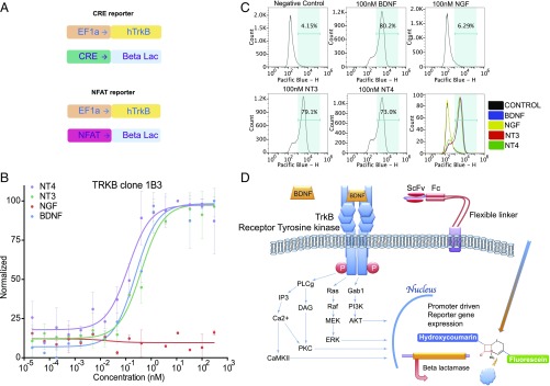
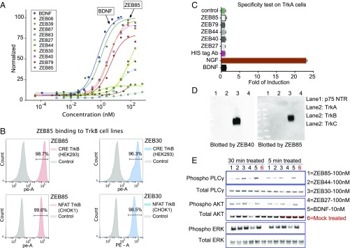
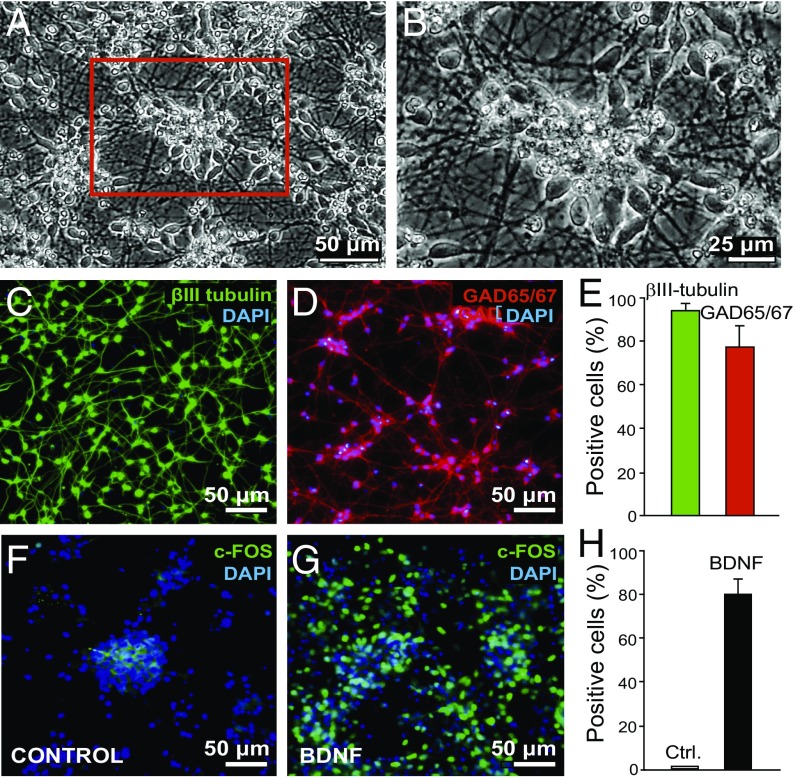
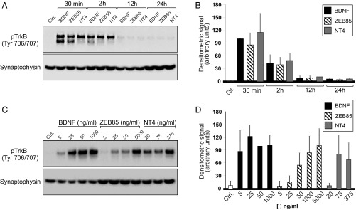
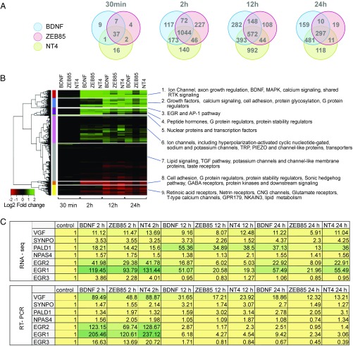
The diverse physiological roles of the neurotrophin family have long prompted exploration of their potential as therapeutic agents for nerve injury and neurodegenerative diseases. To date, clinical trials of one family member, brain-derived neurotrophic factor (BDNF), have disappointingly failed to meet desired endpoints. Contributing to these failures is the fact that BDNF is pharmaceutically a nonideal biologic drug candidate. It is a highly charged, yet is a net hydrophobic molecule with a low molecular weight that confers a short t1/2 in man. To circumvent these shortcomings of BDNF as a drug candidate, we have employed a function-based cellular screening assay to select activating antibodies of the BDNF receptor TrkB from a combinatorial human short-chain variable fragment antibody library. We report here the successful selection of several potent TrkB agonist antibodies and detailed biochemical and physiological characterization of one such antibody, ZEB85. By using a human TrkB reporter cell line and BDNF-responsive GABAergic neurons derived from human ES cells, we demonstrate that ZEB85 is a full agonist of TrkB, comparable in potency to BDNF toward human neurons in activation of TrkB phosphorylation, canonical signal transduction, and mRNA transcriptional regulation.

Keywords: TrkB; agonist; antibody; combinatorial library; membrane tethered.


Conflict of interest statement

Conflict of interest statement: This work was supported by Zebra Biologics. P.S.D. and R.M.L. are affiliated with Zebra Biologics.

Figures

Fig. 1.
Fig. 1.
Reporter cell line construction and characterization. (A) Design of exogenously transduced lentiviral constructs for the reporter cell lines. (B) Concentration–response curves of different ligands on the TrkB reporter line (NFAT). (C) Flow cytometry analysis of the TrkB reporter cell line (NFAT) activation when treated with different ligands. (D) Illustration of the reporter mechanism when activated.
Fig. 2.
Fig. 2.
Characterization of the active antibody clones identified from the functional screening. (A) Concentration–response curve of 10 representative active clones on TrkB reporter cell line. (B) Purified ZEB85 antibody binding to the TrkB reporter cell surface. (C) Representative TrkB agonist clones are not active on TrkA reporter cell line. (D) Western blot of Trk family ectodomains with two TrkB agonist clones. (E) Time-dependent phosphorylation of TrkB receptor downstream signaling molecules when stimulated by agonist ligands.
Fig. 3.
Fig. 3.
H9 hES cells differentiate as a monolayer of inhibitory neurons that are highly responsive to BDNF. (A and B) Phase-contrast microscopy of 30 DIV (days in vitro) neurons. (C) Immunocytochemistry of 30 DIV neurons stained with β-ΙΙΙ tubulin and DAPI. (D) Immunocytochemistry of 30 DIV neurons with GAD65/67 and DAPI. (E) Quantification of C and D. Values are mean ± SD of three independent experiments. (F and G) The 30 DIV neurons were untreated (F) or treated with 50 ng/mL BDNF (G) for 12 h and subsequently fixed and immunostained with DAPI or c-Fos antibody. Quantification is shown in H. Values are mean ± SD of two independent experiments.
Fig. 4.
Fig. 4.
Effect of ZEB85, BDNF, and NT4 on TrkB phosphorylation by 30 DIV H9 neurons. (A) Representative immunoblot of the time course of TrkB phosphorylation. Cultures were treated with 50 ng/mL BDNF, 5 μg/mL ZEB85, or 75 ng/mL NT4 at the indicated time points. Cell lysates were subjected to Western blot analyses for phospho-TrkB or synaptophysin (used as loading control) as indicated in Materials and Methods. (B) Densitometric values (mean ± SE) quantified from the blots of six independent differentiation experiments. (C) Representative blots of the concentration response of TrkB phosphorylation. Cultures were treated with the indicated concentrations of ZEB85, BDNF, or NT4 for 30 min and then subjected to Western blot analysis. (D) Densitometric values (mean ± SE) quantified from the blots of six independent differentiations experiments.
Fig. 5.
Fig. 5.
RNA sequencing on 30 DIV H9 neurons treated with 50 ng/mL of BDNF, 100 ng/mL of NT4, and 5 μg/mL of ZEB85. (A) Venn diagrams illustrating the number of differentially expressed genes under the three treatment conditions (BDNF, ZEB85, NT4) and four time points (30 min, 2 h, 12 h, and 24 h). (B) Heat map of hierarchical cluster analysis with Gene Ontology annotation for the top 1,000 differentially expressed genes; absolute fold change >2 and adjusted P value <0.01. (C) Expression fold changes relative to the control of seven genes obtained with RNA-seq and RT-PCR. Green color indicates higher level of increase, and bright yellow indicates intermediate level of change.
Fig. 6.
Fig. 6.
TrkB phosphorylation in cultured mouse cortical neurons and dendritic outgrowth in mouse retinal explants by TrkB agonists. (A) Phospho-TrkB Western blot of cultured mouse cortical neurons after addition of BDNF (Left) and ZEB85 (Right). Embryonic day 21 mouse cortical neurons were cultured for 7 d. BDNF (100 ng/mL) or ZEB85 (50 μg/mL) was added, and cultures were harvested at 15–240 min and assayed for phospho-TrkB (arrow) by Western blot. Molecular weight markers are on the right. (B) Dendritic field analysis of RGCs in retinal explants after 3 d of treatment with ZEB44, ZEB85, BDNF, or vehicle (control). Neurons were labeled by DiI/DiO DiOlistics. Arrows indicate RGC axons. (Scale bars: 100 μm.) (C) Sholl analysis of dendritic fields of RGCs after TrkB agonist antibody or BDNF treatment. Numbers of RGCs are indicated. +P < 0.05, ZEB44 vs. vehicle (control); #P < 0.05 and ###P < 0.001, ZEB85 vs. vehicle; *P < 0.05, **P < 0.01, and ***P < 0.001, BDNF vs. vehicle, Kruskal–Wallis test. (D) AUC analysis of Sholl plots and (E) quantification of total dendrite length after various agonist treatments. *P < 0.05, **P < 0.01, and ***P < 0.005 vs. vehicle, ANOVA, Tukey post hoc test.

References

    1. Mitre M, Mariga A, Chao MV. Neurotrophin signalling: Novel insights into mechanisms and pathophysiology. Clin Sci (Lond) 2017;131:13–23. - PMC - PubMed
    1. Nagahara AH, Tuszynski MH. Potential therapeutic uses of BDNF in neurological and psychiatric disorders. Nat Rev Drug Discov. 2011;10:209–219. - PubMed
    1. Egan MF, et al. The BDNF val66met polymorphism affects activity-dependent secretion of BDNF and human memory and hippocampal function. Cell. 2003;112:257–269. - PubMed
    1. Yeo GS, et al. A de novo mutation affecting human TrkB associated with severe obesity and developmental delay. Nat Neurosci. 2004;7:1187–1189. - PubMed
    1. Friedel S, et al. Mutation screen of the brain derived neurotrophic factor gene (BDNF): Identification of several genetic variants and association studies in patients with obesity, eating disorders, and attention-deficit/hyperactivity disorder. Am J Med Genet B Neuropsychiatr Genet. 2005;132B:96–99. - PubMed

Publication types

MeSH terms