Review
. 2018 Sep;11(3):475-484.
doi: 10.1007/s12178-018-9510-3.

Management of Pediatric Ankle Fractures

Affiliations
Review

Management of Pediatric Ankle Fractures

Z Deniz Olgun et al. Curr Rev Musculoskelet Med. 2018 Sep.

Abstract

Purpose of review: Summarize classic and recent information regarding the unique subset of ankle fractures in children with open growth plates and share the authors' decision-making and surgical techniques.

Recent findings: Recent research on pediatric ankle fractures has centered on the accurate prediction and prevention of growth arrest following fractures of the distal tibia. Another source of discussion is the necessity and benefit of CT scanning in classification and treatment approach. Pediatric ankle fractures continue to pose clinical challenges for orthopedic surgeons. While open anatomic reduction and internal fixation continue to produce good outcomes for intra-articular fractures, outcomes of physeal injuries are more difficult to predict. More studies are needed to determine which patients may benefit more from surgical treatment of physeal injuries.

Keywords: Distal tibial fracture; Distal tibial physeal fracture; Pediatric ankle fractures; Physeal fracture.


Conflict of interest statement

Conflict of Interest

The authors declare that they have no conflicts of interest.

Human and Animal Rights and Informed Consent

This article does not contain any studies with human or animal subjects performed by any of the authors.
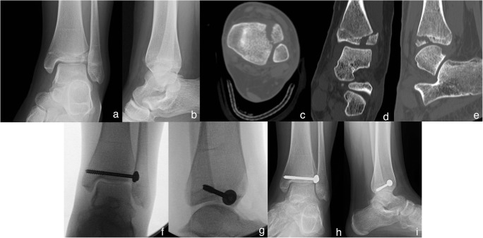
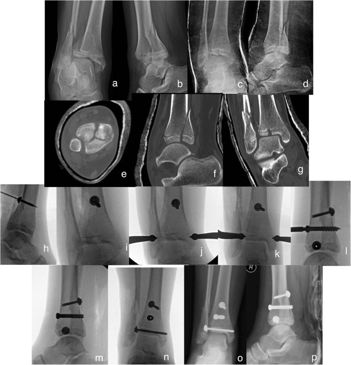
Figures

Fig. 1
Fig. 1
A metaphyseal corner fracture in the process of healing. This fracture was discovered during the non-accidental trauma work-up of a 2-month-old infant who presented with unexplainable lethargy (special thanks to Dr. Amisha Shah, personal collection)
Fig. 2
Fig. 2
A classic Salter-Harris-I fracture of the distal tibia
Fig. 3
Fig. 3
Salter-Harris-II fracture of the distal tibia and fibular shaft fracture in a 12-year-old male (a, b). The patient underwent closed reduction and long-leg casting. Post-reduction radiographs did not allow adequate assessment of the physis (c, d), so a CT scan was performed (e, f). Physeal widening on the sagittal images was measured as 2 mm, and decision was made for conservative treatment. Radiographs at 6 months are not conclusive for continued growth at the physis; however, growth is going to be monitored for 2 years post-injury or until symmetric growth arrest lines are observed (g, h)
Fig. 4
Fig. 4
A 12-year-old female sustained this triplane fracture due to a twisting injury while playing lacrosse. ad AP radiograph reveals a SH-IV injury whereas on lateral a SH-II injury is appreciated, typical for triplane fractures. CT scans revealed more than 2 mm of gapping at the fracture line; for this reason, the patient was indicated for operative fixation. 6.5 cannulated screws were utilized to compress the posterior malleolar fracture first, followed by open reduction of the joint line and fixation of the latter by a 4.5 cannulated screw (h, i). Six months post-injury, fracture is healed and growth plates are closing (j, k). Patient has full range of motion compared to the opposite ankle and has resumed sports at her pre-injury level
Fig. 5
Fig. 5
This 12-year-old female sustained this Tillaux fracture due to a missed step going down the stairs (a, b). The patient was short-leg casted and underwent CT scanning, which revealed unacceptable fracture gap at the joint (ce). Note on CT closure of the medial and posterior areas of the growth plate. She underwent open reduction and fixation with a single cannulated screw (f, g). At her 3-month follow-up, complete physeal closure about the ankle is noted (h, i)
Fig. 6
Fig. 6
This triplane fracture in this 13-year-old female was sustained due to a fall (a, b). She was closed reduced under sedation and casted (c, d), then a CT scan obtained (eg). Due to the proximal displacement of the posterior malleolar piece as noted on CT scan (f), decision was made to proceed with a posterolateral approach to the distal tibia and push fragment distally. An anti-glide screw was placed at the tip of the fracture line in anti-glide fashion (h). This was performed without drilling to allow the screw’s mass to push the fragment downward. Attention was then turned to the joint line without rigid fixation of the posterior piece to avoid over compressing the joint line and failing with reduction of the lateral joint fragment. Several projections were trialed to find this semi-external rotated view where the fracture line was noted to be most prominent (i). A large periarticular fracture clamp was then applied percutaneously and adequate reduction could be seen on fluoroscopy (j). The joint was fixed utilizing a 4.5 cannulated screw which lagged the fracture further (k, i). Finally, the posterior piece was fixed utilizing a 6.5 cannulated screw (l). Final radiographs reveal adequate joint line reduction (mp)

References

    1. Peterson CA, Peterson HA. Analysis of the incidence of injuries to the epiphyseal growth plate. The Journal of Trauma: Injury, Infection, and Critical Care. 1972;12:275–281. doi: 10.1097/00005373-197204000-00002. - DOI - PubMed
    1. Mann DC, Rajmaira S. Distribution of physeal and nonphyseal fractures in 2,650 long-bone fractures in children aged 0-16 years. J Pediatr Orthop. 1990;10:713–716. doi: 10.1097/01241398-199011000-00002. - DOI - PubMed
    1. Mizuta T, Benson WM, Foster BK, Paterson DC, Morris LL. Statistical analysis of the incidence of physeal injuries. J Pediatr Orthop. 1987;7:518–523. doi: 10.1097/01241398-198709000-00003. - DOI - PubMed
    1. Hynes D, O’Brien T. Growth disturbance lines after injury of the distal tibial physis. Their significance in prognosis. J Bone Joint Surg Br. 1988;70:231–3. Twenty-six distal tibial fractures were reviewed and growth disturbance lines were evaluated. A “normal” pattern was seen to occur in patients who did not have growth arrest. - PubMed
    1. O WH, Craig C, Banks HH. Epiphyseal injuries. Pediatr. Clin. North Am. 1974;21:407–422. - PubMed

LinkOut - more resources