. 2019 Mar;74(3):549-559.
doi: 10.1111/all.13559. Epub 2018 Dec 16.

Type 2/Th2-driven inflammation impairs olfactory sensory neurogenesis in mouse chronic rhinosinusitis model

Affiliations

Type 2/Th2-driven inflammation impairs olfactory sensory neurogenesis in mouse chronic rhinosinusitis model

Angela Rouyar et al. Allergy. 2019 Mar.

Abstract

Background: Chronic rhinosinusitis (CRS) with nasal polyps (CRSwNP) is a chronic inflammatory disease often accompanied by impairment of sense of smell. This symptom has been somewhat overlooked, and its relationship to inflammatory cytokines, tissue compression, neuronal loss, and neurogenesis is still unclear.

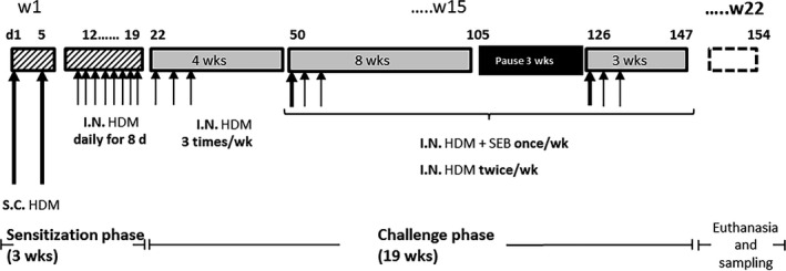
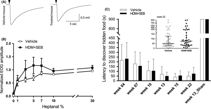
Methods: In order to elucidate potential mechanisms leading to CRS in humans, we have established a type 2/T helper type 2 cell (Th2)-mediated allergic CRS mouse model, based on house dust mite (HDM) and Staphylococcus aureus enterotoxin B (SEB) sensitization. The inflammatory status of the olfactory epithelium (OE) was assessed using histology, biochemistry, and transcriptomics. The sense of smell was evaluated by studying olfactory behavior and recording electro-olfactograms (EOGs).

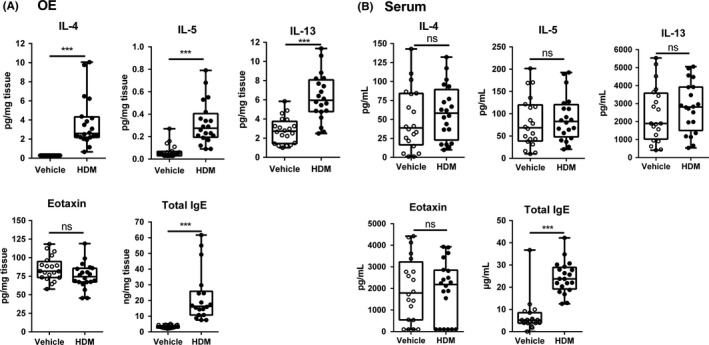
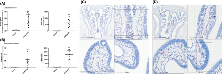
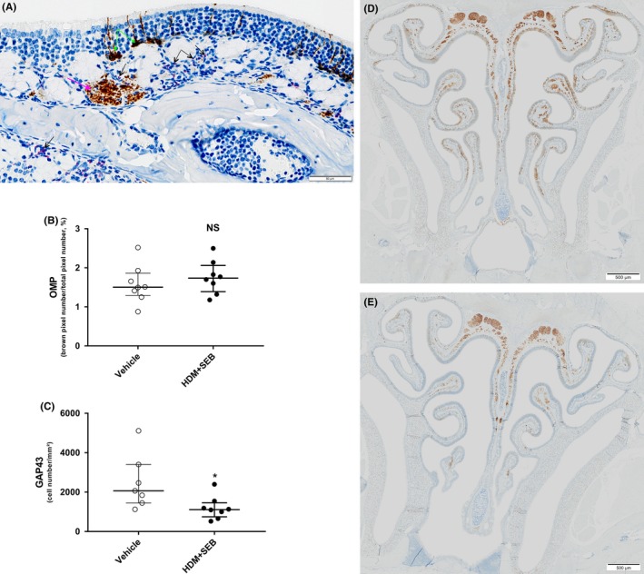
Results: After 22 weeks, a typical type 2/Th2-mediated inflammatory profile was obtained, as demonstrated by increased interleukin (IL)-4, IL-5, and IL-13 in the OE. The number of mast cells and eosinophils was increased, and infiltration of these cells into the olfactory mucosa was also observed. In parallel, transcriptomic and histology analyses indicated a decreased number of immature olfactory neurons, possibly due to decreased renewal. However, the number of mature sensory neurons was not affected and neither the EOG nor olfactory behavior was impaired.

Conclusion: Our mouse model of CRS displayed an allergic response to HDM + SEB administration, including the type 2/Th2 inflammatory profile characteristic of human eosinophilic CRSwNP. Although the sense of smell did not appear to be altered in these conditions, the data reveal the influence of chronic inflammation on olfactory neurogenesis, suggesting that factors unique to humans may be involved in CRSwNP-associated anosmia.

Keywords: Type 2/Th2 inflammation; chronic rhinosinusitis; olfaction; olfactory epithelium; sensory neurogenesis.


Conflict of interest statement

Biton B, Bock M‐D, Classe M, Clément M, Didier M, Fourgous V, Françon D, Gorski R, Guillemot J‐C, Haddad E‐B, Le‐Guern J, Leonetti M, Mikol V, Orsini C, Paul P, Ponsolles C, Remaury A, Roche S, Rocheteau‐Beaujouan L, Rouyar A are Sanofi employees and may hold stock and/or stock options in the company.

Figures

Figure 1
Figure 1
Protocol for allergen HDM sensitization and SEB administration. Two groups of mice of 38 animals were treated either with PBS (control group) or with HDM + SEB (treated group). On Days 1 and 5, S.C. HDM (HDM 100 μg + 200 μg Alum in PBS [100 μL]) was administered, followed by daily I.N. HDM (HDM 20 μg in 40 μL PBS [20 μL/nare]) from Days 12 to 19. I.N. HDM + SEB: 20 μg HDM + SEB 10 ng (20 μL/nare). HDM, house dust mite; I.N., intranasally; PBS, phosphate buffer saline; S.C. subcutaneously; SEB, Staphylococcal enterotoxin type B
Figure 2
Figure 2
Differential expression of cytokines and IgE in (A) OE and (B) serum. Measurement of cytokines, IL‐4, IL‐5, and IL‐13, and eotaxin (CCL11) by Bio‐Plex multiplex and IgE by ELISA in OE of 20 controls (vehicle) and 20 HDM + SEB‐treated mice (HDM). All individual data are expressed in box‐and‐whisker plots showing the median values with interquartile range and minimum and maximum values. Statistical analysis was performed using a Wilcoxon‐Mann‐Whitney U test, and P values below < 0.05 were considered statistically significant. ***< 0.001 vs controls. ELISA, enzyme‐linked immunosorbent assay; HDM, house dust mite; IL, interleukin; ns, not significant; OE, olfactory epithelium
Figure 3
Figure 3
Eosinophils and mast cell counts in (A) olfactory mucosa and in (B) respiratory mucosa. Eosinophil infiltration was evaluated using Sirius red staining and quantified by pixel count (number of red pixels/total number of pixels) and expressed as a percentage in an olfactory or respiratory area previously defined. Mast cells were stained based on IHC using mast cell tryptase antibody. Positive cells were counted as a number of cells per squared millimeter. Graphs represent median with interquartile range of eight mice (plots) for each group. Statistical differences between groups were assessed using the Wilcoxon‐Mann‐Whitney U test, and P values below < 0.05 were considered statistically significant. (C) Sirius red staining of a vehicle animal at low magnification with higher magnification inset. (D) Sirius red staining of an HDM + SEB animal at low magnification with higher magnification inset. Pink circles indicate areas of eosinophil infiltration. ***< 0.001 vs controls. HDM, house dust mite; IHC, immunohistochemistry; SEB, Staphylococcal enterotoxin type B
Figure 4
Figure 4
(A) Distribution of GAP43‐positive immature cells and interaction between eosinophils and nerve bundles within olfactory mucosa of HDM + SEB exposed mouse (IHC GAP43 combined with Sirius red staining); black arrows point to eosinophils stained by Sirius red, pink arrow points to a nerve bundle stained with GAP43 antibody, and green arrows point to GAP43‐positive immature olfactory neurons. (B) Number of mature olfactory neurons and (C) immature olfactory neurons in OE. Mature olfactory neurons were stained by IHC using OMP antibody and quantified by pixel count (brown pixels/total pixel number) and expressed as a percentage. Immature olfactory neurons were stained by IHC using GAP43 antibody and counted as a number of cells per squared millimeter. Graphs represent median with interquartile range of 7‐8 mice (plots) for each group. Statistical differences between groups were assessed using the Wilcoxon‐Mann‐Whitney U test, and P values below < 0.05 were considered statistically significant. (D) GAP43 IHC at low magnification in a vehicle animal. (E) GAP43 IHC at low magnification in an HDM + SEB animal. *< 0.05 vs controls. GAP43, growth‐associated protein 43; HDM, house dust mite; IHC, immunohistochemistry; NS, not significant; OE, olfactory epithelium; OMP, olfactory marker protein; SEB, Staphylococcal enterotoxin type B
Figure 5
Figure 5
Characterization of sense of smell (A) typical recordings of 10% heptanal‐evoked EOGs of OSNs from vehicle‐treated and HDM + SEB‐treated mice. Black arrows indicate odorant (heptanal 10%) stimulation (100 ms). (B) EOG responses to increasing odorant percentage in vehicle and HDM + SEB groups. Values represent the means of peak amplitudes ± SEM (n = 7‐8/group), normalized to the response to 100% heptanal. (C) Buried food test. Bar graph represents the time course of median latency to discover a hidden food ± MAD in the vehicle and HDM + SEB‐treated groups (n = 38/group). In the Week 13_Sham experiment, mice had to discover a food pellet without odorant. The inset shows the latency distribution in Week 22 for both groups. Graphs represent median with interquartile range of 38 mice (plots) for each group. EOG, electro‐olfactograms; HDM, house dust mite; MAD, median absolute deviation; OSN, olfactory sensory neurons; SEB, Staphylococcal enterotoxin type B

References

    1. Hastan D, Fokkens WJ, Bachert C, et al. Chronic rhinosinusitis in Europe–an underestimated disease. A GA(2)LEN study. Allergy. 2011;66:1216‐1223. - PubMed
    1. Schleimer RP. Immunopathogenesis of chronic rhinosinusitis and nasal polyposis. Annu Rev Pathol. 2017;12:331‐357. - PMC - PubMed
    1. Hulse KE, Stevens WW, Tan BK, Schleimer RP. Pathogenesis of nasal polyposis. Clin Exp Allergy. 2015;45:328‐346. - PMC - PubMed
    1. Fokkens WJ, van Drunen C, Georgalas C, Ebbens F. Role of fungi in pathogenesis of chronic rhinosinusitis: the hypothesis rejected. Curr Opin Otolaryngol Head Neck Surg. 2012;20:19‐23. - PubMed
    1. Bachert C, Claeys SEM, Tomassen P, van Zele T, Zhang N. Rhinosinusitis and asthma: a link for asthma severity. Curr Allergy Asthma Rep. 2010;10:194‐201. - PubMed

Publication types

MeSH terms