. 2018 Sep;109(9):2811-2821.
doi: 10.1111/cas.13733. Epub 2018 Sep 3.

Identification of ENTPD8 and cytidine in pancreatic cancer by metabolomic and transcriptomic conjoint analysis

Affiliations

Identification of ENTPD8 and cytidine in pancreatic cancer by metabolomic and transcriptomic conjoint analysis

Yong An et al. Cancer Sci. 2018 Sep.

Abstract

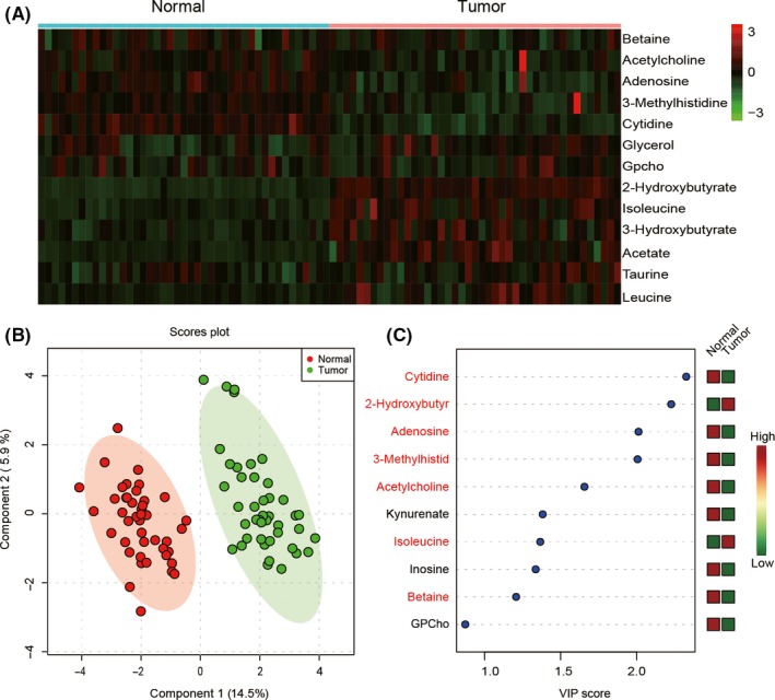
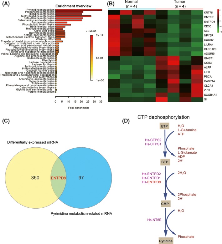
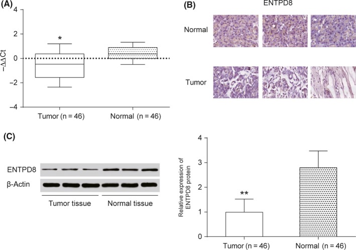
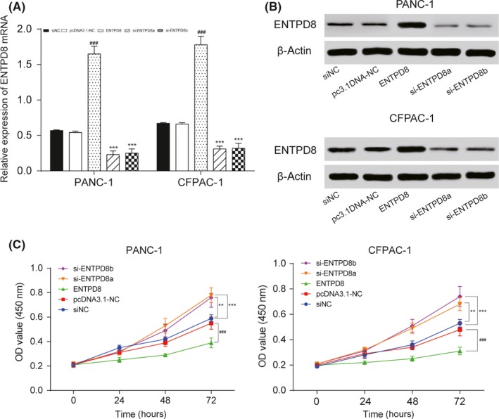
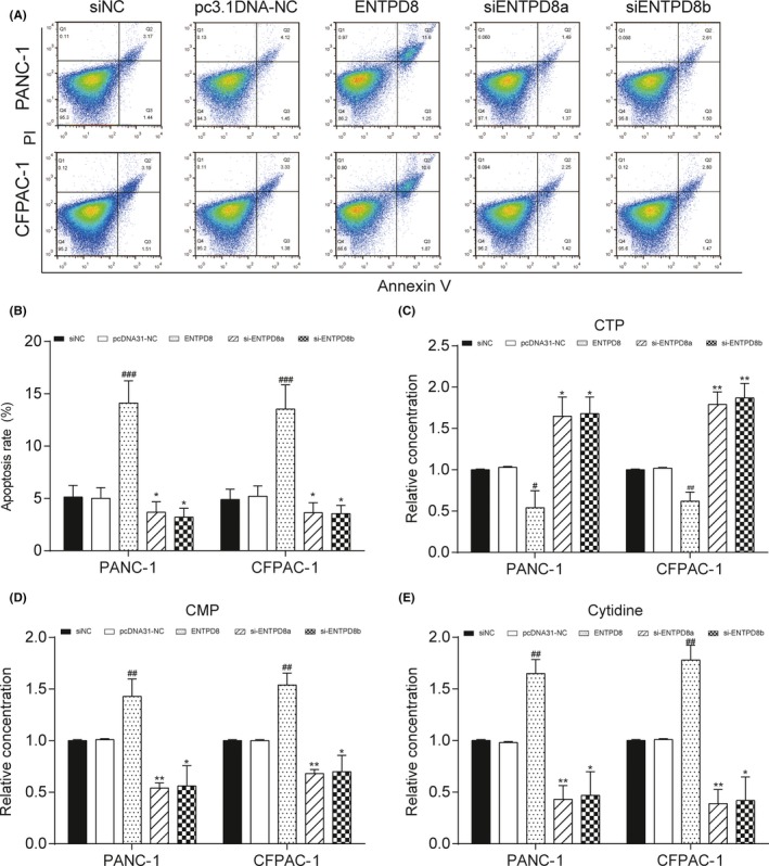
To identify metabolic pathways that were perturbed in pancreatic cancer (PC), we investigated gene-metabolite networks by integration of metabolomic and transcriptomic. In this research, we undertook the metabolomic study of 43 paired human PC samples, aiming to identify key metabolic alterations in PC. We also carried out in vitro experiments to validate that the key metabolite cytidine and its related gene ENTPD8 played an important role in PC cell proliferation. We screened out 13 metabolites differentially expressed in PC tissue (PCT) by liquid chromatography/mass spectrometry analysis on 34 metabolites, and the partial least square discrimination analysis results revealed that 9 metabolites among them were remarkably altered in PCT compared to adjacent noncancerous tissue (variable importance in projection >1, P < .05). Among the 9 metabolites, 7 might be potential biomarkers. The most significantly enriched metabolic pathway was pyrimidine metabolism. We analyzed 351 differentially expressed genes from The Cancer Genome Atlas and intersected them with Kyoto Encyclopedia of Genes and Genomes metabolic pathways. We found that ENTPD8 had a gene-metabolite association with cytidine in the CTP dephosphorylation pathway. We verified by in vitro experiments that the CTP dephosphorylation pathway was changed in PCT compared with adjacent noncancerous tissue. ENTPD8 was downregulated in PCT, causing a reduction in cytidine formation and hence weakened CTP dephosphorylation in pyrimidine metabolism.

Keywords: metabolic pathway; metabolite; metabolomic; pancreatic cancer; transcriptomic.


Figures

Figure 1
Figure 1
Metabolic biomarker analysis in pancreatic cancer tissue (Tumor) and adjacent nontumor tissue (Normal). A, 13 altered metabolites according to liquid chromatography/mass spectrometry analysis visualized by a heat map. B, Partial least square discrimination analysis (PLSDA) model showed that tumor and normal samples were clearly separated in the x‐axis direction. Each point represents an independent sample, and the ellipse represents the 95% confidence interval. C, 10 metabolites with the highest variable importance in projection (VIP) scores of PC1 were screened out by the PLSDA model. Seven potential biomarkers were shown in red
Figure 2
Figure 2
Enriched metabolomic pathway analysis in pancreatic cancer tissue. A, Overview of metabolite sets enrichment. P‐value represents the enrichment level; length of bar represents the amount of metabolites. B, Heat map of the top 10 upregulated and downregulated mRNAs in pancreatic cancer tissue. Data were acquired from The Cancer Genome Atlas. C, Venn diagram of differentially expressed mRNAs and pyrimidine metabolism related mRNAs. D, CTP dephosphorylation in pyrimidine metabolism involves metabolic biomarker cytidine and its related gene ENTPD8
Figure 3
Figure 3
Expression level of ENTPD8 in pancreatic cancer tissue. A, Quantitative RTPCR was undertaken to measure the relative expression level of ENTPD8 mRNA in tumor tissues and normal tissues. B, Immunohistochemical assay shows that ENTPD8 appears positive in the normal cell nucleus. C, Western blot analysis to measure ENTPD8 protein expression level. *< .05; **P < .01, indicates statistical significance compared with negative group
Figure 4
Figure 4
Effects of ENTPD8 on proliferation and protein expression of PANC‐1 and CFPAC‐1 pancreatic cancer cells. A, After transfection, the relative expression level of ENTPD8 was measured by quantitative RTPCR. B, After transfection, the relative expression level of ENTPD8 protein was measured by Western blot analysis, and its expression was upregulated in the ENTPD8 group and downregulated in the si‐ENTPD8 group. C, CCK‐8 assay shows that cell viability in the ENTPD8 group was inhibited. **< .01, ***< .001 indicated statistical significance compared with the negative (NC) group. ### < .001 indicates statistical significance compared with pcDNA3.1‐NC group
Figure 5
Figure 5
Effect of ENTPD8 on apoptosis and metabolites in two pancreatic cancer cell lines. A,B, Flow cytometry assay shows that ENTPD8 promoted cell apoptosis in both PANC‐1 and CFPAC‐1 cells. C‐E, Relative metabolite concentrations of CTP (C), cytidine monophosphate (CMP) (D) and cytidine (E) in cell lines. *< .05, **< .01 indicates statistical significance compared with the negative (NC) group. # < .05, ## < .01, ### < .001 indicates statistical significance compared with pcDNA3.1‐NC group

References

    1. Siegel RL, Miller KD, Jemal A. Cancer statistics, 2016. CA Cancer J Clin. 2016;66:7‐30. - PubMed
    1. Duell EJ, Lucenteforte E, Olson SH, et al. Pancreatitis and pancreatic cancer risk: a pooled analysis in the International Pancreatic Cancer Case–Control Consortium (PanC4). Ann Oncol. 2012;23:2964‐2970. - PMC - PubMed
    1. Zhang H, Wang Y, Gu X, Zhou J, Yan C. Metabolomic profiling of human plasma in pancreatic cancer using pressurized capillary electrochromatography. Electrophoresis. 2011;32:340‐347. - PubMed
    1. Auslander N, Yizhak K, Weinstock A, et al. A joint analysis of transcriptomic and metabolomic data uncovers enhanced enzyme‐metabolite coupling in breast cancer. Sci Rep. 2016;6:29662. - PMC - PubMed
    1. Farshidfar F, Weljie AM, Kopciuk KA, et al. A validated metabolomic signature for colorectal cancer: exploration of the clinical value of metabolomics. Br J Cancer. 2016;115:848‐857. - PMC - PubMed

Substances