. 2018 Sep;11(5):1454-1465.
doi: 10.1038/s41385-018-0051-2. Epub 2018 Jul 9.

The JAK inhibitor ruxolitinib reduces inflammation in an ILC3-independent model of innate immune colitis

Affiliations

The JAK inhibitor ruxolitinib reduces inflammation in an ILC3-independent model of innate immune colitis

A M Overstreet et al. Mucosal Immunol. 2018 Sep.

Abstract

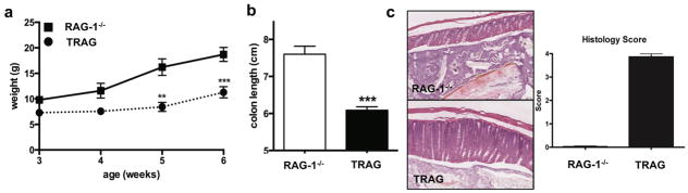
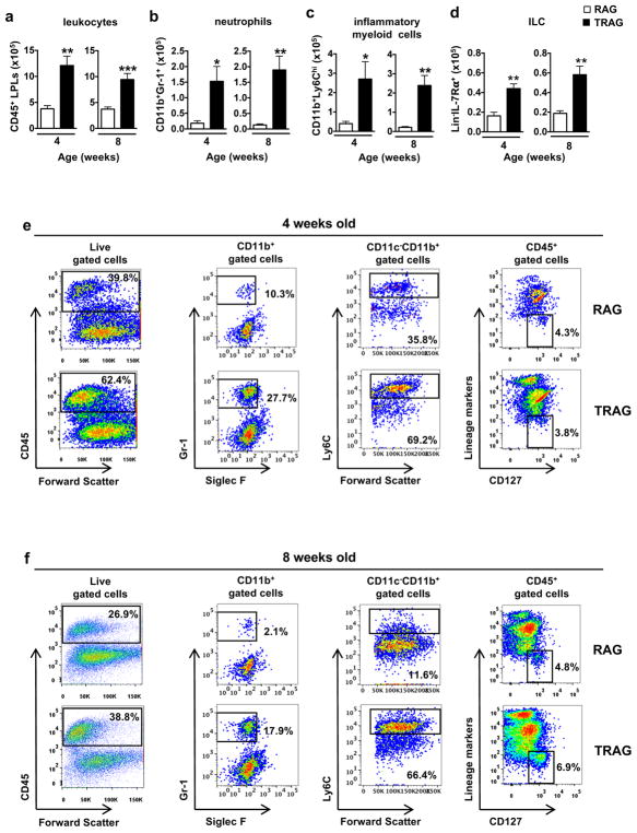
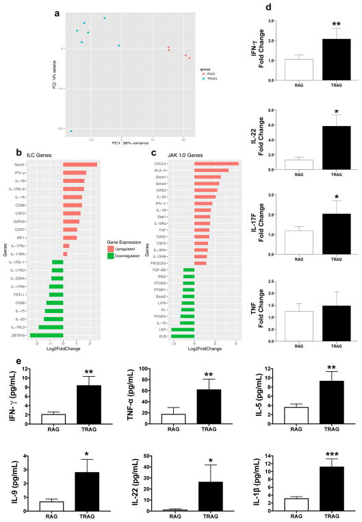
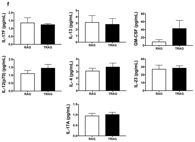
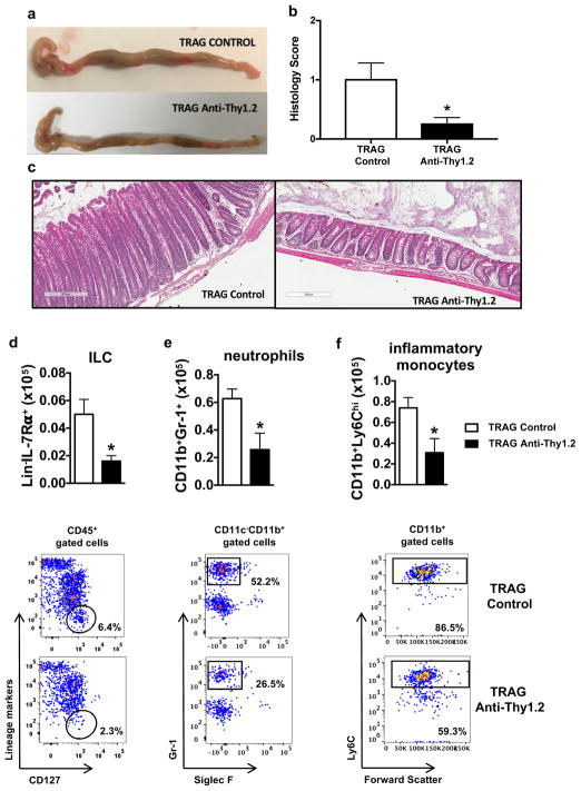
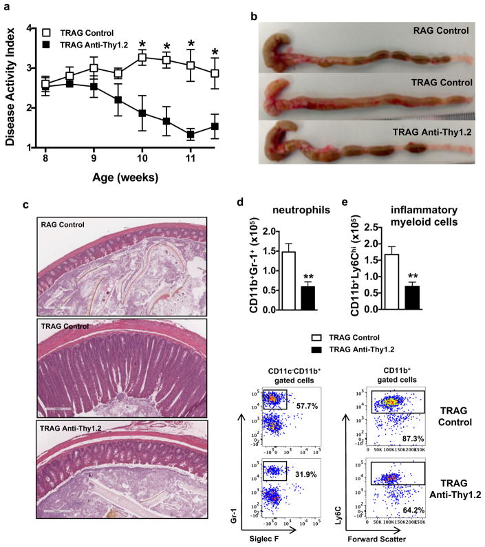
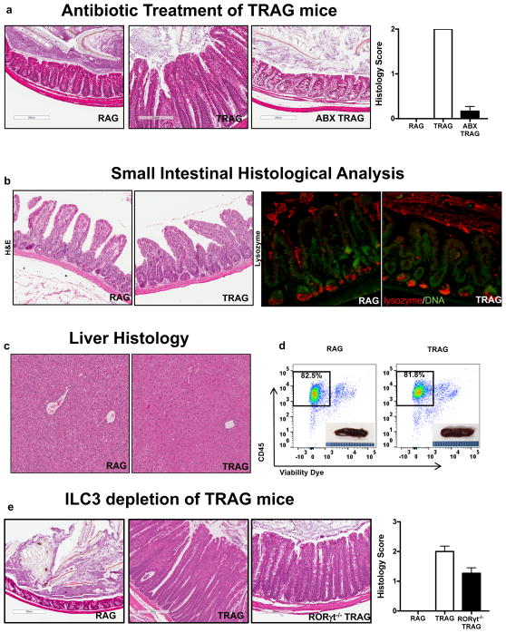
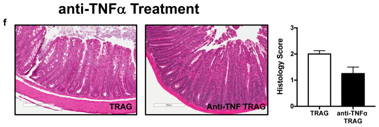
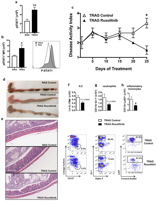
Innate immunity contributes to the pathogenesis of inflammatory bowel disease (IBD). However, the mechanisms of IBD mediated by innate immunity are incompletely understood and there are limited models of spontaneous innate immune colitis to address this question. Here we describe a new robust model of colitis occurring in the absence of adaptive immunity. RAG1-deficient mice expressing TNFAIP3 in intestinal epithelial cells (TRAG mice) spontaneously developed 100% penetrant, early-onset colitis that was limited to the colon and dependent on intestinal microbes but was not transmissible to co-housed littermates. TRAG colitis was associated with increased mucosal numbers of innate lymphoid cells (ILCs) and depletion of ILC prevented colitis in TRAG mice. ILC depletion also therapeutically reversed established colitis in TRAG mice. The colitis in TRAG mice was not prevented by interbreeding to mice lacking group 3 ILC nor by depletion of TNF. Treatment with the JAK inhibitor ruxolitinib ameliorated colitis in TRAG mice. This new model of colitis, with its predictable onset and colon-specific inflammation, will have direct utility in developing a more complete understanding of innate immune mechanisms that can contribute to colitis and in pre-clinical studies for effects of therapeutic agents on innate immune-mediated IBD.


Conflict of interest statement

The authors declare no conflict of interest

Figures

Figure 1
Figure 1
Spontaneous innate immune mediated colitis in TRAG mice. Mice were monitored for health and the onset of colitis by (a) body weight vs. age; and following euthanasia at 6 weeks of age for (b) colon length and (c) histology. **p<0.01, ***p<0.005 (n > 5 mice/group).
Figure 2
Figure 2
Colons of TRAG mice have increased inflammatory cells. The mucosa of TRAG mice contained increased total numbers of (a) CD45+ leukocytes including (b) neutrophils, (c) inflammatory myeloid cells (Ly6Chi) and (d) innate lymphoid cells (ILC). (e,f) representative FACS plots showing gating of innate cells from LPLs of mice at 4 and 8 weeks of age. Comparisons were made using unpaired student’s t test. *p<0.05, **p<0.01, ***p<0.001. Data represent 2 independent experiments, mean ± SEM (n = 4 mice/group/experiment).
Figure 3
Figure 3
Gene expression and cytokine profile of TRAG colitis. (a) Principal component analysis of gene expression in TRAG vs. RAG1−/− colonic mucosa. (b,c) Molecular drivers of the differential gene expressions patterns identified using Ingenuity Pathway Analysis of genes associated with colitis in TRAG mice implicates the (b) ILC and (c) JAK1/2 pathway. (d) Quantitative PCR showing increased expression of INFγ, IL22 and IL17, but not TNF, in the mucosa of TRAG mice. (e) Cytokine secretion by LPLs isolated from RAG1−/− vs. TRAG mucosa showing increased secretion of IFNγ, TNF, IL-5, IL-9, IL-22 and IL-1β from LPLs of TRAG mice. (f) cytokines detected but not statistically different between TRAG and RAG1−/− LPLs included IL-17F, IL-17A, GM-CSF, IL-23, IL-4, IL-12 and IL-13. (Mann-Whitney) *p<0.05, **p<0.01, ***p<0.001 (n= 7–10 mice/group (except TRAG array group where n=4))
Figure 3
Figure 3
Gene expression and cytokine profile of TRAG colitis. (a) Principal component analysis of gene expression in TRAG vs. RAG1−/− colonic mucosa. (b,c) Molecular drivers of the differential gene expressions patterns identified using Ingenuity Pathway Analysis of genes associated with colitis in TRAG mice implicates the (b) ILC and (c) JAK1/2 pathway. (d) Quantitative PCR showing increased expression of INFγ, IL22 and IL17, but not TNF, in the mucosa of TRAG mice. (e) Cytokine secretion by LPLs isolated from RAG1−/− vs. TRAG mucosa showing increased secretion of IFNγ, TNF, IL-5, IL-9, IL-22 and IL-1β from LPLs of TRAG mice. (f) cytokines detected but not statistically different between TRAG and RAG1−/− LPLs included IL-17F, IL-17A, GM-CSF, IL-23, IL-4, IL-12 and IL-13. (Mann-Whitney) *p<0.05, **p<0.01, ***p<0.001 (n= 7–10 mice/group (except TRAG array group where n=4))
Figure 4
Figure 4
Prophylactic anti-Thy 1.2 reduces ILCs and ameliorates colitis in TRAG mice. IP injections of anti-Thy 1.2 (1 mg) were administered to TRAG mice from 4–6 weeks of age every three days. Anti-Thy1.2 treatment reduced colitis in TRAG mice as indicated by (a) colon length and gross morphology and (b,c) histology. TRAG mice treated with anti-Thy1.2 antibody prior to the development of colitis displayed reduced numbers of colonic mucosal (d) ILC, (e) neutrophils and (f) inflammatory myeloid cells, compared to control TRAG mice. (Mann-Whitney for (b); T test for other data) *p<0.05, p<0.005. Data represent 2 independent experiments, mean ± SEM (n = 8–10 mice/group).
Figure 5
Figure 5
Therapeutic anti-Thy 1.2 treatment reverses established colitis in TRAG mice. Anti-Thy1.2 (1 mg/mouse; i.p.) treatments were administered every three days beginning at 8 weeks of age for 21 days. (a) Disease activity showing clinical improvement in Thy1.2 treated TRAG mice. After treatment reduced colitis was evident by (b) gross morphology of the colon and (c) histology. Colonic LPLs were assessed for total numbers of (d) neutrophils and (e) inflammatory myeloid cells. (for (a) 2-way ANOVA with sidak multiple comparison. T test for other data) *p<0.05, ** p<0.01. Data represent of 2 independent experiments, mean ± SEM (n = 5 mice/group).
Figure 6
Figure 6
TRAG colitis is microbially driven, restricted to the colon, and independent of group 3 ILCs. (a) Histology and histological cores showing suppression of colitis in antibiotic treated TRAG mice (ABX TRAG). Lack of small bowel and systemic inflammation in TRAG mice. (b) Histology showing absence of ileitis (upper panel H&E) and preservation of Paneth cells (lower panel red=lysozyme) in the terminal ileum and (c) lack of hepatitis in liver of RAG1−/− or TRAG mice. (d) Flow cytometry and total cell counts of spleen showing lack of expansion of CD45+ leukocytes in the primary immune tissue of TRAG mice (inset: picture of spleens). (e) Histology showing that colitis is not prevented in TRAG × Rorc−/− mice and (f) in TRAG mice treated with anti-TNF antibody.
Figure 6
Figure 6
TRAG colitis is microbially driven, restricted to the colon, and independent of group 3 ILCs. (a) Histology and histological cores showing suppression of colitis in antibiotic treated TRAG mice (ABX TRAG). Lack of small bowel and systemic inflammation in TRAG mice. (b) Histology showing absence of ileitis (upper panel H&E) and preservation of Paneth cells (lower panel red=lysozyme) in the terminal ileum and (c) lack of hepatitis in liver of RAG1−/− or TRAG mice. (d) Flow cytometry and total cell counts of spleen showing lack of expansion of CD45+ leukocytes in the primary immune tissue of TRAG mice (inset: picture of spleens). (e) Histology showing that colitis is not prevented in TRAG × Rorc−/− mice and (f) in TRAG mice treated with anti-TNF antibody.
Figure 7
Figure 7
The JAK1/2 inhibitor ruxolitinib reduced colitis in TRAG mice. Mice were gavaged twice daily (60mg/kg) from 3 to 7 weeks of age. (a) Flow cytometry indicating that there are more phospho-STAT1+ Thy1.2 cells that also (b) have a higher MFI for phospho-STAT1 in LPLs from TRAG mice compared to those of RAG1−/− mice. Clinical improvement following ruxolitinib treatment was indicated by (c) disease activity (d) gross colon morphology, and (e) histology. (f–h) LPL counts show a significant reduction of inflammatory monocytes and a trend of decreased leukocytes, ILCs, and neutrophils in the colonic mucosa of ruxolitinib treated mice. (for (c) 2-way ANOVA with Sidak multiple comparison test. T test for other data) *p<0.05, p<0.01. Data represent mean ± SEM (n = 3–4 mice/group).

References

    1. Strober W, Fuss IJ, Blumberg RS. The immunology of mucosal models of inflammation. Annu Rev Immunol. 2002;20:495–549. - PubMed
    1. Artis D, Spits H. The biology of innate lymphoid cells. Nature. 2015;517:293–301. - PubMed
    1. Geremia A, et al. IL-23-responsive innate lymphoid cells are increased in inflammatory bowel disease. J Exp Med. 2011;208:1127–1133. - PMC - PubMed
    1. Buonocore S, et al. Innate lymphoid cells drive interleukin-23-dependent innate intestinal pathology. Nature. 2010;464:1371–1375. - PMC - PubMed
    1. Boone DL, et al. The ubiquitin-modifying enzyme A20 is required for termination of Toll-like receptor responses. Nat Immunol. 2004;5:1052–1060. - PubMed

Publication types

MeSH terms

LinkOut - more resources