. 2018 Jun 22:9:1433.
doi: 10.3389/fimmu.2018.01433. eCollection 2018.

Alternative Pathway Is Essential for Glomerular Complement Activation and Proteinuria in a Mouse Model of Membranous Nephropathy

Affiliations

Alternative Pathway Is Essential for Glomerular Complement Activation and Proteinuria in a Mouse Model of Membranous Nephropathy

Wentian Luo et al. Front Immunol. .

Abstract

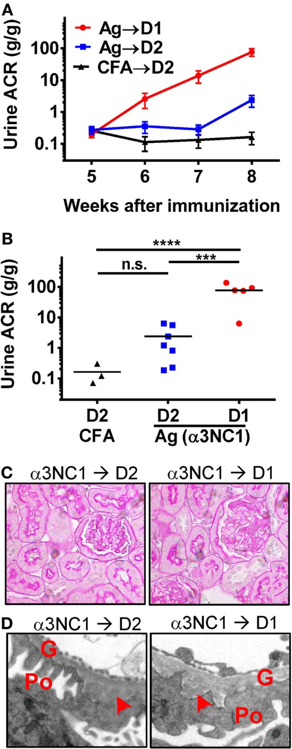
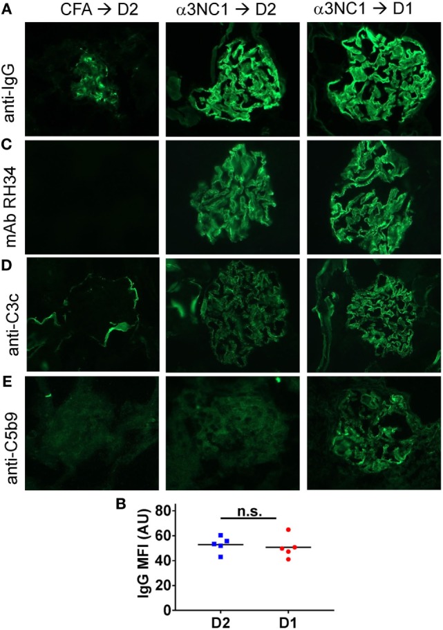
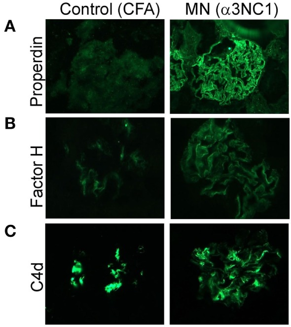
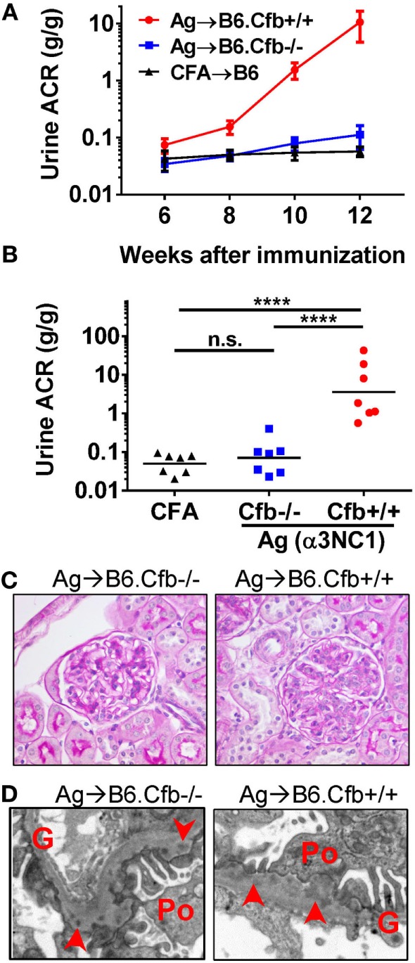
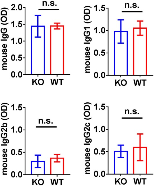
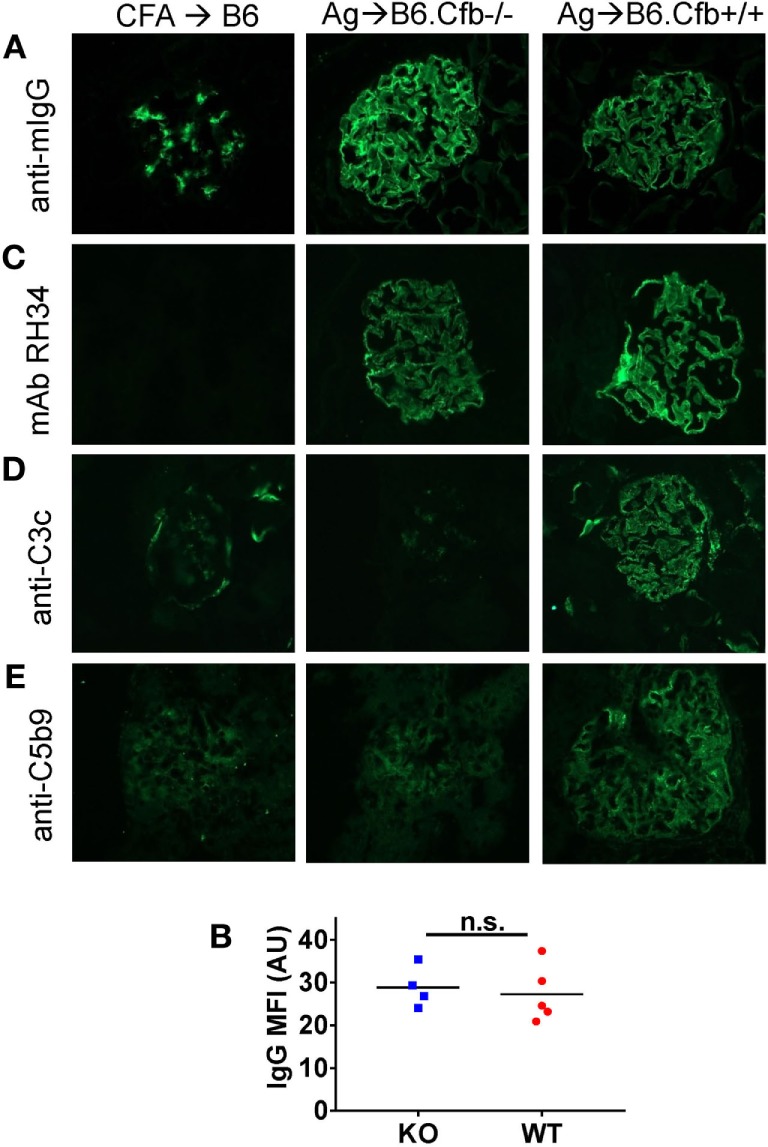
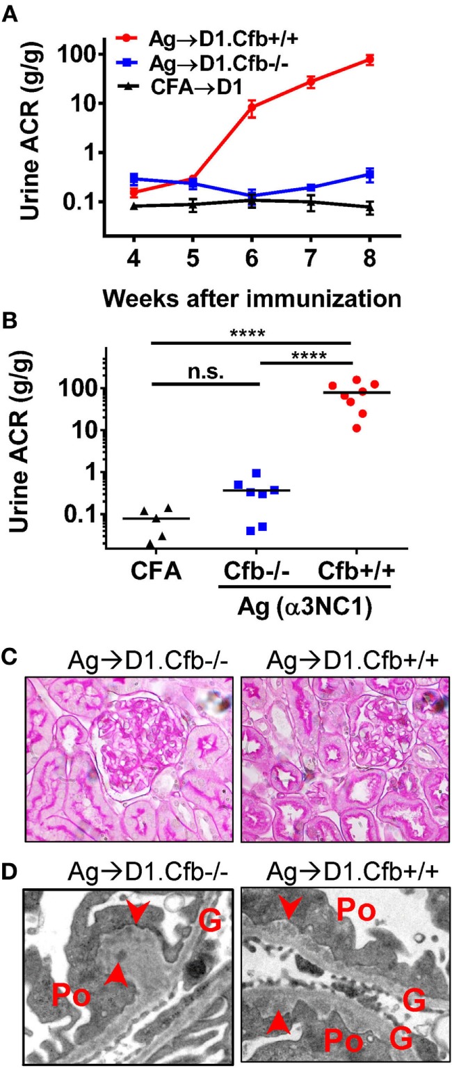
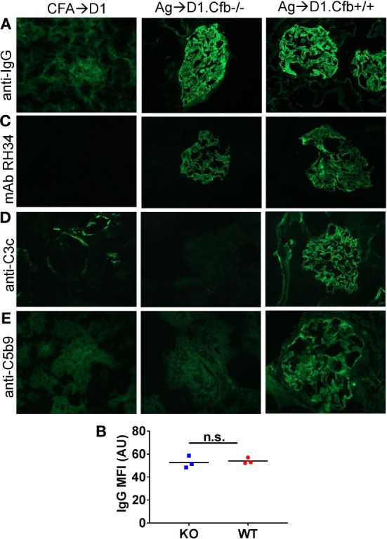
Membranous nephropathy is an immune kidney disease caused by IgG antibodies that form glomerular subepithelial immune complexes. Proteinuria is mediated by complement activation, as a result of podocyte injury by C5b-9, but the role of specific complement pathways is not known. Autoantibodies-mediating primary membranous nephropathy are predominantly of IgG4 subclass, which cannot activate the classical pathway. Histologic evidence from kidney biopsies suggests that the lectin and the alternative pathways may be activated in membranous nephropathy, but the pathogenic relevance of these pathways remains unclear. In this study, we evaluated the role of the alternative pathway in a mouse model of membranous nephropathy. After inducing the formation of subepithelial immune complexes, we found similar glomerular IgG deposition in wild-type mice and in factor B-null mice, which lack a functional alternative pathway. Unlike wild-type mice, mice lacking factor B did not develop albuminuria nor exhibit glomerular deposition of C3c and C5b-9. Albuminuria was also reduced but not completely abolished in C5-deficient mice. Our results provide the first direct evidence that the alternative pathway is necessary for pathogenic complement activation by glomerular subepithelial immune complexes and is, therefore, a key mediator of proteinuria in experimental membranous nephropathy. This knowledge is important for the rational design of new therapies for membranous nephropathy.

Keywords: albuminuria; alternative pathway; complement C5; factor B; glomerulonephritis; membrane attack complex; membranous nephropathy; mouse models.


Figures

Figure 1
Figure 1
C5 deficiency protects against albuminuria in experimental MN. (A) Time course of urinary albumin-to-creatinine ratio (ACR) in D1 mice (circles) and D2 mice (squares) immunized with α3NC1 (N = 5–7 per group). Control D2 mice (triangles) were immunized with adjuvant alone (N = 3). Shown are means and SEM. (B) Scatterplot depicting the final ACR values (at week 8). The significance of differences among groups was analyzed by one-way ANOVA with Dunnett’s correction for multiple comparisons. ***P < 0.001, ****P < 0.0001. n.s., not significant. (C) Light microscopy shows normal appearance of glomeruli and tubules in α3NC1-immunized D2 and D1 mice (periodic acid–Schiff staining; original magnification 400×). (D) Transmission electron microscopy shows subepithelial electron dense deposits (arrowhead), areas of glomerular basement membrane (G) thickening, and effacement of podocyte (Po) foot processes. Original magnification 7,500×.
Figure 2
Figure 2
Analysis of glomerular immune complexes and complement deposition in D1 and D2 mice immunized with α3NC1. Kidneys were collected at week 8 post-immunization from adjuvant immunized D2 mice (left), α3NC1-immunized D2 mice (middle), and α3NC1-immunized D1 mice (right). (A) Direct immunofluorescence staining shows mouse IgG binding along the glomerular basement membrane (GBM) in α3NC1-immunized D1 and D2 mice. Control mice show non-specific mesangial deposition of mouse IgG. (B) Mean fluorescence intensity (MFI) of IgG staining, expressed in arbitrary units (AU), was compared in α3NC1-immunized D2 and D1 mice. The difference was not significant (n.s.) by t test. (C) Indirect immunofluorescence staining with mAb RH34 shows GBM deposition of exogenous antigen in all α3NC1-immunized mice, which is absent in adjuvant-immunized mice. (D) Direct immunofluorescence staining reveals C3c deposition along the capillary loops in α3NC1-immunized D1 and D2 mice. In control mice, C3c staining is positive in the Bowman’s capsule and kidney tubules. (E) Indirect immunofluorescence shows capillary loop staining for C5b-9 in α3NC1-immunized D1 mice. A weak background of non-specific immunofluorescence is observed in adjuvant-immunized and α3NC1-immunized D2 mice. Original magnification 400×.
Figure 3
Figure 3
Histologic evidence of AP activation in experimental MN. Glomerular deposition of properdin (A), factor H (B), and C4d (C) was evaluated by indirect immunofluorescence staining of frozen kidney sections from α3NC1-immunized D1 mice and adjuvant-immunized control mice. Properdin and factor H staining along the capillary loops was found in α3NC1-immunized D1 mice, but not in adjuvant-immunized mice. Mesangial C4d staining was found in all mice, while weaker segmental staining for C4d along capillary loops was also present in α3NC1-immunized mice.
Figure 4
Figure 4
B6.Cfb−/− mice immunized with α3NC1 are protected against albuminuria despite developing subepithelial immune complexes. (A) Time course of urinary albumin-to-creatinine ratio (ACR) in B6.Cfb+/+ mice (circles) and B6.Cfb−/− mice (squares) immunized with α3NC1 (N = 7 per group). Control B6 mice (triangles), including both genotypes, were immunized with adjuvant alone (N = 7). Shown are means and SEM. (B) Scatterplot depicts the ACR values at the endpoint of this experiment (week 12). The significance of differences among groups was analyzed by one-way ANOVA with Dunnett’s correction for multiple comparisons. ***P < 0.001, ****P < 0.0001. n.s., not significant. (C) Morphology of kidneys from α3NC1-immunized B6.Cfb−/− and B6.Cfb+/+ mice appears normal by light microscopy (periodic acid–Schiff staining, original magnification 400×). (D) Transmission electron microscopy shows subepithelial electron dense deposits (arrowhead), expansion of the glomerular basement membrane (G), and podocyte (Po) foot process effacement. Original magnification 7,500×.
Figure 5
Figure 5
Analysis of circulating anti-α3NC1 mouse IgG antibodies. The levels of circulating mouse IgG, IgG1, IgG2b, and IgG2c antibodies binding to rh-α3NC1 were measured by indirect ELISA. Mouse sera were diluted 1/5,000 for total IgG, 1/2,000 for IgG1, and 1/500 for IgG2b and IgG2c. The significance of differences between Cfb−/− (KO) and Cfb+/+ (WT) mice was evaluated by t-test (n.s., not significant).
Figure 6
Figure 6
Analysis of glomerular immune complexes and complement deposition in B6 mice immunized with α3NC1. Kidneys from adjuvant-immunized B6 mice (left), α3NC1-immunized B6.Cfb−/− mice (middle), and α3NC1-immunized B6.Cfb−/− mice (right) were collected at week 12 after the initial immunization. (A) Immunofluorescence staining for mouse IgG shows glomerular basement membrane (GBM) staining in α3NC1-immunized B6.Cfb−/− and B6.Cfb+/+ mice. Adjuvant-immunized mice show non-specific mesangial IgG deposition. (B) Mean fluorescence intensity (MFI) of IgG staining, expressed in arbitrary units (AU), was compared in α3NC1-immunized B6.Cfb−/− and B6.Cfb+/+ mice. Significance was evaluated by t test (n.s., not significant). (C) Staining with mAb RH34 shows GBM deposition of exogenous antigen in α3NC1 immunized B6.Cfb−/− and B6.Cfb+/+ mice, which is absent in control mice. (D) Direct immunofluorescence staining reveals C3c deposition in a capillary loop pattern in α3NC1-immunized B6.Cfb+/+ mice. C3c staining is absent in B6.Cfb−/− mice. (E) Indirect immunofluorescence shows C5b-9 deposition along the GBM in α3NC1-immunized B6.Cfb+/+ mice, but not B6.Cfb−/− mice. Original magnification 400×.
Figure 7
Figure 7
D1.Cfb−/− mice immunized with α3NC1 are protected against albuminuria despite developing subepithelial immune complexes. (A) Time course of urinary albumin-to-creatinine ratio (ACR) in D1.Cfb+/+ mice (circles) and D1.Cfb−/− mice (squares) immunized with α3NC1 (N = 7–8 per group). Control D1 mice (triangles), including both genotypes, were immunized with adjuvant alone (N = 5). Shown are means and SEM. (B) Scatterplot depicts the ACR values at the last time point (week 8). The significance of differences among groups was analyzed by one-way ANOVA with Dunnett’s correction multiple comparisons. ***P < 0.001, ****P < 0.0001, n.s., not significant. (C) Morphology of kidneys from α3NC1-immunized D1.Cfb−/− (left) and D1.Cfb+/+ mice (right) appeared normal by light microscopy (periodic acid–Schiff staining, original magnification 400×). (D) Transmission electron microscopy shows subepithelial electron dense deposits (arrowhead), thickening of the glomerular basement membrane (G), and podocyte (Po) foot process effacement. Original magnification 15,000×.
Figure 8
Figure 8
Analysis of glomerular immune complexes and complement deposition in D1 mice immunized with α3NC1. Kidneys were collected from adjuvant immunized D1 mice (left), α3NC1-immunized D1.Cfb−/− mice (middle), and α3NC1-immunized D1.Cfb−/− mice (right) at week 8 after the initial immunization. (A) Immunofluorescence staining for mouse IgG shows glomerular basement membrane (GBM) staining in α3NC1-immunized D1.Cfb−/− and D1.Cfb+/+ mice. Adjuvant-immunized control mice show non-specific mesangial IgG deposition. (B) Mean fluorescence intensity (MFI) of IgG staining, expressed in arbitrary units (AU), was compared in α3NC1-immunized D1.Cfb−/− and D2.Cfb+/+ mice. The difference was not significant (n.s.) by t test. (C) Staining with mAb RH34 shows the GBM deposition of exogenous antigen in both groups of α3NC1-immunized mice, which is absent in control mice. (D) Direct immunofluorescence staining reveals capillary loop deposition of C3c in α3NC1-immunized D1.Cfb+/+ mice, while C3c staining is absent in D1.Cfb−/− mice. (E) Indirect immunofluorescence shows C5b-9 deposition along the GBM in α3NC1-immunized D1.Cfb+/+ mice but not D1.Cfb−/− mice. Original magnification 400×.
Figure 9
Figure 9
In vitro complement activation by glomerular immune complexes from α3NC1-immunized Cfb−/− mice. Immunofluorescence analysis of C3c deposition onto kidneys cryosections from α3NC1-immunized D1.Cfb−/− mice (left) and control non-immunized wild-type DBA/1 mice (right), after incubation with 33% normal mouse serum in buffer containing Ca2+ and Mg2+ (A), Mg2+ and EGTA (B), or EDTA (C). Complement pathways active in each buffer are indicated on the left, according to the key shown at the bottom.
Figure 10
Figure 10
Decreased glomerular basement membrane (GBM) staining by anti-heparan sulfate mAb JM403 in experimental MN. (A) Immunofluorescence staining with anti-heparan sulfate mAb JM403 revealed intense staining in the GBM of control mice, which was markedly decreased in α3NC1-immunized B6.Cfb+/+ mice. (B) GBM staining for agrin core protein was similar in both groups.

References

    1. Beck LH, Jr, Salant DJ. Membranous nephropathy: from models to man. J Clin Invest (2014) 124(6):2307–14.10.1172/JCI72270 - DOI - PMC - PubMed
    1. Cattran DC, Brenchley PE. Membranous nephropathy: integrating basic science into improved clinical management. Kidney Int (2017) 91(3):566–74.10.1016/j.kint.2016.09.048 - DOI - PubMed
    1. Beck LH, Jr, Bonegio RG, Lambeau G, Beck DM, Powell DW, Cummins TD, et al. M-type phospholipase A2 receptor as target antigen in idiopathic membranous nephropathy. N Engl J Med (2009) 361(1):11–21.10.1056/NEJMoa0810457 - DOI - PMC - PubMed
    1. Tomas NM, Beck LH, Jr, Meyer-Schwesinger C, Seitz-Polski B, Ma H, Zahner G, et al. Thrombospondin type-1 domain-containing 7A in idiopathic membranous nephropathy. N Engl J Med (2014) 371(24):2277–87.10.1056/NEJMoa1409354 - DOI - PMC - PubMed
    1. Debiec H, Lefeu F, Kemper MJ, Niaudet P, Deschenes G, Remuzzi G, et al. Early-childhood membranous nephropathy due to cationic bovine serum albumin. N Engl J Med (2011) 364(22):2101–10.10.1056/NEJMoa1013792 - DOI - PubMed