. 2018 Apr 12:9:30-40.
doi: 10.1016/j.omto.2018.04.002. eCollection 2018 Jun 29.

Enhanced Control of Oncolytic Measles Virus Using MicroRNA Target Sites

Affiliations

Enhanced Control of Oncolytic Measles Virus Using MicroRNA Target Sites

Mathias Felix Leber et al. Mol Ther Oncolytics. .

Abstract

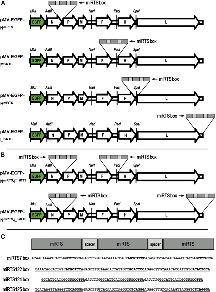
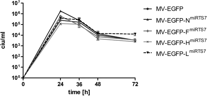
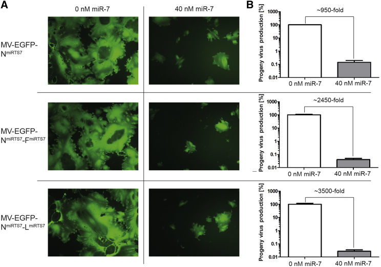
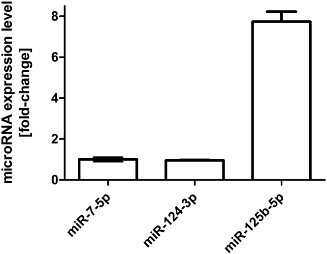
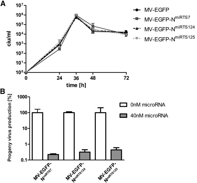
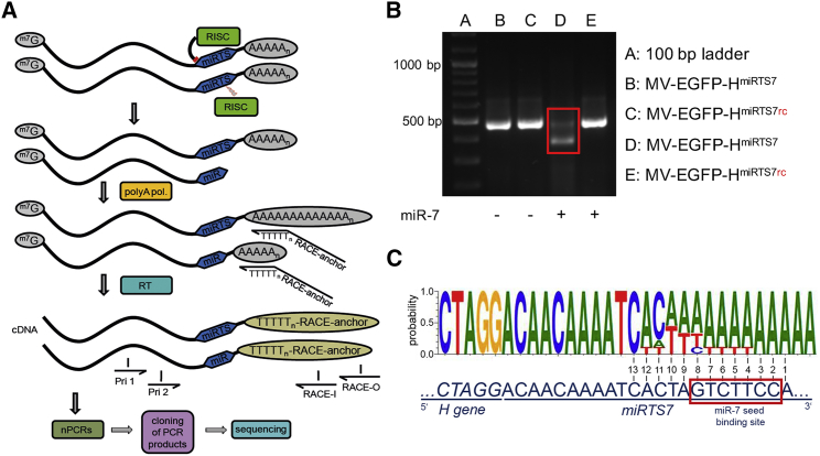
Measles viruses derived from the live-attenuated Edmonton-B vaccine lineage are currently investigated as novel anti-cancer therapeutics. In this context, tumor specificity and oncolytic potency are key determinants of the therapeutic index. Here, we describe a systematic and comprehensive analysis of a recently developed post-entry targeting strategy based on the incorporation of microRNA target sites (miRTS) into the measles virus genome. We have established viruses with target sites for different microRNA species in the 3' untranslated regions of either the N, F, H, or L genes and generated viruses harboring microRNA target sites in multiple genes. We report critical importance of target-site positioning with proximal genomic positions effecting maximum vector control. No relevant additional effect of six versus three miRTS copies for the same microRNA species in terms of regulatory efficiency was observed. Moreover, we demonstrate that, depending on the microRNA species, viral mRNAs containing microRNA target sites are directly cleaved and/or translationally repressed in presence of cognate microRNAs. In conclusion, we report highly efficient control of measles virus replication with various miRTS positions for development of safe and efficient cancer virotherapy and provide insights into the mechanisms underlying microRNA-mediated vector control.

Keywords: measles virus; microRNA target sites; microRNAs; oncolytic viruses; post-entry targeting; vector engineering; vector safety.


Figures

Figure 1
Figure 1
Genome Structure of Recombinant Measles Viruses (A) Genome structure of engineered viruses encoding EGFP (enhanced GFP) in an additional transcription unit (ATU) upstream of the N gene as well as a synthetic microRNA target site (miRTS) box in the 3′ UTR of the NFH, or L gene. (B) Genome structure of engineered viruses encoding EGFP (enhanced GFP) in an additional transcription unit (ATU) upstream of the N gene as well as synthetic miRTS boxes in the 3′ UTRs of the N and F or N and L genes. (C) Sequences of four different miRTS boxes complementary to miR-7-5p, miR-122-5p, miR-124-3p, and miR-125b-5p, respectively. miRTS are underlined and separated by nonunderlined spacer nucleotides. The miRTS portions that are complementary to the microRNA seed sequences are indicated in bold print.
Figure 2
Figure 2
Replication Kinetics of MV-EGFP-NmiRTS7/-FmiRTS7/-HmiRTS7/-LmiRTS7 in Vero Cells One-step growth curves of MV-EGFP-NmiRTS7/-FmiRTS7/-HmiRTS7/-LmiRTS7 and MV-EGFP control virus in the producer cell line Vero. Cells were infected at an MOI of 3 and scraped into their medium at 24, 36, 48, 72, and 96 hr post-infection to determine virus progeny titers in cell infectious units (ciu). Error bars represent standard deviation of three technical replicates per sample.
Figure 3
Figure 3
Functional Analysis and Comparison of Different miRTS Box Positions Vero cells were transfected with 0 or 40 nM miR-7-5p mimics and subsequently infected with MV-EGFP-NmiRTS7/-FmiRTS7/-HmiRTS7/-LmiRTS7 or MV-EGFP at an MOI of 0.03. (A) Fluorescence and phase contrast microscopy images 34 hr post-infection, ×50 magnification. (B) Progeny virus determination. Thirty-six hours post-infection, cells were scraped into their medium. Virus progeny titers were determined and are shown with normalization to progeny virus titers in absence of transfected microRNA. Error bars represent SD of three technical replicates per sample. (C) Cell viability of all samples was determined at 96 hr post-infection using a colorimetric (XTT) assay. Error bars represent SD of three technical replicates per sample. (D) Total RNA was isolated 32 hr post infection, subjected to RT-PCR, and cDNA was used for PCR. Gene-specific primer pairs corresponding to regions up- and downstream of the miRTS box insertion sites within the N, F, H, and L ORF, respectively, were used (upper gel), while β-actin-specific primers were used as an input control (lower gel). RT-PCR products were subjected to agarose gel electrophoresis.
Figure 4
Figure 4
Regulation Efficiency of Single versus Double miRTS Box Viruses Vero cells were transfected with 0 or 40 nM microRNA mimics and subsequently infected with MV-EGFP-NmiRTS7, MV-EGFP-NmiRTS7-FmiRTS7, or MV-EGFP-NmiRTS7-LmiRTS7 at an MOI of 0.03. (A) Forty-one hours post-infection, fluorescence microscopy images were acquired (×100 magnification). (B) Forty-two hours post-infection, cells were scraped into their medium. Virus progeny titers were determined and are shown with normalization to progeny virus titers in absence of transfected microRNA. Error bars represent SD of three technical replicates per sample.
Figure 5
Figure 5
MicroRNA Expression Levels in Human Brain Expression levels of miR-124-3p and miR-125b-5p were determined by qRT-PCR in commercially available human brain total RNA relative to the expression level of miR-7-5p. MicroRNA levels were quantified using a serial dilution standard curve, and fold difference was calculated relative to miR-7-5p. Error bars represent SD of three technical replicates per sample.
Figure 6
Figure 6
Characterization of MV-EGFP-NmiRTS124 and -NmiRTS125 (A) One-step growth curves of MV-EGFP-NmiRTS7/-NmiRTS124/-NmiRTS125 and MV-EGFP control virus in Vero cells. Infection was performed at an MOI of 3. Cells were scraped into their medium at 24, 36, 48, and 72 hr post-infection, and virus progeny titers were determined. Error bars represent SD of three technical replicates per sample. (B) Progeny virus production in the absence or presence of cognate microRNAs. Thirty-eight hours post-infection, cells were scraped in their medium. Virus progeny titers were determined and are shown with normalization to progeny virus titers in absence of transfected microRNA. Error bars represent SD of three technical replicates per sample.
Figure 7
Figure 7
Mechanism of microRNA-Based Vector Control To elucidate the mechanism of microRNA-based vector control, a 3′ RACE (rapid amplification of cDNA ends) followed by sequencing approach was applied. Vero cells were transfected with miR-7-5p mimics and subsequently infected with MV-EGFP-HmiRTS7 or MV-EGFP-HmiRTS7rc at an MOI of 0.03. Thirty-five hours p.i., total RNA was isolated. (A) Schematic depiction of the RACE procedure. Total RNA was poly(A) tailed, and cDNA synthesis was performed using a RACE anchor primer. The RACE anchor primer contains a poly-T sequence complementary to the poly(A) tail of poly(A)-tailed RNAs, which is preceded by a nucleotide other than T (V) in order to position the primer at the beginning of the poly(A) sequence of the template. Two reactions of a nested PCR with primers complementary to regions upstream of the miRTS box (Pri 1, Pri 2) and regions within the RACE anchor sequence (RACE-I, RACE-O) were performed. Products of the second reaction of the nested PCR were then subjected to gel electrophoresis and bands containing PCR products of interest were cloned for subsequent Sanger sequencing. (B) Gel electrophoresis image showing products of the second reaction of the nested PCR. PCR products from samples transfected with miR-7-5p and infected with MV-EGFP-HmiRTS7 (red box) were gel purified, cloned, and subjected to sequencing. (C) Sequencing result of cloned RACE-PCR fragments from H mRNA containing a miRTS7 box in presence of miR-7-5p. The upper sequence is the consensus cleavage sequence of the nine RACE-PCR fragments that showed cleavage within the first miRTS. The lower sequence shows the uncleaved sequence of the miRTS for miR-7-5p for comparison. Nucleotides are numbered from 3′ to 5′ starting at the 3′ end of the first microRNA target sequence with the miR-7-5p seed sequence indicated by a red box.

References

    1. Kelly E., Russell S.J. History of oncolytic viruses: genesis to genetic engineering. Mol. Ther. 2007;15:651–659. - PubMed
    1. Vacchelli E., Eggermont A., Sautès-Fridman C., Galon J., Zitvogel L., Kroemer G., Galluzzi L. Trial watch: oncolytic viruses for cancer therapy. OncoImmunology. 2013;2:e24612. - PMC - PubMed
    1. Pol J., Bloy N., Obrist F., Eggermont A., Galon J., Cremer I., Erbs P., Limacher J.M., Preville X., Zitvogel L. Trial Watch: oncolytic viruses for cancer therapy. OncoImmunology. 2014;3:e28694. - PMC - PubMed
    1. Pol J., Buqué A., Aranda F., Bloy N., Cremer I., Eggermont A., Erbs P., Fucikova J., Galon J., Limacher J.M. Trial watch—oncolytic viruses and cancer therapy. OncoImmunology. 2015;5:e1117740. - PMC - PubMed
    1. Pol J., Kroemer G., Galluzzi L. First oncolytic virus approved for melanoma immunotherapy. OncoImmunology. 2015;5:e1115641. - PMC - PubMed