

Brain Metabolism Alterations Induced by Pregnancy Swimming Decreases Neurological Impairments Following Neonatal Hypoxia-Ischemia in Very Immature Rats
- PMID: 29988536
- PMCID: PMC6026645
- DOI: 10.3389/fneur.2018.00480
Brain Metabolism Alterations Induced by Pregnancy Swimming Decreases Neurological Impairments Following Neonatal Hypoxia-Ischemia in Very Immature Rats
Abstract
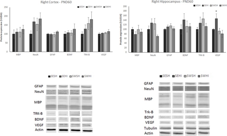
Introduction: Prematurity, through brain injury and altered development is a major cause of neurological impairments and can result in motor, cognitive and behavioral deficits later in life. Presently, there are no well-established effective therapies for preterm brain injury and the search for new strategies is needed. Intra-uterine environment plays a decisive role in brain maturation and interventions using the gestational window have been shown to influence long-term health in the offspring. In this study, we investigated whether pregnancy swimming can prevent the neurochemical metabolic alterations and damage that result from postnatal hypoxic-ischemic brain injury (HI) in very immature rats. Methods: Female pregnant Wistar rats were divided into swimming (SW) or sedentary (SE) groups. Following a period of adaptation before mating, swimming was performed during the entire gestation. At postnatal day (PND3), rat pups from SW and SE dams had right common carotid artery occluded, followed by systemic hypoxia. At PND4 (24 h after HI), the early neurochemical profile was measured by 1H-magnetic resonance spectroscopy. Astrogliosis, apoptosis and neurotrophins protein expression were assessed in the cortex and hippocampus. From PND45, behavioral testing was performed. Diffusion tensor imaging and neurite orientation dispersion and density imaging were used to evaluate brain microstructure and the levels of proteins were quantified. Results: Pregnancy swimming was able to prevent early metabolic changes induced by HI preserving the energetic balance, decreasing apoptotic cell death and astrogliosis as well as maintaining the levels of neurotrophins. At adult age, swimming preserved brain microstructure and improved the performance in the behavioral tests. Conclusion: Our study points out that swimming during gestation in rats could prevent prematurity related brain damage in progeny with high translational potential and possibly interesting cost-benefits. HIGHLIGHTS - Prematurity is a major cause of neurodevelopmental impairments;- Swimming during pregnancy reduces brain damage after HI injury;- Pregnancy is an important but underestimated preventive window.
Keywords: brain; hypoxia-ischemia; magnetic resonance imaging; neuroprotection; pregnancy swimming; prematurity.
Figures


Similar articles
-
Dose-Dependent Neuroprotective Effects of Bovine Lactoferrin Following Neonatal Hypoxia-Ischemia in the Immature Rat Brain.Nutrients. 2021 Oct 29;13(11):3880. doi: 10.3390/nu13113880. Nutrients. 2021. PMID: 34836132 Free PMC article.
-
Previous adaptation triggers distinct molecular pathways and modulates early and long-term neuroprotective effects of pregnancy swimming preventing neonatal hypoxia-ischemia damage in rats.Brain Res. 2020 Apr 15;1733:146722. doi: 10.1016/j.brainres.2020.146722. Epub 2020 Feb 8. Brain Res. 2020. PMID: 32045594
-
Pregnancy swimming prevents early brain mitochondrial dysfunction and causes sex-related long-term neuroprotection following neonatal hypoxia-ischemia in rats.Exp Neurol. 2021 May;339:113623. doi: 10.1016/j.expneurol.2021.113623. Epub 2021 Jan 30. Exp Neurol. 2021. PMID: 33529673
-
Metabolic Alterations in Developing Brain After Injury: Knowns and Unknowns.Neurochem Res. 2015 Dec;40(12):2527-43. doi: 10.1007/s11064-015-1600-7. Epub 2015 Jul 7. Neurochem Res. 2015. PMID: 26148530 Free PMC article. Review.
-
Why considering sexual differences is necessary when studying encephalopathy of prematurity through rodent models.Eur J Neurosci. 2020 Jul;52(1):2560-2574. doi: 10.1111/ejn.14664. Epub 2020 Jan 20. Eur J Neurosci. 2020. PMID: 31885096 Review.
Cited by
-
Early Neuroprotective Effects of Bovine Lactoferrin Associated with Hypothermia after Neonatal Brain Hypoxia-Ischemia in Rats.Int J Mol Sci. 2023 Oct 25;24(21):15583. doi: 10.3390/ijms242115583. Int J Mol Sci. 2023. PMID: 37958562 Free PMC article.
-
Dose-Dependent Neuroprotective Effects of Bovine Lactoferrin Following Neonatal Hypoxia-Ischemia in the Immature Rat Brain.Nutrients. 2021 Oct 29;13(11):3880. doi: 10.3390/nu13113880. Nutrients. 2021. PMID: 34836132 Free PMC article.
-
Mild Neonatal Brain Hypoxia-Ischemia in Very Immature Rats Causes Long-Term Behavioral and Cerebellar Abnormalities at Adulthood.Front Physiol. 2019 Jun 5;10:634. doi: 10.3389/fphys.2019.00634. eCollection 2019. Front Physiol. 2019. PMID: 31231232 Free PMC article.
-
Nutritional Intervention for Developmental Brain Damage: Effects of Lactoferrin Supplementation in Hypocaloric Induced Intrauterine Growth Restriction Rat Pups.Front Endocrinol (Lausanne). 2019 Feb 8;10:46. doi: 10.3389/fendo.2019.00046. eCollection 2019. Front Endocrinol (Lausanne). 2019. PMID: 30800096 Free PMC article.
References
LinkOut - more resources
Full Text Sources
Other Literature Sources