

Neurotrophic keratitis: current challenges and future prospects
- PMID: 29988739
- PMCID: PMC6029608
- DOI: 10.2147/EB.S117261
Neurotrophic keratitis: current challenges and future prospects
Abstract
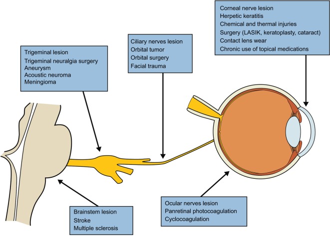
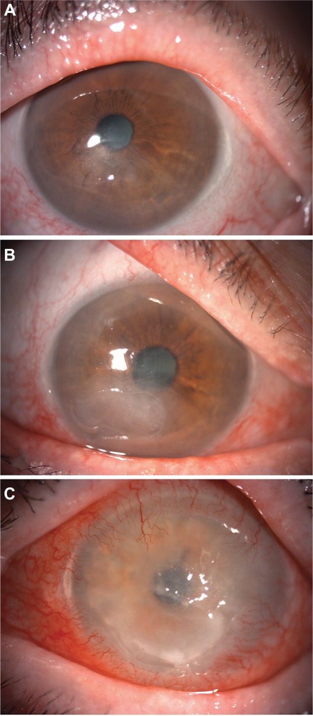
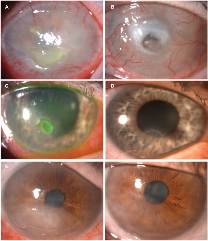
Neurotrophic keratitis (NK) is a degenerative corneal disease caused by damage of trigeminal corneal innervation, which leads to spontaneous epithelial breakdown and corneal ulceration. The impairment of corneal sensory innervation causes the reduction of both protective reflexes and trophic neuromodulators that are essential for the vitality, metabolism, and wound healing of ocular surface tissues. A wide range of ocular and systemic conditions, including herpetic keratitis, ocular chemical burns, corneal surgery, diabetes, multiple sclerosis, and neurosurgical procedures, can cause NK by damaging trigeminal innervation. Diagnosis of NK requires careful investigation of any ocular and systemic condition associated with the disease, complete ocular surface examination, and quantitative measurement of corneal sensitivity. The clinical stages of NK range from corneal epithelial alterations (stage 1) to persistent epithelial defect (stage 2) and ulcer (stage 3), which may progress to corneal perforation. Management of NK is based on clinical severity, and the aim of the therapy is to halt the progression of corneal damage and promote epithelial healing. Although several medical and surgical treatments have been proposed, no therapies are currently available to restore corneal sensitivity, and thus, NK remains difficult and challenging to treat. The purpose of this review is to summarize available evidence on the pathogenesis, diagnosis, and treatment of NK. Novel medical and surgical therapies including the topical administration of nerve growth factor and corneal neurotization are also described.
Keywords: corneal nerves; neurotrophic corneal ulcer; neurotrophic keratitis.
Conflict of interest statement
Disclosure The authors report no conflicts of interest in this work.
Figures
References
-
- Mackie IA. Neuroparalytic keratitis. In: Fraunfelder F, Roy FH, Meyer SM, editors. Current Ocular Therapy. Philadelphia, PA, USA: WB Saunders; 1995.
-
- Bonini S, Lambiase A, Rama P, Caprioglio G, Aloe L. Topical treatment with nerve growth factor for neurotrophic keratitis. Ophthalmology. 2000;107(7):1347–1351. - PubMed
-
- Hsu HY, Modi D. Etiologies, quantitative hypoesthesia, and clinical outcomes of neurotrophic keratopathy. Eye Contact Lens. 2015;41(5):314–317. - PubMed
-
- Muller LJ, Marfurt CF, Kruse F, Tervo TM. Corneal nerves: structure, contents and function. Exp Eye Res. 2003;76(5):521–542. - PubMed
-
- Rozsa AJ, Beuerman RW. Density and organization of free nerve endings in the corneal epithelium of the rabbit. Pain. 1982;14(2):105–120. - PubMed
Publication types
LinkOut - more resources
Full Text Sources
Other Literature Sources
