. 2018 Feb 14:9:1-12.
doi: 10.1016/j.omto.2018.02.001. eCollection 2018 Jun 29.

Oncolytic Immunotherapy for Bladder Cancer Using Coxsackie A21 Virus

Affiliations

Oncolytic Immunotherapy for Bladder Cancer Using Coxsackie A21 Virus

Nicola E Annels et al. Mol Ther Oncolytics. .

Abstract

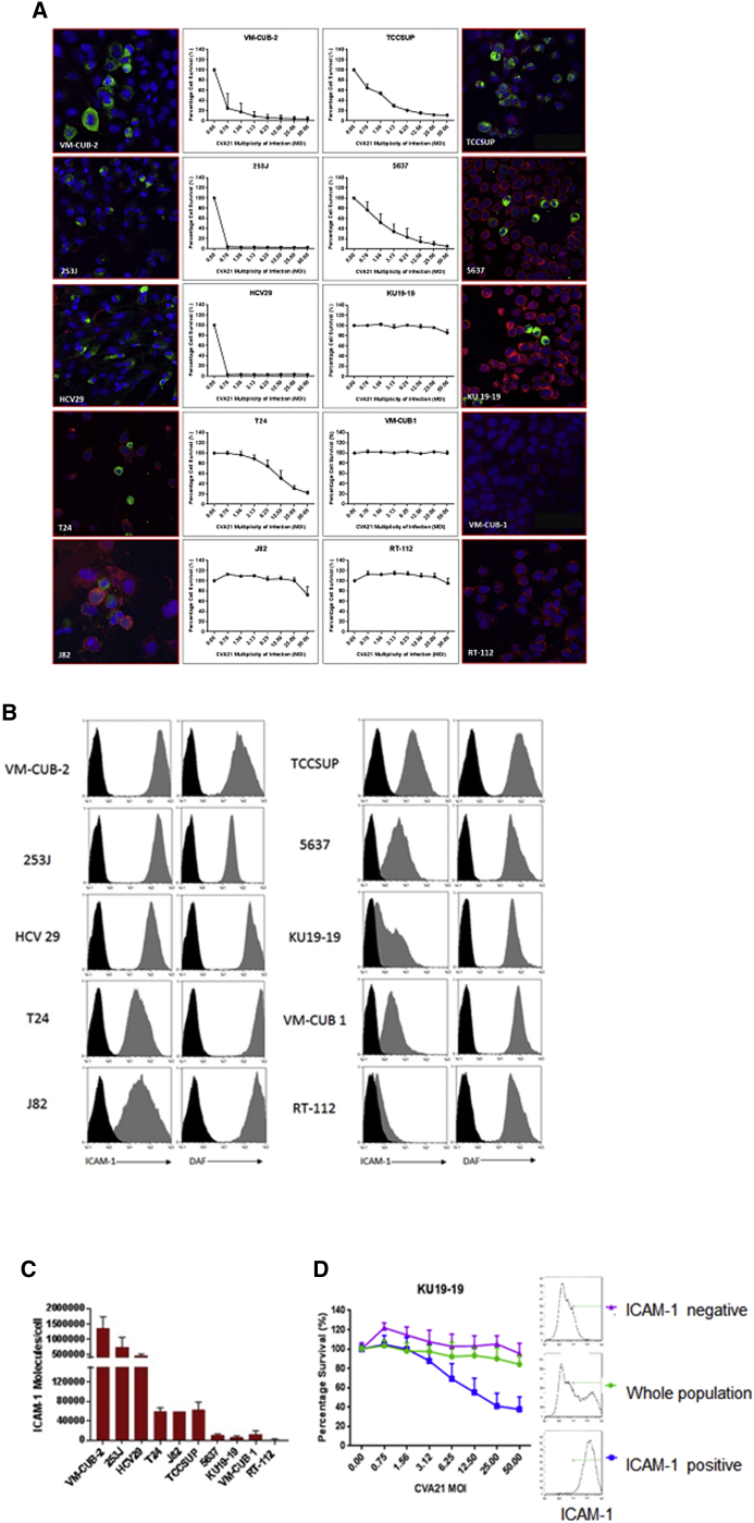
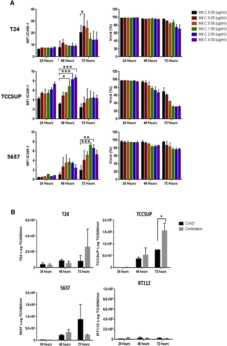
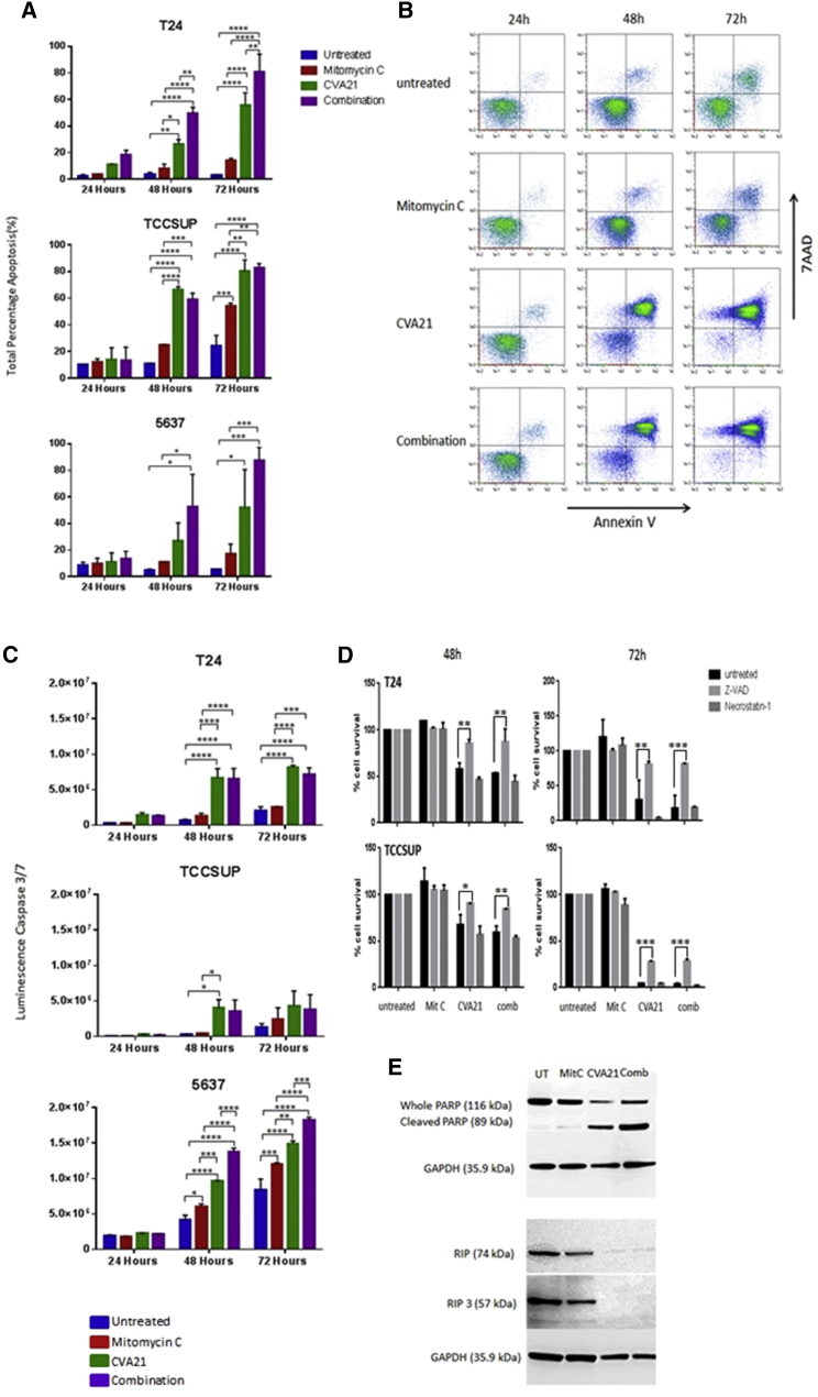
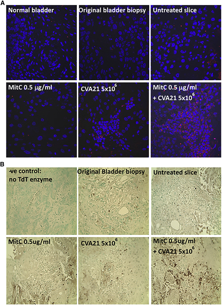
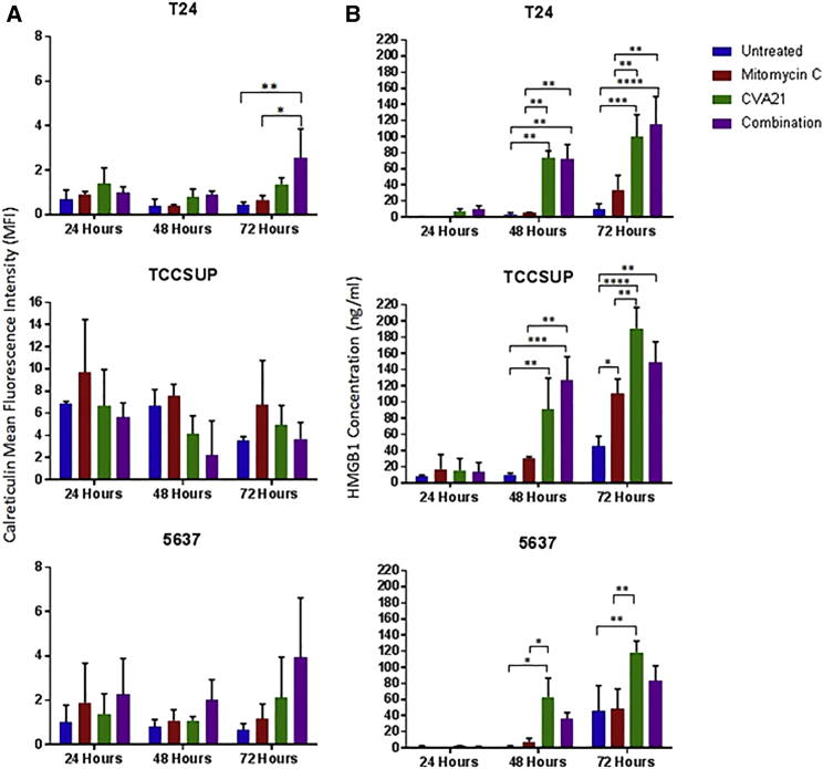
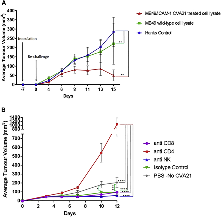
As a clinical setting in which local live biological therapy is already well established, non-muscle invasive bladder cancer (NMIBC) presents intriguing opportunities for oncolytic virotherapy. Coxsackievirus A21 (CVA21) is a novel intercellular adhesion molecule-1 (ICAM-1)-targeted immunotherapeutic virus. This study investigated CVA21-induced cytotoxicity in a panel of human bladder cancer cell lines, revealing a range of sensitivities largely correlating with expression of the viral receptor ICAM-1. CVA21 in combination with low doses of mitomycin-C enhanced CVA21 viral replication and oncolysis by increasing surface expression levels of ICAM-1. This was further confirmed using 300-μm precision slices of NMIBC where levels of virus protein expression and induction of apoptosis were enhanced with prior exposure to mitomycin-C. Given the importance of the immunogenicity of dying cancer cells for triggering tumor-specific responses and long-term therapeutic success, the ability of CVA21 to induce immunogenic cell death was investigated. CVA21 induced immunogenic apoptosis in bladder cancer cell lines, as evidenced by expression of the immunogenic cell death (ICD) determinant calreticulin, and HMGB-1 release and the ability to reject MB49 tumors in syngeneic mice after vaccination with MB49 cells undergoing CVA21 induced ICD. Such CVA21 immunotherapy could offer a potentially less toxic, more effective option for the treatment of bladder cancer.

Keywords: bladder cancer; coxsackievirus A21; intercellular adhesion molecule-1.


Figures

Figure 1
Figure 1
Susceptibility of Bladder Cancer Cell Lines to CVA21 Infection and Expression Profile of Surface ICAM-1 and DAF on Bladder Cancer Cell Lines (A) Monolayer cultures of human bladder cancer cells were challenged with increasing multiplicities of CVA21 and assessed for cell survival at 72 hr post-infection, with live cells being quantified by MTS assay. Confocal images of human bladder cancer cell lines 24 hr post-CVA21 infection are shown (green, CVA21 viral proteins; red, wheat germ agglutinin; blue, nuclei stained with TO-PRO-3). Magnification 40× is shown. (B) Surface expression of ICAM-1 and DAF on bladder cancer cell lines was determined by flow cytometry. Cell lines were incubated with the relevant PE-conjugated isotype control antibody (black histogram), anti-DAF monoclonal antibody, or anti-ICAM-1 monoclonal antibody (gray histogram). (C) Absolute numbers of ICAM-1 molecules on bladder cancer cells were determined by QuantiBRITE PE analysis. (D) KU19-19 bladder cancer cells were stained with an anti-ICAM-1-PE antibody and sorted into ICAM-1-positive or negative populations using magnetic enrichment of PE-positive cells. Together with the whole unsorted KU19-19 population, the different fractions were challenged with increasing multiplicities of CVA21 and assessed for cell survival at 72 hr post-infection, with live cells being quantified by MTS assay.
Figure 2
Figure 2
The Effects of Mitomycin-C on Bladder Cancer Cells and Viral Replication (A) Low doses of Mitomycin-C upregulate ICAM-1 protein expression with minimal cytotoxic effects. Bladder cancer cell lines were either left untreated or treated with Mitomycin-C at increasing doses for one hour before determining the mean fluorescence intensity (MFI) of ICAM-1 expression by flow cytometry after 24, 48, and 72 hr. The cells were also assessed for any cytotoxicity using the live/dead cell discriminator, ViViD. Data represent averaged results from two independent experiments, and two-way ANOVA statistical analysis was performed. *p < 0.05; **p < 0.01; ***p < 0.001; ****p < 0.0001. (B) Effect of Mitomycin-C on CVA21 replication in bladder cancer cell lines is shown. Bladder cancer cell lines were treated with or without 0.5 μg/mL Mitomycin-C for 1 hr before infecting with CVA21 at MOI of 11.44 for T24, 1.0 for TCCSUP, 1.8 for 5637, and 25 for RT112. Supernatants were harvested 24, 48, and 72 hr after infection, and viral titers were measured by standard plaque assays on SKMEL-28 cell monolayers. Graphs represent pooled data from two independent experiments. *p < 0.05.
Figure 3
Figure 3
CVA21-Induced Cell Death of Bladder Cancer Cell Lines (A) Determination of levels of apoptosis in three bladder cancer cell lines left untreated or treated with Mitomycin-C, CVA21, or the combination at 24, 48, and 72 hr post-treatment by means of annexin-V- and 7AAD-based flow cytometry. Graphs represent pooled data from two independent experiments, and the statistical significance of the treatment group comparisons was analyzed using two-way ANOVA; *p < 0.05; **p < 0.01; ***p < 0.001; ****p < 0.0001. (B) Representative dot plots recorded for TCCSUP cells following the different treatments at 24, 48, and 72 hr post-treatment are shown. (C) Apoptosis (quantified via caspase 3/7 activity) in three bladder cancer cell lines left untreated or treated with Mitomycin-C, CVA21, or the combination at 48 and 72 hr post-treatment is shown. Graphs represent pooled data from two independent experiments, and the statistical significance of the treatment group comparisons was analyzed using two-way ANOVA; *p < 0.05; **p < 0.01; ***p < 0.001; ****p < 0.0001. (D) Increase in percentage cell survival was seen in CVA21 and combination-treated cells with Z-VAD-FMK, apoptosis inhibitor, whereas the necroptosis inhibitor, necrostatin-1, failed to increase cell survival in bladder cancer cell lines treated with CVA21 alone or in combination with Mitomycin-C. Graphs represent pooled data from two independent experiments, and the statistical significance of the treatment group comparisons was analyzed using two-way ANOVA; *p < 0.05; **p < 0.01; ***p < 0.001; ****p < 0.0001. (E) Western blot analysis demonstrating absence of RIP1 and RIP3 necroptosis markers but upregulation of cleaved PARP in TCCSUP cells 72 hr post-CVA21 infection is shown.
Figure 4
Figure 4
Enhanced Cytotoxicity of Mitomycin-C and CVA21 on Bladder Cancer Tumor Slices Precision-cut slicing of resection material from a patient with squamous bladder cancer was performed using a vibratome. The 300-μm slices were either left untreated or treated with Mitomycin-C, CVA21, or the combination and then cultured for 48 hr. Slices were then fixed, paraffin embedded, and 4-μm sections prepared. These sections together with a section from the original biopsy were stained for (A) presence of viral protein (red) and the nuclear stain TO-PRO-3 (blue) and (B) evidence of apoptosis using the TumorTACS in situ apoptosis detection kit (apoptotic cells are identified by the dark brown staining). Magnification 40× is shown.
Figure 5
Figure 5
Selective Induction of HMGB1 and Calreticulin by CVA21 Infection of Bladder Cancer Cells The bladder cancer cell lines, T24, TCCSUP, and 5637, were either left untreated or treated with Mitomycin-C (0.5 μg/mL), CVA21, or the combination. Cells and supernatants were harvested at 24, 48, and 72 hr time points. Cell surface calreticulin (CRT) exposure was determined by FACS analysis of the cells (A) whereas HMGB1 accumulation was determined by ELISA analysis of supernatants (B). Graphs represent pooled data from two independent experiments, and the statistical significance of the treatment group comparisons was analyzed using two-way ANOVA; *p < 0.05; **p < 0.01; ***p < 0.001; ****p < 0.0001.
Figure 6
Figure 6
CVA21-Induced ICD Effectively Vaccinates Mice (A) MB49 cells transfected with human ICAM-1 were treated with or without CVA21 for 24 hr, washed, and lysates prepared. These lysates along with HANKS media as a control were injected subcutaneously into one flank of immunocompetent syngeneic C57BL/6 mice (8 per group). One week later, mice were re-challenged with living MB49 cells, which were inoculated subcutaneously (s.c.) into the contralateral flank. Tumor incidence and growth were routinely monitored. Graphs represent pooled data from two independent experiments, and the means ± SDs of 8 mice per group are shown. Two-way ANOVA statistical analysis was performed. *p < 0.05; **p < 0.01; ***p < 0.001; ****p < 0.0001. (B) Depletion of CD8+, CD4+, and NK cells in C57BL/6 mice vaccinated with MB49/ICAM-1 lysates is shown. Depletions were conducted by intraperitoneal injections of purified mAbs 1 day before re-challenge with viable MB49 cells as well as throughout the duration of the experiment. Control mice had been vaccinated with wild-type MB49 cell lysate before re-challenge with viable MB49 cells and received no immune-cell-depleting antibodies. Tumor incidence and growth were monitored, and the statistical significance of the intergroup comparisons of tumor volumes was analyzed using two-way ANOVA. *p < 0.05; **p < 0.01; ***p < 0.001; ****p < 0.0001.

References

    1. Cancer Research UK (2016). Bladder cancer statistics. http://www.cancerresearchuk.org/health-professional/cancer-statistics/st....
    1. Aldousari S., Kassouf W. Update on the management of non-muscle invasive bladder cancer. Can. Urol. Assoc. J. 2010;4:56–64. - PMC - PubMed
    1. Fuge O., Vasdev N., Allchorne P., Green J.S.A. Immunotherapy for bladder cancer. Res. Rep. Urol. 2015;7:65–79. - PMC - PubMed
    1. Sanofi Pasteur (2016). Sanofi Pasteur statement on discontinuation of BCG. http://www.sanofipasteur.ca/node/50701.
    1. Davies, B. (2016). Sanofi shuts down bladder cancer drug production: inevitable drug shortage to harm patients. https://www.forbes.com/sites/benjamindavies/2016/11/17/sanofi-shuts-down....